

Imaging
 Thoracic imaging in COVID-19
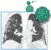
Typical findings include bilateral multifocal parenchymal opacities, predominantly in the peripheral and lower zones.
- Rhinosinusitis and the role of imaging
Imaging should only be used in complicated sinus infections, in recurrent or chronic sinus disease, or in surgical planning.
- Mönckeberg medial sclerosis
A man with chronic kidney disease presents with pain and ulceration of the fingers.
- Vertebral fractures after denosumab cessation
The patient had a 6-month history of lumbar and dorsal back pain, initially suspected to be facet arthrosis.
- Denosumab cessation
Bone loss is rapid if patients miss injections. Clinicians can take extra measures to ensure patients get appropriate treatment.
- Page kidney after a renal biopsy
A patient presents with flank pain and worsening hypertension and renal function 3 weeks after percutaneous renal biopsy.
- Infective endocarditis: Don’t forget the ICE
A recent article did not mention intracardiac echocardiography.
- In reply: Infective endocarditis: Don’t forget the ICE
Echocardiography relies on finding an anatomic abnormality, whereas 18FDG-PET is a functional examination.