Article Text

Abstract
OBJECTIVE To give a comprehensive review of transverse myelopathy (TM), a rare but serious condition reported in 1–2% of patients with systemic lupus erythematosus (SLE).
METHODS 14 patients with SLE and TM were evaluated and 91 additional cases published in the English and German literature reviewed.
RESULTS TM presented either as the initial manifestation or within five years of the diagnosis of SLE. Most patients presented with a detectable sensory deficit at the thoracic level. In our 14 patients, 22% of the patients showed complete neurological recovery, whereas in the total patient population of 105 (our cases plus those reviewed in the literature), complete recovery was observed in 50%, partial recovery in 29% and no improvement or deterioration in 21%. Treatment with intravenous methylprednisolone followed by cyclophosphamide seemed to be most effective. Seventy per cent of the total patient population had abnormal magnetic resonance imaging findings. In our group of 14 patients, those with higher disease activity (measured by the SLAM) at onset of TM were treated more aggressively (for example, with plasmapheresis and intravenous pulse cyclophosphamide). TM in our patients was associated with antiphospholipid antibodies in 43% of the cases as compared with 64% of the total patient population. Optic neuritis occurred in 48% of the total patient population with SLE and TM, suggesting an association.
CONCLUSIONS TM in SLE is a poorly understood entity. Outcome might be more favourable than previously suggested. There is an association of TM with antiphospholipid antibodies in SLE patients. Treatment including intravenous cyclophosphamide may improve the final outcome. This report emphasises the need for multicentre trials to establish guidelines for optimal treatment.
- systemic lupus erythematosus
- transverse myelopathy
Statistics from Altmetric.com
Systemic lupus erythematosus (SLE) is a multisystem autoimmune disease with protean manifestations. Neuropsychiatric manifestations are present in up to 60% of patients with SLE with psychosis, seizures, headaches and cerebrovascular accidents (CVA) being most common.1 ,2 Transverse myelopathy (TM), a process involving the entire thickness of the spinal cord, is a rare event with an estimated incidence of 1.34/million in the general population.3 Potential causes of TM include multiple sclerosis, viral infections (herpes simplex, influenza, Epstein-Barr virus), immunisations (smallpox, influenza) and intoxication (baclofen, penicillins, lead). The usually dramatic presentation with rapidly progressive symptoms involving motor, sensory and autonomic functions including loss of bowel and bladder control makes TM a medical emergency. The pathophysiological mechanism of TM in SLE is uncertain, although vasculitis and arterial thrombosis resulting in ischaemic cord necrosis have been suggested.4 The fact that TM in SLE is rare, having been reported in only 1–2% of patients,1renders the study of this serious entity difficult. We performed a retrospective review of 14 patients with SLE and TM who were seen at our medical centres and also provided a concise review of the literature.
Methods
Out of a total population of approximately 600 patients with SLE seen in two academic institutions, 14 patients with TM were identified. All patients met the diagnostic criteria for SLE as proposed in the American College of Rheumatology revised classification criteria for SLE.5 TM was diagnosed by clinical manifestation indicative of a spinal cord lesion with associated sensory or motor deficits, bladder/bowel dysfunction, or any combination thereof.
Patient data were collected from inpatient and outpatient medical records at Albert Einstein Medical Center and Thomas Jefferson University Hospital. It included clinical manifestations of SLE and TM, clinical course and laboratory data such as serology, antiphospholipid antibodies and magnetic resonance imaging (MRI) findings. Disease activity was determined using the Systemic Lupus Activity Measure (SLAM).6 Patients were divided into three treatment groups: group A received intravenous methylprednisolone (MP) 1 g daily for three days followed by oral prednisone (n=5); group B received intravenous MP 1 g daily for three days followed by intravenous cyclophosphamide (CP) up to 1 g/m2 body surface area (n=4); group C received intravenous MP 1 g daily for three days followed by five days of plasmapheresis with four exchanges of plasma/day and intravenous CP as in group B (n=4). All patients were treated with tapering doses of oral corticosteroids.
Thirty five publications of case reports and case series from the English and German literature were reviewed. These publications dated from 1972 to 1999 and included 91 cases.4 ,7-39Demographic data as well as data on presentation, treatment and outcome were extracted from the literature. Patients in studies before 1982 were diagnosed as having SLE according to the preliminary criteria for the classification of SLE from 1972.40 In total, we report on 105 cases of TM in patients with SLE.
Results
Table 1 shows the demography of our patient population compared with the total patient population (our patients plus the patients from the literature). The age of our patient group was slightly older on average compared with the total patient population (demographic data of seven patients from the literature were not available). There were nine male patients (9%) with TM identified in the total patient population. This is consistent with the percentage of male patients encountered in the general population of patients with SLE. In most patients, TM presented either as the initial manifestation of SLE or within five years of the diagnosis of SLE. In our patients, nine (64%) of the patients already had a diagnosis of SLE before the first episode of TM compared with 54 (54%) in the total patient population. Seventy nine per cent of the total patient population had only one episode of TM during the period of observation, 14% of patients had two episodes and 7% had more than two episodes.
Patient details
The clinical and laboratory features of our patients are shown in tables 2 and 3, respectively. Most of our patients had a detectable sensory deficit at the thoracic level with T7 being the most common. Three patients (patients 1, 3 and 11) did not present with a level of sensory deficit. It is of interest to note that the MRI of patient 1 was consistent with TM although a sensory level was not detectable. The most common levels of involvement in the total population were T5–T8.
Clinical features of our patients
Laboratory features of our patients
It has been postulated that patients with TM frequently suffer from other neurological manifestations of SLE.10 ,37 Three (21%) of our patients also suffered from optic neuritis. This number is lower that that in the total patient population, where 27 (48%) of TM patients also had optic neuritis. Unfortunately in only 55 of the 105 cases was the subject of optic neuritis considered. Although cranial neuropathies are present in 3–16% of SLE patients, the exact incidence of optic neuritis is unknown but it probably is much less frequent than in patients with SLE and TM. Other neurological manifestations in our patients included psychosis/depression (three patients), cerebrovascular accident (two patients), seizures (one patient) and pseudotumour cerebri (one patient).
Several reports indicate that there is a significant association of TM and the presence of antiphospholipid antibodies (aPL).19 ,26 ,38 ,41 In our patients, six of 11 patients (55%) tested were positive for either anticardiolipin antibodies (aCL) or lupus anticoagulant (LAC). In only 64 of the 105 total patient population were data on aPL available. Forty one (64%) of these 64 patients had aPL present during their episodes of TM. The prevalence of aPL in SLE (including asymptomatic patients) is approximately 30–50%.42 The incidence of aPL in SLE patients with TM is somewhat higher than that in the general SLE patient population.
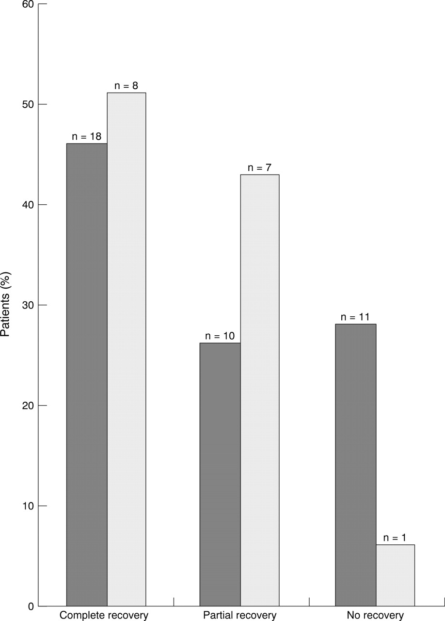
MRI is considered the diagnostic method of choice to confirm TM.18 ,43 ,44 MRI results were available in 55 of the 105 total patient population. Seven (54%) of our patients had a MRI consistent with TM, while 39 (70%) of all 55 patients had abnormal MRI findings. Figure 1 shows a representative MRI (from patient 2) with increased signal intensity of the mid to lower thoracic spinal cord and atrophy (attenuation) of the spinal cord. We attempted to correlate patient outcome with MRI findings in the total patient population (fig2). Complete resolution occurred at approximately the same frequency in patients with abnormal MRI (46 %) and normal MRI (51%). However, 28% of the patients with an abnormal MRI remained paraplegic whereas only 6% of the patients with a normal MRI remained paraplegic. This indicates that more patients with an abnormal MRI had an unfavourable outcome.

MRI from one of our patients (patient 2) showing increased signal intensity of the mid to lower thoracic spinal cord and atrophy (attenuation) of the spinal cord.
MRI results compared with outcome. Data from patients with an abnormal MRI (n=39) are shown in black columns, those of patients with a normal MRI (n=16) in gray columns.
In our patients only three (22%) patients had complete resolution of their symptoms whereas nine (64%) patients remained unchanged and two (14%) patients had partial recovery (table 4). However, the overall outcome in the total patient population with SLE and TM was more favourable. Of these 105 patients, outcome data were available in 86 cases. In one study,19 treatment and outcome were not considered and several other patients died early in the course of the disease. Complete recovery occurred in 50%, partial recovery in 29% and no improvement or deterioration was observed in 21%.
Disease activity, treatment and outcome of our patients with SLE and TM
As TM in SLE is a very rare manifestation, treatment guidelines for this entity have not been developed. In the older studies,9 ,12 ,15 most patients were treated with intravenous corticosteroids alone, whereas more recently some centres prefer a more aggressive approach with intravenous corticosteroids plus intravenous CP.21 ,32 ,45 ,46Plasmapheresis has been used to complement this treatment regimen,25 and it was also performed on some of our patients. It is difficult to draw conclusions on the different forms of treatment for TM from literature reports presented over a long time span. We therefore attempted to identify tendencies with regards to outcome for different treatment regimens. We observed that treatment with intravenous MP alone led to significant recovery. The use of intravenous MP followed by intravenous CP seemed to be more effective than intravenous MP alone. Additional treatment with plasmapheresis in patients that did not improve with intravenous MP and intravenous CP did not seem to improve outcome further. No unusual features could be identified in the patients who did not respond to treatment.
It would be of interest to correlate the activity of SLE at onset of TM with the patients' outcome. Unfortunately, the calculation of the SLAM from the cases derived from the literature was not possible because of the lack of important clinical data. In our patient group, patients with a higher SLAM score were generally treated more aggressively (table 4). However, more aggressive treatment was not associated with better clinical outcome, as all patients in treatment group C remained paraplegic.
Discussion
TM is a rare, but serious complication of SLE. Previous reports indicated a prevalence of about 1–2%.1 This was confirmed in our patient population where TM occurred in about 2% of patients. This paper is the first attempt to analyse all cases of TM in patients with SLE published in the literature. We include the presentation of TM in 14 patients with SLE from two academic centres as well as 91 cases reported in the published literature.
There have been conflicting data in the literature about the timing of TM compared with the onset of SLE. From our study, TM occurring as the initial manifestation of SLE is rather common (39% of the total patient population). In 42% of the total patient population analysed, TM presented within the first five years of a diagnosis with SLE. The prominent presentation of TM in SLE seems to be a sensory level, predominantly in the thoracic area.
Surprisingly, the outcome of TM in SLE in the total patient population was favourable. This is in contrast with several reports,8 ,30 ,43 ,47 including our own patients where the overall outcome was generally unfavourable. It has been postulated30 that aggressive treatment early in the course of the disease is crucial for a favourable response. This could not be confirmed in our patients, where most were treated soon after the onset of symptoms. Furthermore, outcome in general could not be related to disease activity. The evaluation of the total patient population of 105 cases indicates that the clinical manifestations of TM may improve with the use of intravenous MP, but that results are even more favourable if intravenous MP is followed by intravenous CP. It is unclear whether plasmapheresis has any additional therapeutic benefit. In our patients, those with higher SLE disease activity were treated with a more aggressive regimen. This, however, was not associated with a better outcome. As there are no therapeutic guidelines for TM in SLE, clinicians are probably inclined to use more aggressive treatment in patients that present with severe neurological deficits and multisystem disease. It remains to be seen whether plasmapheresis has any role in the treatment of TM in the future.
It is of interest that the prevalence of aPL seems to be higher in SLE patients with TM compared with SLE patients in general. Lavalleet al 19 report 10 of 11 patients with TM and SLE as having aPL. Of the 64 out of a total of 105 patients tested for aPL, 64% of all patients tested were positive for aPL. This might be important for the aetiology of TM, as spinal cord necrosis secondary to arterial thrombosis might be a pathological factor. Unfortunately, necropsy material from patients with TM and SLE is limited. In two studies,8 ,9 necrosis of the spinal cord was present at necropsy, but thrombosis of the spinal vessels was not seen. A spinal vessel thrombosis might explain the predominance of TM in the thoracic spine. The blood supply of the spinal cord relies on three vessels: the anterior median longitudinal arterial trunk and a pair of posterolateral trunks near the posterior nerve rootlets. The longitudinal arterial trunks are largest in the cervical and lumbar regions and are much smaller in the thoracic region. If there were vascular compromise secondary to thrombosis, the thoracic cord would be most vulnerable because of the limited blood supply. The early use of anticoagulation in these patients might be important for the outcome of the disease. This aspect of TM in SLE has not been studied. We identified six patients with either LAC or aCL in our patients. Only two of these patients (patients 7 and 11, respectively), were treated with anticoagulation before the onset of TM. Both patients remained paraplegic. Three more patients received anticoagulation once their LAC/aCL status was known. Of these, two remained paraplegic and one had a residual sensory deficit. Furthermore, only aCL and LAC are routinely studied in SLE patients. It is possible that other antibodies with hypercoagulable properties that are not routinely tested, play a part in the disease process. Recently, anti-annexin V,48anti-activated protein C49 and anti-oxidised low density lipoprotein antibodies50 have been associated with arterial and venous thrombosis.
Also of interest is the association of optic neuritis with TM in patients with SLE. The association of TM and optic neuritis was first described by Devic in 1894.51 It has been postulated that optic neuritis in SLE is associated with aPL.26 In our patients, there was no association of aPL with optic neuritis or any other neurological manifestations.
In summary, we have provided the clinical features, treatment and outcome of 14 cases of SLE related TM as well as the review of 91 additional cases previously described in the literature. Although we attempted to report as completely as possible on the 105 cases that were available for review, important data were often not available in the literature reports. This pertains especially to the disease activity of SLE. Our references date back to 1972, when certain diagnostic studies (for example, MRI, antiphospholipid antibody tests) or treatment options (for example, intravenous CP) were not available. Furthermore, there are several studies in the non-English literature that could not be included in our review. While the overall outcome of TM in SLE seems to be better than previously thought, the optimal treatment regimen of this condition is not certain although patients seem to benefit from intravenous CP in addition to intravenous MP with tapering doses or oral corticosteroids. Controlled studies and perhaps the creation of a registry for SLE patients with TM could help to develop an effective treatment strategy and shed more light on this rare but serious condition.
Acknowledgments
We thank Dr Ioannis Tassiulas for a critical review of the manuscript.
References
Footnotes
Funding: this work was supported in part by the Lupus Foundation of Philadelphia, the Gilbert Memorial Trust and the Pearl Lisker Memorial Research Foundation.