
Main menu

  • Home
  • Content
    • Current Issue
    • Ahead of Print
    • Past Issues
    • Supplements
    • Article Type
  • Specialty
    • Articles by Specialty
  • CME/MOC
    • Articles
    • Calendar
  • Info For
    • Manuscript Submission
    • Authors & Reviewers
    • Subscriptions
    • About CCJM
    • Contact Us
    • Media Kit
  • Conversations with Leaders
  • Conference Coverage
    • Kidney Week 2024
    • CHEST 2024
    • ACR Convergence 2023
    • Kidney Week 2023
    • ObesityWeek 2023
    • IDWeek 2023
    • CHEST 2023
    • MDS 2023
    • IAS 2023
    • ACP 2023
    • AAN 2023
    • ACC / WCC 2023
    • AAAAI Meeting 2023
    • ACR Convergence 2022
    • Kidney Week 2022
    • AIDS 2022
  • Other Publications
    • www.clevelandclinic.org

User menu

  • Register
  • Log in

Search

  • Advanced search
Cleveland Clinic Journal of Medicine
  • Other Publications
    • www.clevelandclinic.org
  • Register
  • Log in
Cleveland Clinic Journal of Medicine

Advanced Search

  • Home
  • Content
    • Current Issue
    • Ahead of Print
    • Past Issues
    • Supplements
    • Article Type
  • Specialty
    • Articles by Specialty
  • CME/MOC
    • Articles
    • Calendar
  • Info For
    • Manuscript Submission
    • Authors & Reviewers
    • Subscriptions
    • About CCJM
    • Contact Us
    • Media Kit
  • Conversations with Leaders
  • Conference Coverage
    • Kidney Week 2024
    • CHEST 2024
    • ACR Convergence 2023
    • Kidney Week 2023
    • ObesityWeek 2023
    • IDWeek 2023
    • CHEST 2023
    • MDS 2023
    • IAS 2023
    • ACP 2023
    • AAN 2023
    • ACC / WCC 2023
    • AAAAI Meeting 2023
    • ACR Convergence 2022
    • Kidney Week 2022
    • AIDS 2022
The Clinical Picture

Vulvar and gluteal manifestations of Crohn disease

Anna Camille Moreno, DO, NCMP, Oluwatosin Goje, MD, MSCR, FACOG, Melissa Peck Piliang, MD, FAAD and Pelin Batur, MD, NCMP, CCD
Cleveland Clinic Journal of Medicine October 2019, 86 (10) 645-646; DOI: https://doi.org/10.3949/ccjm.86a.19062
Anna Camille Moreno
Duke Women’s Health Associates, Department of OB/GYN, Duke University Medical Center; Assistant Professor, Duke University, Raleigh, NC
  • Find this author on Google Scholar
  • Find this author on PubMed
  • Search for this author on this site
Oluwatosin Goje
Center for Gynecologic Infectious Disease; Director, Ob/Gyn & Women’s Health Institute, Cleveland Clinic
  • Find this author on Google Scholar
  • Find this author on PubMed
  • Search for this author on this site
Melissa Peck Piliang
Dermatology and Anatomic Pathology; Vice Chair, Education; Associate Program Director, Dermatology Residency; Associate Program Director, Dermatopathology Fellowship, Clevel and ClinicWomen’s Health Institute, A8-406, Cleveland Clinic
  • Find this author on Google Scholar
  • Find this author on PubMed
  • Search for this author on this site
Pelin Batur
Ob/Gyn & Women’s Health Institute, Cleveland Clinic; Associate Professor of Medicine, Cleveland Clinic Lerner College of Medicine of Case Western Reserve University Cleveland, OH; Steering Committee, Women’s Preventive Services Initiative, American College of Obstetricians and Gynecologists and US Department of Health and Human Services, Health Resources & Services Administration; Clinical Guideline Committee of the American College of Physicians
Roles: Deputy Editor
  • Find this author on Google Scholar
  • Find this author on PubMed
  • Search for this author on this site
  • For correspondence: [email protected]

A 37-year-old woman presented with recurring painful swelling and erythema of the vulva over the last year. Despite a series of negative vaginal cultures, she was prescribed multiple courses of antifungal and antibacterial treatments, while her symptoms continued to worsen. She had no other relevant medical history except for occasional diarrhea and abdominal cramping, which were attributed to irritable bowel syndrome.

On examination, she had symmetric edema and erythema of the vulva (Figure 1). Closer inspection revealed a nonulcerated, slightly friable nodule of approximately 4 mm on her right labium minus. A biopsy of this region demonstrated multiple noncaseating granulomas and mixed inflammatory infiltrates. An acid-fast stain for mycobacteria was negative. Vulvar skin ultrasonography demonstrated fistulas and increased dermal thickness with altered subcutaneous tissue. She was encouraged to undergo colonoscopy, which showed findings suggestive of Crohn disease.

Figure 1
  • Download figure
  • Open in new tab
  • Download powerpoint
Figure 1

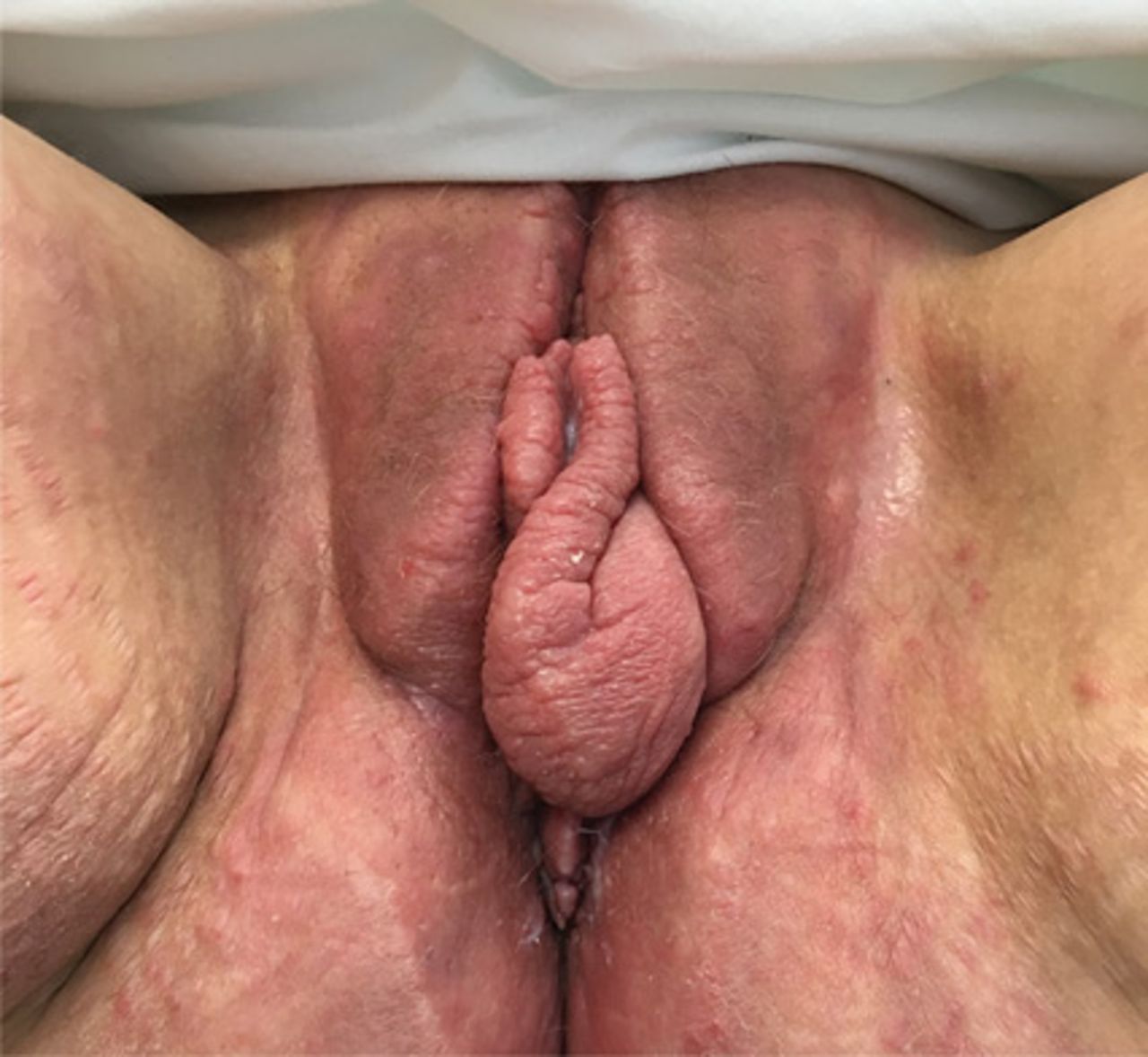
Symmetric edema and erythema of the vulva.

CROHN DISEASE OUTSIDE THE GASTROINTESTINAL TRACT

Crohn disease primarily affects the gastrointestinal tract but is associated with extraintestinal manifestations (in the oral cavity, eyes, skin, and joints) in up to 45% of patients.1

The most common mucocutaneous manifestations are granulomatous lesions that extend directly from the gastrointestinal tract, including perianal and peristomal skin tags, fistulas, and perineal ulcerations. In most cases, the onset of cutaneous manifestations follows intestinal disease, but vulvar Crohn disease may precede gastrointestinal symptoms in approximately 25% of patients, with the average age at onset in the mid-30s.1

The pathogenesis of vulvar Crohn disease remains unclear. One theory involves production of immune complexes from the gastrointestinal tract and a possible T-lymphocyte-mediated type IV hypersensitivity reaction.2

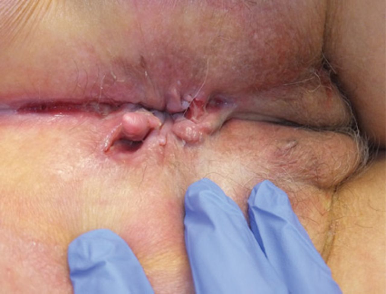
The most commonly reported symptoms include pain, dyspareunia, pruritus, and discharge.1 The classic findings, found in 50% of cases, include perianal and peristomal skin tags, fistulae, perineal ulcers, linear ulcers (resembling knife cuts), abscesses, and fissures.3 Figure 2 shows a linear ulcer in the gluteal cleft, with sharply demarcated borders and resembling a knife cut, in another patient treated at our institution. Associated perianal fissures are also seen, in addition to vulvar edema.4

The diagnosis of vulvar Crohn disease should be considered in a patient who has vulvar pain, edema, and ulcerations not otherwise explained, whether or not gastrointestinal Crohn disease is present. The diagnosis is established with clinical history and characteristic histopathology on biopsy. Multiple biopsies may be needed, and early endoscopy is recommended to establish the diagnosis. The histologic features include noncaseating and nonnecrotizing granulomatous dermatitis or vulvitis with occasional reports of eosinophilic infiltrates and necrobiosis.5,6 An imaging study such as ultrasonography is sometimes used to differentiate between a specific cutaneous manifestation of Crohn disease and its complications such as perianal fistula or abscess.

Figure 2
  • Download figure
  • Open in new tab
  • Download powerpoint
Figure 2

A linear ulcer with sharply demarcated borders in the gluteal cleft in another patient.

Clinical vulvar lesions are nonspecific, and those of Crohn disease are frequently mistaken for infectious, inflammatory, or traumatic vulvitis. Diagnostic biopsy for histologic analysis is warranted.

  • © 2019 The Cleveland Clinic Foundation. All Rights Reserved.

REFERENCES

    1. Andreani SM,
    2. Ratnasingham K,
    3. Dang HH,
    4. Gravante G,
    5. Giordano P
    . Crohn’s disease of the vulva. Int J Surg 2010; 8(1):2–5. doi: 10.1016/j.ijsu.2009.09.012
    1. Siroy A,
    2. Wasman J
    . Metastatic Crohn disease: a rare cutaneous entity. Arch Pathol Lab Med 2012; 136(3):329–332 doi: 10.5858/arpa.2010-0666-RS
    1. Foo WC,
    2. Papalas JA,
    3. Robboy SJ,
    4. Selim MA
    . Vulvar manifestations of Crohn’s disease. Am J Dermatopathol 2011; 33(6):588–593. doi: 10.1097/DAD.0b013e31820a2635
    1. Amankwah Y,
    2. Haefner H
    . Vulvar edema. Dermatol Clin 2010; 28(4):765–777. doi: 10.1016/j.det.2010.08.001
    1. Emanuel PO,
    2. Phelps RG
    . Metastatic Crohn’s disease: a histopathologic study of 12 cases. J Cutan Pathol 2008; 35(5):457–461. doi: 10.1111/j.1600-0560.2007.00849.x
    1. Hackzell-Bradley M,
    2. Hedblad MA,
    3. Stephansson EA
    . Metastatic Crohn’s disease: report of 3 cases with special reference to histopathologic findings. Arch Dermatol 1996; 132(8):928–932. doi: 10.1001/archderm.132.8.928

Navigate

  • Current Issue
  • Past Issues
  • Supplements
  • Article Type
  • Specialty
  • CME/MOC Articles
  • CME/MOC Calendar
  • Media Kit

Authors & Reviewers

  • Manuscript Submission
  • Authors & Reviewers
  • Subscriptions
  • About CCJM
  • Contact Us
  • Cleveland Clinic Center for Continuing Education
  • Consult QD

Share your suggestions!

Copyright © 2025 The Cleveland Clinic Foundation. All rights reserved. The information provided is for educational purposes only. Use of this website is subject to the website terms of use and privacy policy. 

Powered by HighWire