
Main menu

  • Home
  • Content
    • Current Issue
    • Ahead of Print
    • Past Issues
    • Supplements
    • Article Type
  • Specialty
    • Articles by Specialty
  • CME/MOC
    • Articles
    • Calendar
  • Info For
    • Manuscript Submission
    • Authors & Reviewers
    • Subscriptions
    • About CCJM
    • Contact Us
    • Media Kit
  • Conversations with Leaders
  • Conference Coverage
    • Kidney Week 2024
    • CHEST 2024
    • ACR Convergence 2023
    • Kidney Week 2023
    • ObesityWeek 2023
    • IDWeek 2023
    • CHEST 2023
    • MDS 2023
    • IAS 2023
    • ACP 2023
    • AAN 2023
    • ACC / WCC 2023
    • AAAAI Meeting 2023
    • ACR Convergence 2022
    • Kidney Week 2022
    • AIDS 2022
  • Other Publications
    • www.clevelandclinic.org

User menu

  • Register
  • Log in

Search

  • Advanced search
Cleveland Clinic Journal of Medicine
  • Other Publications
    • www.clevelandclinic.org
  • Register
  • Log in
Cleveland Clinic Journal of Medicine

Advanced Search

  • Home
  • Content
    • Current Issue
    • Ahead of Print
    • Past Issues
    • Supplements
    • Article Type
  • Specialty
    • Articles by Specialty
  • CME/MOC
    • Articles
    • Calendar
  • Info For
    • Manuscript Submission
    • Authors & Reviewers
    • Subscriptions
    • About CCJM
    • Contact Us
    • Media Kit
  • Conversations with Leaders
  • Conference Coverage
    • Kidney Week 2024
    • CHEST 2024
    • ACR Convergence 2023
    • Kidney Week 2023
    • ObesityWeek 2023
    • IDWeek 2023
    • CHEST 2023
    • MDS 2023
    • IAS 2023
    • ACP 2023
    • AAN 2023
    • ACC / WCC 2023
    • AAAAI Meeting 2023
    • ACR Convergence 2022
    • Kidney Week 2022
    • AIDS 2022
The Clinical Picture

Hampton hump in acute pulmonary embolism

Sovik De Sirkar, MD, Joshua Newman, MD, Sorcha Allen, MB, BCh, BAO, Ahmed Elkaryoni, MD, Alexandru Marginean, MD and Amir Darki, MD, MSc
Cleveland Clinic Journal of Medicine May 2022, 89 (5) 236-238; DOI: https://doi.org/10.3949/ccjm.89a.21058
Sovik De Sirkar
Department of Internal Medicine, Loyola University Medical Center, Maywood, IL
  • Find this author on Google Scholar
  • Find this author on PubMed
  • Search for this author on this site
  • For correspondence: [email protected]
Joshua Newman
Loyola University Medical Center, Maywood, IL
  • Find this author on Google Scholar
  • Find this author on PubMed
  • Search for this author on this site
Sorcha Allen
Loyola University Medical Center, Maywood, IL
  • Find this author on Google Scholar
  • Find this author on PubMed
  • Search for this author on this site
Ahmed Elkaryoni
Loyola University Medical Center, Maywood, IL
  • Find this author on Google Scholar
  • Find this author on PubMed
  • Search for this author on this site
Alexandru Marginean
Loyola University Medical Center, Maywood, IL
  • Find this author on Google Scholar
  • Find this author on PubMed
  • Search for this author on this site
Amir Darki
Associate Professor of Medicine–Cardiology, Program Director–Interventional Cardiology, Interventional Cardiologist, and Director, Pulmonary Embolism Response Team, Loyola University Medical Center, Maywood, IL
  • Find this author on Google Scholar
  • Find this author on PubMed
  • Search for this author on this site

A 50-year-old woman with a medical history significant for childhood asthma presented to the emergency department with a 3-week history of worsening dyspnea and cough with bilateral lower-extremity swelling, left-side swelling greater than right-side swelling.

On presentation, her heart rate was 121 beats per minute, blood pressure was 197/133 mm Hg, and respiratory rate was 32 breaths per minute with oxygen saturation of 96% on room air. Physical examination was notable for tachycardia and normal S1 and S2 heart sounds without murmurs, rubs, or gallops. Breath sounds were normal bilaterally. Venous Doppler ultrasonography of the left lower extremity revealed acute distal deep vein thrombosis of the posterior tibial and peroneal veins.

Laboratory evaluation revealed the following:

White blood cell count of 13.1 × 10⁹/L (reference range 3.5–10.5) with neutrophilic predominance

  • Hemoglobin of 9.8 g/dL (reference range 11.5–15.5)

  • Platelet count of 588 × 109/L (reference range 150–400)

  • D-dimer of 2,669 ng/mL (reference range < 500).

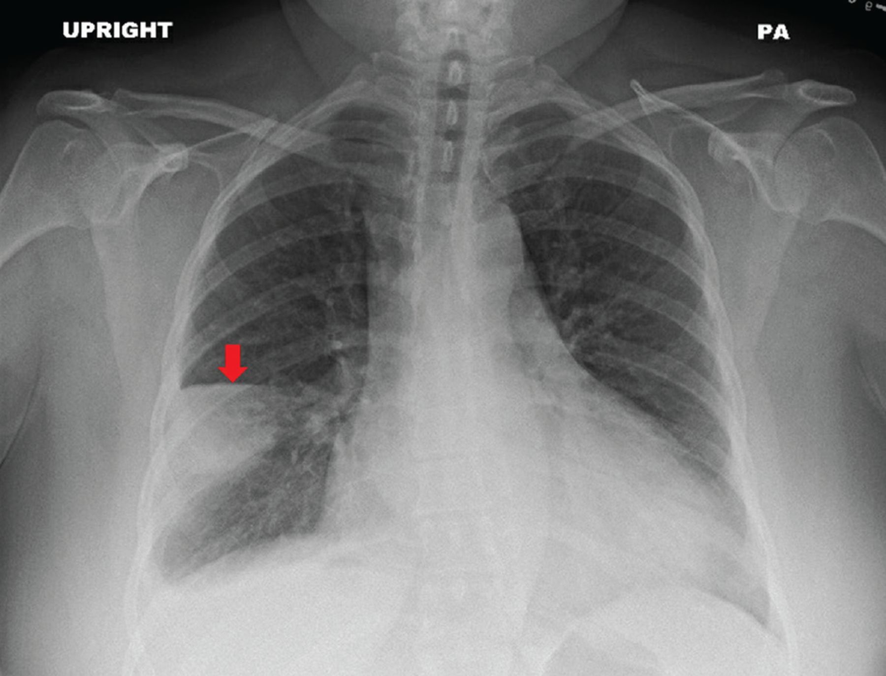
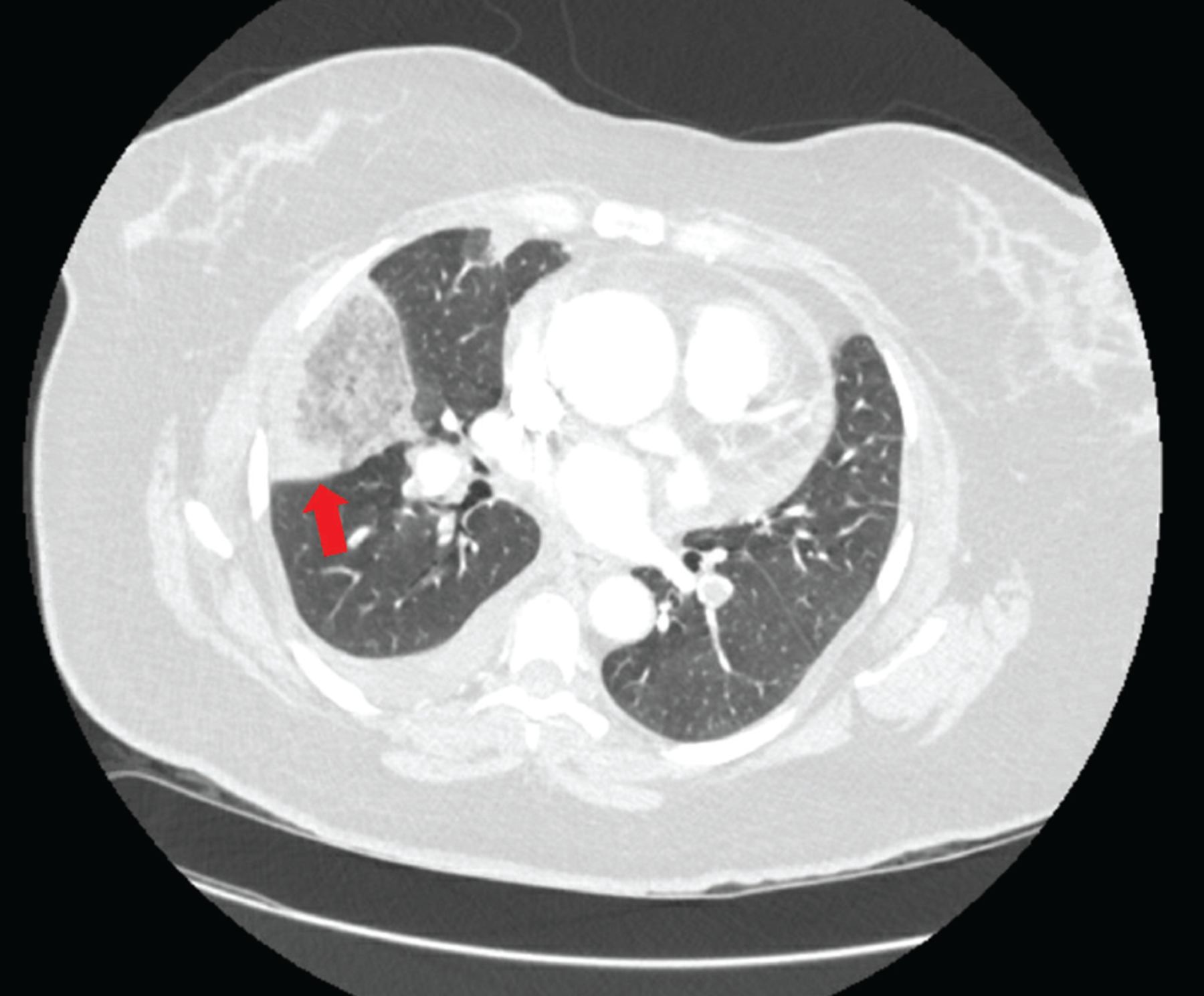
Chest radiography revealed a wedge-shaped opacity in the right lower lobe (Figure 1) concerning for pulmonary infarction. The patient subsequently underwent computed tomographic pulmonary angiography that revealed bilateral segmental and sub-segmental filling defects consistent with acute pulmonary embolism and corresponding opacities in the right and left lower lobes consistent with pulmonary infarction (Figure 2). She was admitted to the hospital, and systemic anti-coagulation was initiated. She ultimately did well and was discharged home. At follow-up 1 month later, her dyspnea had resolved.

Figure 1
  • Download figure
  • Open in new tab
  • Download powerpoint
Figure 1

Posterior-anterior (PA) view of a chest radiograph demonstrated a wedge-shaped opacity (arrow) in the right middle lobe consistent with Hampton hump.

Figure 2
  • Download figure
  • Open in new tab
  • Download powerpoint
Figure 2

Contrast-enhanced axial computed tomographic angiography of the chest in a lung window demonstrated a wedge-shaped opacity (arrow) in the right middle lobe.

HAMPTON HUMP AND PULMONARY INFARCTION

Chest radiography is the initial test of choice when evaluating patients presenting with dyspnea because it is inexpensive, widely available, and can be quickly performed at the bedside. A peripherally located wedge-shaped opacity on chest radiography is referred to as Hampton hump (Figure 1), first described in 1940 by Hampton and Castleman,1,2 who performed an autopsy series to demonstrate the site of opacities seen on chest radiography in patients with pulmonary embolism compared with pulmonary infarction seen at autopsy.

Hampton hump is modestly specific for the diagnosis of pulmonary embolism but lacks sensitivity. In a study evaluating radiographs of patients in the multicenter Prospective Investigation of Pulmonary Embolism Diagnosis trial,3 Hampton hump had a sensitivity of 22% and a specificity of 82%. Computed tomographic pulmonary angiography remains the gold-standard for establishing a diagnosis of pulmonary embolism, with a sensitivity of 89% and a specificity of 95%.4

Pulmonary infarction occurs when blood vessel occlusion results in mismatch of oxygen supply and demand and subsequent hypoxia. This triggers a cascade of pathologic processes culminating in tissue necrosis.5 Pulmonary embolism is a common cause of pulmonary infarction, with an estimated annual incidence of 115 per 100,000 people in the United States.6 The true incidence of subsequent pulmonary infarction is variable. In patients diagnosed with pulmonary embolism, pulmonary infarction has been reported in 15% to 31% of patients on follow-up autopsy and in 9% to 36% of patients on computed tomography.5,7,8

The lungs receive a dual supply of oxygenated blood from the bronchial and pulmonary arteries. In cases of proximal pulmonary embolism, pulmonary infarction is not typically seen owing to the presence of dual circulation.9 However, with more distal pulmonary artery occlusions, a sudden influx of collateral blood flow into small-caliber vessels and increased vascular permeability result in intra-alveolar hemorrhage and infarction.5

Pulmonary infarction: Presentation, risk factors, clinical significance

Clinically, pulmonary infarction can present silently or with any combination of chest pain, syncope, cough, and dyspnea. Significant risk factors include smoking, chronic obstructive pulmonary disease, malignancy, shock, and distal small-artery occlusions.5,8 Advanced age has also historically been considered a risk factor, but recent findings suggest younger patients are at highest risk because of a less-evolved collateral system and higher endogenous nitric oxide levels that produce more vascular anastomoses and influx of bronchial flow.10,11

Little is known about the exact clinical significance of pulmonary infarction. According to limited data, mortality and pulmonary embolism recurrence do not significantly differ among patients with acute pulmonary embolism and ensuing pulmonary infarction compared with those without infarction.12 Long-term consequences such as persistent dyspnea, pleuritic pain, postpulmonary embolism syndrome, and chronic thromboembolic pulmonary hypertension are not well known and should be a focal point of further investigation.

In patients presenting with dyspnea, peripheral wedge-shaped opacity on chest radiography should raise suspicion for pulmonary infarction, warranting further evaluation to diagnose pulmonary embolism.

DISCLOSURES

The authors report no relevant financial relationships which, in the context of their contributions, could be perceived as a potential conflict of interest.

  • Copyright © 2022 The Cleveland Clinic Foundation. All Rights Reserved.

REFERENCES

    1. Hampton A,
    2. Castleman B
    . Correlation of postmortem chest tele-roentgenograms with autopsy findings with special reference to pulmonary embolism and infarction. AJR Am J Roentgenol 1940; 43: 305–326.
    1. McGrath BM,
    2. Groom AG
    . Images in clinical medicine. Hampton’s hump. N Engl J Med 2013; 368(23):2219. doi:10.1056/NEJMicm1204417
    1. Worsley DF,
    2. Alavi A,
    3. Aronchick JM,
    4. Chen JT,
    5. Greenspan RH,
    6. Ravin CE
    . Chest radiographic findings in patients with acute pulmonary embolism: observations from the PIOPED study. Radiology 1993; 189(1):133–136. doi:10.1148/radiology.189.1.8372182
    1. Hogg K,
    2. Brown G,
    3. Dunning J, et al
    . Diagnosis of pulmonary embolism with CT pulmonary angiography: a systematic review. Emerg Med J 2006; 23(3):172–178. doi:10.1136/emj.2005.029397
    1. Kaptein FHJ,
    2. Kroft LJM,
    3. Hammerschlag G, et al
    . Pulmonary infarction in acute pulmonary embolism. Thromb Res 2021; 202:162–169. doi:10.1016/j.thromres.2021.03.022
    1. Wendelboe AM,
    2. Raskob GE
    . Global burden of thrombosis: epidemiologic aspects. Circ Res 2016; 118(9):1340–1347. doi:10.1161/CIRCRESAHA.115.306841
    1. Freiman DG,
    2. Suyemoto J,
    3. Wessler S
    . Frequency of pulmonary thromboembolism in man. N Engl J Med 1965; 272:1278–1280. doi:10.1056/NEJM196506172722406
    1. Tsao MS,
    2. Schraufnagel D,
    3. Wang NS
    . Pathogenesis of pulmonary infarction. Am J Med 1982; 72(4):599–606. doi:10.1016/0002-9343(82)90458-2
    1. Bruzzi JF,
    2. Rémy-Jardin M,
    3. Delhaye D,
    4. Teisseire A,
    5. Khalil C,
    6. Rémy J
    . Multi-detector row CT of hemoptysis. Radiographics 2006; 26(1): 3–22. doi:10.1148/rg.261045726
    1. Islam M,
    2. Filopei J,
    3. Frank M, et al
    . Pulmonary infarction secondary to pulmonary embolism: an evolving paradigm. Respirology 2018. doi:10.1111/resp.13299
    1. Torregrossa AC,
    2. Aranke M,
    3. Bryan NS
    . Nitric oxide and geriatrics: implications in diagnostics and treatment of the elderly. J Geriatr Cardiol 2011; 8(4):230–242. doi:10.3724/SP.J.1263.2011.00230
    1. Cha SI,
    2. Shin KM,
    3. Lee J, et al
    . Clinical relevance of pulmonary infarction in patients with pulmonary embolism. Thromb Res 2012; 130(3):e1–e5. doi:10.1016/j.thromres.2012.03.012

Navigate

  • Current Issue
  • Past Issues
  • Supplements
  • Article Type
  • Specialty
  • CME/MOC Articles
  • CME/MOC Calendar
  • Media Kit

Authors & Reviewers

  • Manuscript Submission
  • Authors & Reviewers
  • Subscriptions
  • About CCJM
  • Contact Us
  • Cleveland Clinic Center for Continuing Education
  • Consult QD

Share your suggestions!

Copyright © 2025 The Cleveland Clinic Foundation. All rights reserved. The information provided is for educational purposes only. Use of this website is subject to the website terms of use and privacy policy. 

Powered by HighWire