
Main menu

  • Home
  • Content
    • Current Issue
    • Ahead of Print
    • Past Issues
    • Supplements
    • Article Type
  • Specialty
    • Articles by Specialty
  • CME/MOC
    • Articles
    • Calendar
  • Info For
    • Manuscript Submission
    • Authors & Reviewers
    • Subscriptions
    • About CCJM
    • Contact Us
    • Media Kit
  • Conversations with Leaders
  • Conference Coverage
    • Kidney Week 2024
    • CHEST 2024
    • ACR Convergence 2023
    • Kidney Week 2023
    • ObesityWeek 2023
    • IDWeek 2023
    • CHEST 2023
    • MDS 2023
    • IAS 2023
    • ACP 2023
    • AAN 2023
    • ACC / WCC 2023
    • AAAAI Meeting 2023
    • ACR Convergence 2022
    • Kidney Week 2022
    • AIDS 2022
  • Other Publications
    • www.clevelandclinic.org

User menu

  • Register
  • Log in

Search

  • Advanced search
Cleveland Clinic Journal of Medicine
  • Other Publications
    • www.clevelandclinic.org
  • Register
  • Log in
Cleveland Clinic Journal of Medicine

Advanced Search

  • Home
  • Content
    • Current Issue
    • Ahead of Print
    • Past Issues
    • Supplements
    • Article Type
  • Specialty
    • Articles by Specialty
  • CME/MOC
    • Articles
    • Calendar
  • Info For
    • Manuscript Submission
    • Authors & Reviewers
    • Subscriptions
    • About CCJM
    • Contact Us
    • Media Kit
  • Conversations with Leaders
  • Conference Coverage
    • Kidney Week 2024
    • CHEST 2024
    • ACR Convergence 2023
    • Kidney Week 2023
    • ObesityWeek 2023
    • IDWeek 2023
    • CHEST 2023
    • MDS 2023
    • IAS 2023
    • ACP 2023
    • AAN 2023
    • ACC / WCC 2023
    • AAAAI Meeting 2023
    • ACR Convergence 2022
    • Kidney Week 2022
    • AIDS 2022
The Clinical Picture

Dancing sternal wires: A radiologic sign of sternal dehiscence

Mohamed M. Gad, MD and Amit Gupta, MD
Cleveland Clinic Journal of Medicine February 2019, 86 (2) 87-88; DOI: https://doi.org/10.3949/ccjm.86a.18091
Mohamed M. Gad
Research Fellow, Department of Cardiovascular Medicine, Heart and Vascular Institute, Cleveland Clinic
  • Find this author on Google Scholar
  • Find this author on PubMed
  • Search for this author on this site
Amit Gupta
Clinical Assistant Professor, Department of Radiology, Division of Cardiothoracic Imaging, University Hospitals Cleveland Medical Center, Case Western Reserve University, Cleveland, OH
  • Find this author on Google Scholar
  • Find this author on PubMed
  • Search for this author on this site
  • For correspondence: [email protected]

A 52-year-old man underwent coronary artery bypass grafting, which was complicated by cardiac tamponade requiring repeat sternotomy with mediastinal washout (Figure 1). On postoperative day 4, plain imaging revealed a midline sternal lucency and a slight leftward deviation of the second-from-the-lowest sternal wire (Figure 2), subtle signs suggesting sternal dehiscence. Those signs were missed initially, as no clinical signs of dehiscence were evident.

Figure 1
  • Download figure
  • Open in new tab
  • Download powerpoint
Figure 1

Radiography immediately after surgery showed cardiac tamponade, which led to repeat sternotomy with mediastinal washout.

Figure 2
  • Download figure
  • Open in new tab
  • Download powerpoint
Figure 2

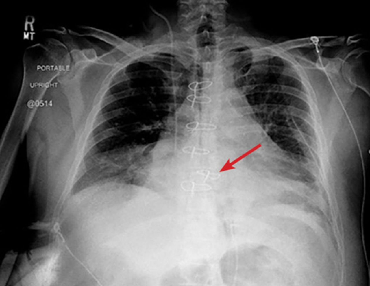
On postoperative day 4, midline sternal lucency and a slight leftward deviation of the second- from-the-lowest sternal wire (arrow)—early signs of dehiscence—resulted in mild loss of alignment.

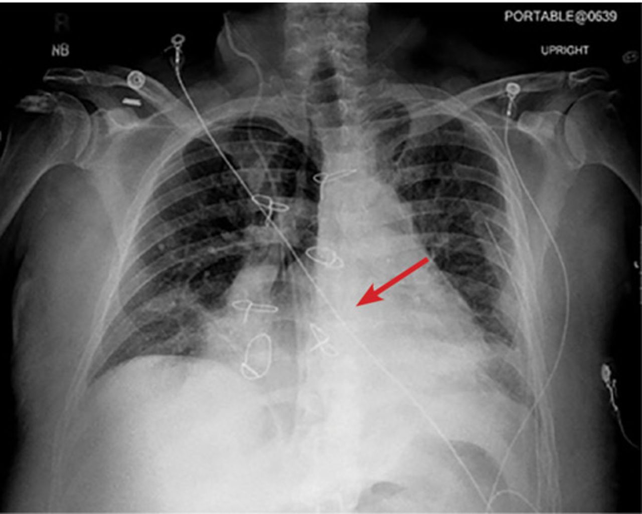
The next day, routine radiography showed widely separated sternal wires (Figure 3), indicating significant progression of sternal dehiscence. The patient subsequently underwent open reduction and internal fixation of the sternum.

Figure 3
  • Download figure
  • Open in new tab
  • Download powerpoint
Figure 3

On postoperative day 5, the widely separated sternal wires indicated advanced sternal dehiscence.

STERNAL DEHISCENCE

Sternal dehiscence is a rare but serious complication of sternotomy.1 In most cases of dehiscence, the sternal wires malfunction, leading to separation of sternal fragments. Lack of proper alignment of the sternum impairs bone healing, and the loose fragments of bone and wire pose a danger of puncturing the heart, making sternal dehiscence a surgical emergency.2

Physical examination may reveal tenderness to palpation, but findings that are more characteristic are an audible click and rocking of the sternum with coughing or forced chest movements.3

Plain chest radiography can clearly show early signs of sternal dehiscence; however, physicians rarely scrutinize the films for wire placement. Subtle signs include loss of sternal alignment with shifting of the segments and central sternal lucency. Gross signs start to appear when 2 or more wires are displaced; these signs are dramatic and rarely missed.

Loss of alignment and central sternal lucency are the earliest radiographic signs of dehiscence. Awareness of early subtle signs can lead to prompt diagnosis and treatment to prevent progression to gross sternal dehiscence.

  • Copyright © 2019 The Cleveland Clinic Foundation. All Rights Reserved.

REFERENCES

    1. Olbrecht VA,
    2. Barreiro CJ,
    3. Bonde PN,
    4. et al
    . Clinical outcomes of noninfectious sternal dehiscence after median sternotomy. Ann Thorac Surg 2006; 82(3):902–907. doi:10.1016/j.athoracsur.2006.04.058
    1. Efthymiou CA,
    2. Kay PH,
    3. Nair UR
    . Repair of spontaneous right ventricular rupture following sternal dehiscence. A novel technique. Interact Cardiovasc Thorac Surg 2010; 10(1):12–13. doi:10.1510/icvts.2009.217810
    1. Santarpino G,
    2. Pfeiffer S,
    3. Concistré G,
    4. Fischlein T
    . Sternal wound dehiscence from intense coughing in a cardiac surgery patient: could it be prevented? G Chir 2013; 34(4):112–113. pmid:23660161

Navigate

  • Current Issue
  • Past Issues
  • Supplements
  • Article Type
  • Specialty
  • CME/MOC Articles
  • CME/MOC Calendar
  • Media Kit

Authors & Reviewers

  • Manuscript Submission
  • Authors & Reviewers
  • Subscriptions
  • About CCJM
  • Contact Us
  • Cleveland Clinic Center for Continuing Education
  • Consult QD

Share your suggestions!

Copyright © 2025 The Cleveland Clinic Foundation. All rights reserved. The information provided is for educational purposes only. Use of this website is subject to the website terms of use and privacy policy. 

Powered by HighWire