


A 57-year-old man with a 20-year history of type 2 diabetes mellitus presented with a 1-year history of itchy rashes on both knees. He reported trying to treat the rashes with over-the-counter topical steroids on and off for the preceding 6 months.
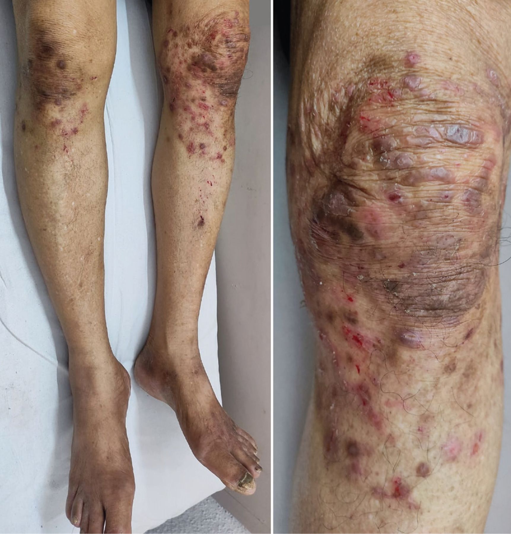
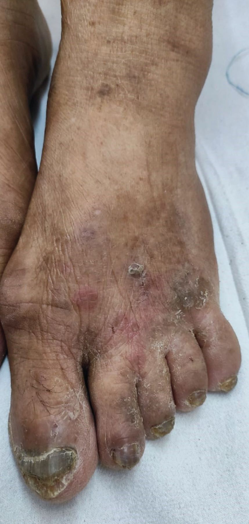
Physical examination revealed well- to ill-defined erythematous plaques with overlying papules and pustules in an annular pattern on both knees (Figure 1). Yellow-black discoloration, distal onycholysis, and dystrophy was noted on all of his toenails (Figure 2). Microscopic study of a potassium hydroxide preparation of scrapings from the active borders of the lesions on his knee was positive for dermatophytes.

Erythematous plaques with discontinuous annular borders and scaling on both knees, and a close-up view of the plaque on the left knee.

Ill-defined scaly plaque on the left foot with nail changes, including discoloration.
Based on the clinical features, history of steroid use, and positive potassium hydroxide mount, a diagnosis of tinea incognito was made. The patient was treated with oral itraconazole 200 mg daily for 1 month and luliconazole cream for 2 months, resulting in clinical improvement.
ABOUT TINEA INCOGNITO
Tinea incognito is an atypical form of dermatophyte infection that results from local immune suppression from systemic or topical corticosteroids. Clinically, the lesions lack the well-defined border, central clearing, and scaling classically associated with dermatophytosis. Use of corticosteroids may further suppress inflammatory signs, making tinea incognito lesions appear less erythematous.
Trichophyton rubrum is the most common causative organism, followed by Trichophyton mentagrophytes and Epidermophyton floccosum.1 Extensive atypical and drug-resistant cases have been reported due to Trichophyton indotineae.
Majocchi granuloma is a rare form of deep fungal folliculitis that occurs in longstanding dermatophytosis and is characterized by perifollicular papules, pustules, or nodules, often appearing within an erythematous plaque on the extremities or face.2 Although the presence of follicular papules and pustules in our patient could indicate progression to Majocchi granuloma, a conclusive diagnosis of this condition can be made only through histopathology. Areas of atrophy within the lesion may be seen due to chronic corticosteroid use.
Given the varied clinical presentations of tinea incognito, a broad differential diagnosis is essential, as it may mimic conditions such as eczema, psoriasis, lichenoid dermatitis, or blistering diseases.1
Diagnosis and treatment
The 2 key issues in managing tinea incognito are proper diagnosis and prevention. Diagnosis can be delayed because of the atypical appearance of the lesions, but careful examination for scaly erythematous edges aids in identification. Although newer diagnostic modalities such as dermoscopy and confocal laser scanning microscopy have become available, a potassium hydroxide mount remains an important modality for providing evidence of dermatophyte infection.3 A fungal culture should be ordered in chronic or recurrent dermatophytosis and in cases where antifungal resistance is suspected. Emerging tools like polymerase chain reaction and matrix-assisted laser desorption ionization–time-of-flight mass spectrometry offer greater sensitivity.3
Treatment modalities include promptly stopping steroid use and starting topical and oral antifungals. For mild or localized tinea infections, topical antifungals like terbinafine, clotrimazole, and miconazole are the first line of treatment, applied twice daily for several weeks.3 For severe or widespread infections, or those involving hair-bearing areas including Majocchi granuloma, oral antifungals such as terbinafine, itraconazole, or fluconazole are preferred, with treatment typically lasting several weeks to months, depending on the severity and response. Longer duration of treatment with itraconazole is required in drug-resistant cases.4
THE BOTTOM LINE
Diagnosing tinea incognito can be challenging due to its nonspecific clinical presentation. Poor response to prolonged steroid use, followed by a rebound increase in erythema, papules, and scaling when steroids are discontinued should prompt a reassessment of the diagnosis. It is essential for clinicians to recognize that tinea incognito can mimic other skin conditions and to have a low threshold for performing a potassium hydroxide mount in suspected cases, especially if topical steroids are the planned treatment.
DISCLOSURES
The authors report no relevant financial relationships which, in the context of their contributions, could be perceived as a potential conflict of interest.
- Copyright © 2025 The Cleveland Clinic Foundation. All Rights Reserved.
In this issue


Jump to section
Related Articles
Cited By...
- No citing articles found.