
Main menu

  • Home
  • Content
    • Current Issue
    • Ahead of Print
    • Past Issues
    • Supplements
    • Article Type
  • Specialty
    • Articles by Specialty
  • CME/MOC
    • Articles
    • Calendar
  • Info For
    • Manuscript Submission
    • Authors & Reviewers
    • Subscriptions
    • About CCJM
    • Contact Us
    • Media Kit
  • Conversations with Leaders
  • Conference Coverage
    • Kidney Week 2024
    • CHEST 2024
    • ACR Convergence 2023
    • Kidney Week 2023
    • ObesityWeek 2023
    • IDWeek 2023
    • CHEST 2023
    • MDS 2023
    • IAS 2023
    • ACP 2023
    • AAN 2023
    • ACC / WCC 2023
    • AAAAI Meeting 2023
    • ACR Convergence 2022
    • Kidney Week 2022
    • AIDS 2022
  • Other Publications
    • www.clevelandclinic.org

User menu

  • Register
  • Log in

Search

  • Advanced search
Cleveland Clinic Journal of Medicine
  • Other Publications
    • www.clevelandclinic.org
  • Register
  • Log in
Cleveland Clinic Journal of Medicine

Advanced Search

  • Home
  • Content
    • Current Issue
    • Ahead of Print
    • Past Issues
    • Supplements
    • Article Type
  • Specialty
    • Articles by Specialty
  • CME/MOC
    • Articles
    • Calendar
  • Info For
    • Manuscript Submission
    • Authors & Reviewers
    • Subscriptions
    • About CCJM
    • Contact Us
    • Media Kit
  • Conversations with Leaders
  • Conference Coverage
    • Kidney Week 2024
    • CHEST 2024
    • ACR Convergence 2023
    • Kidney Week 2023
    • ObesityWeek 2023
    • IDWeek 2023
    • CHEST 2023
    • MDS 2023
    • IAS 2023
    • ACP 2023
    • AAN 2023
    • ACC / WCC 2023
    • AAAAI Meeting 2023
    • ACR Convergence 2022
    • Kidney Week 2022
    • AIDS 2022
The Clinical Picture

Bilateral earlobe creases and coronary artery disease

Abhishek Sharma, MD, Chukwudi Obiagwu, MD and Ekaterina Sikorskaya, MD
Cleveland Clinic Journal of Medicine November 2016, 83 (11) 786-787; DOI: https://doi.org/10.3949/ccjm.83a.15160
Abhishek Sharma
Division of Cardiology, Department of Medicine, State University of New York, Downstate Medical Center, Brooklyn, NY
  • Find this author on Google Scholar
  • Find this author on PubMed
  • Search for this author on this site
  • For correspondence: [email protected]
Chukwudi Obiagwu
Department of Medicine, Maimonides, Medical Center, Brooklyn, NY
  • Find this author on Google Scholar
  • Find this author on PubMed
  • Search for this author on this site
Ekaterina Sikorskaya
Department of Medicine, The Brooklyn, Hospital Center, Brooklyn, NY
  • Find this author on Google Scholar
  • Find this author on PubMed
  • Search for this author on this site
  • Article
  • Figures & Data
  • Info & Metrics
  • PDF
Loading

A 70-year-old man with hypertension and hypercholesterolemia presented to the emergency department after the acute onset of substernal, pressure-like chest pain while climbing a flight of stairs. His physical examination was normal, but he was noted to have bilateral diagonal earlobe creases (the Frank sign) (Figure 1), considered by some to indicate risk of coronary artery disease.1–5

FIGURE 1
  • Download figure
  • Open in new tab
  • Download powerpoint
FIGURE 1

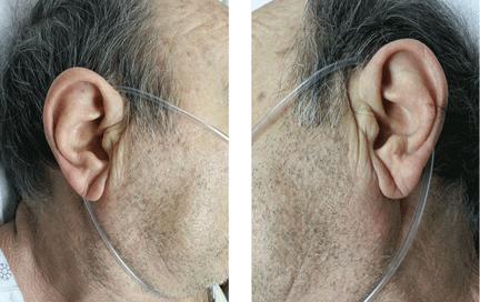
Earlobe creases (the Frank sign) were noted bilaterally.

Electrocardiography showed atrial fibrillation with a ventricular rate of 149 beats per minute, ST-segment elevation in leads V1 and aVR, and ST-segment depression in leads V3 to V6, II, III, and aVF.

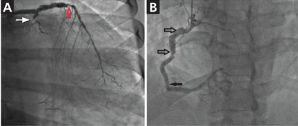
Urgent coronary arteriography showed severe coronary artery disease (Figure 2). Left ventriculography showed an ejection fraction of 50% with mild anterior wall hypokinesis. A drug-eluting stent was placed in the mid-left anterior descending artery. The patient tolerated the procedure well, and his chest pain resolved afterward.

FIGURE 2
  • Download figure
  • Open in new tab
  • Download powerpoint
FIGURE 2

In A, coronary angiography showed 80% stenosis of the mid-left anterior descending artery (red arrow), chronic total occlusion of the left circumflex artery (white arrow), and in B, mild to moderate diffuse atherosclerosis of the right coronary artery (arrows).

A STILL-UNCLEAR ASSOCIATION

Sanders T. Frank, in 1973, first described a diagonal wrinkle-like line on the earlobe as a sign of coronary artery disease.1 Subsequently, autopsy studies suggested that deep bilateral earlobe creases could be an important sign of coronary atherosclerosis.2 Diagonal earlobe creases have been shown to be independently associated with increased prevalence, extent, and severity of coronary artery disease.3,4 They are also associated with major adverse cardiovascular events4 and ischemic stroke.5 The mechanism linking diagonal earlobe creases and atherosclerotic disease is not yet clear.

This patient’s presentation and evaluation remind us that bilateral earlobe creases may be useful to include in the clinical examination of patients with suspected coronary artery disease and may facilitate early recognition of disease in a patient at high risk.

  • Copyright © 2016 The Cleveland Clinic Foundation. All Rights Reserved.

REFERENCES

  1. ↵
    1. Frank ST
    . Aural sign of coronary-artery disease. N Engl J Med 1973; 289:327–328.
    OpenUrlPubMed
  2. ↵
    1. Patel V,
    2. Champ C,
    3. Andrews PS,
    4. Gostelow BE,
    5. Gunasekara NP,
    6. Davidson AR
    . Diagonal earlobe creases and atheromatous disease: a postmortem study. J R Coll Phys Lond 1992; 26:274–277.
    OpenUrlPubMed
  3. ↵
    1. Kaukola S,
    2. Manninen V,
    3. Valle M,
    4. Halonen PI
    . Ear-lobe crease and coronary atherosclerosis. Lancet 1979; 2:1377.
    OpenUrlPubMed
  4. ↵
    1. Shmilovich H,
    2. Cheng VY,
    3. Rajani R,
    4. et al
    . Relation of diagonal ear lobe crease to the presence, extent, and se verity of coronary artery disease determined by coronary computed tomography angiography. Am J Cardiol 2012; 109:1283–1287.
    OpenUrlCrossRefPubMed
  5. ↵
    1. Zapata-Wainberg G,
    2. Vivancos J
    . Images in clinical medicine: bilateral earlobe creases. N Engl J Med 2013; 368:e32.
    OpenUrlCrossRefPubMed
PreviousNext
Back to top

In this issue

Cleveland Clinic Journal of Medicine: 83 (11)
Cleveland Clinic Journal of Medicine
Vol. 83, Issue 11
1 Nov 2016
  • Table of Contents
  • Table of Contents (PDF)
  • Index by author
Print
Download PDF
Article Alerts
Sign In to Email Alerts with your Email Address
Email Article

Thank you for your interest in spreading the word on Cleveland Clinic Journal of Medicine.

NOTE: We only request your email address so that the person you are recommending the page to knows that you wanted them to see it, and that it is not junk mail. We do not capture any email address.

Enter multiple addresses on separate lines or separate them with commas.
Bilateral earlobe creases and coronary artery disease
(Your Name) has sent you a message from Cleveland Clinic Journal of Medicine
(Your Name) thought you would like to see the Cleveland Clinic Journal of Medicine web site.
CAPTCHA
This question is for testing whether or not you are a human visitor and to prevent automated spam submissions.
Citation Tools
Bilateral earlobe creases and coronary artery disease
Abhishek Sharma, Chukwudi Obiagwu, Ekaterina Sikorskaya
Cleveland Clinic Journal of Medicine Nov 2016, 83 (11) 786-787; DOI: 10.3949/ccjm.83a.15160

Citation Manager Formats

  • BibTeX
  • Bookends
  • EasyBib
  • EndNote (tagged)
  • EndNote 8 (xml)
  • Medlars
  • Mendeley
  • Papers
  • RefWorks Tagged
  • Ref Manager
  • RIS
  • Zotero
Share
Bilateral earlobe creases and coronary artery disease
Abhishek Sharma, Chukwudi Obiagwu, Ekaterina Sikorskaya
Cleveland Clinic Journal of Medicine Nov 2016, 83 (11) 786-787; DOI: 10.3949/ccjm.83a.15160
Twitter logo Facebook logo Mendeley logo
  • Tweet Widget
  • Facebook Like
  • Linkedin Share Button

Jump to section

  • Article
    • A STILL-UNCLEAR ASSOCIATION
    • REFERENCES
  • Figures & Data
  • Info & Metrics
  • PDF

Related Articles

  • No related articles found.
  • PubMed
  • Google Scholar

Cited By...

  • No citing articles found.
  • Google Scholar

More in this TOC Section

  • Tinea incognito
  • Prolonged venous filling time and dependent rubor in a patient with peripheral artery disease
  • Sarcoidosis with diffuse purplish erythematous plaques on the hands
Show more The Clinical Picture

Similar Articles

Subjects

  • Cardiology
  • Emergency Medicine

Navigate

  • Current Issue
  • Past Issues
  • Supplements
  • Article Type
  • Specialty
  • CME/MOC Articles
  • CME/MOC Calendar
  • Media Kit

Authors & Reviewers

  • Manuscript Submission
  • Authors & Reviewers
  • Subscriptions
  • About CCJM
  • Contact Us
  • Cleveland Clinic Center for Continuing Education
  • Consult QD

Share your suggestions!

Copyright © 2025 The Cleveland Clinic Foundation. All rights reserved. The information provided is for educational purposes only. Use of this website is subject to the website terms of use and privacy policy. 

Powered by HighWire