


Article Figures & Data
Figures
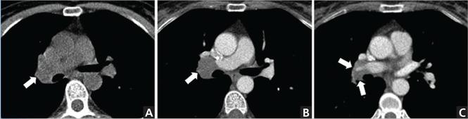
- FIGURE 1
In a patient with colon cancer undergoing a workup for metastases, axial CT without contrast (A) shows prominence of the right hilar region (arrow). Axial CT with contrast enhancement obtained subsequently (B and C) shows that this abnormality corresponds to right hilar lymphadenopathy partially encasing the right pulmonary artery (arrows).
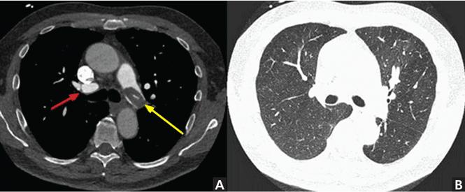
- FIGURE 2
In a 79-year-old patient with chronic thromboembolic pulmonary hypertension, CT angiography of the pulmonary artery (A) shows weblike (red arrow) and partially calcified filling defects (yellow arrow), as well as diffuse mild mosaic attenuation of lung parenchyma (B).
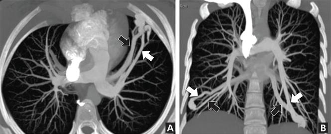
- FIGURE 3
CT pulmonary angiography with intravenous contrast in a patient being evaluated for arteriovenous malformation. Maximum-intensity projection images reconstructed in the axial (A) and coronal (B) planes show bilateral arteriovenous malformations with corresponding feeding arteries (white arrows) and draining veins (black arrows).
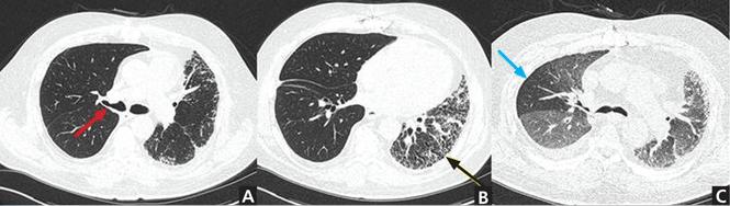
- FIGURE 4
CT without contrast in a patient with a history of interstitial lung disease and right lung transplant shows the patent but partially narrowed anastomotic site of the right bronchus (A) (red arrow). In B, the native left lung is small, with evidence of bronchiectasis, bronchiolectasis, and areas of honeycombing (black arrow). In C, the transplanted lung is notable for areas of air trapping in the right upper lobe on expiratory images (blue arrow), which is associated with central airway narrowing.

Tables
Intravenous contrast required
Malignancy workup and follow-up may require contrast to delineate hila, chest wall, vascular margins, or recurrent or residual tumor
CT pulmonary angiography for pulmonary embolism
CT pulmonary venography for superior vena cava syndrome
CT pulmonary angiography for arteriovenous malformation Suspected or known malignancy or infection of chest wall or mediastinumIntravenous contrast usually not required
Solitary pulmonary nodule a
Lung screening with low-dose CT
Pulmonary parenchyma, to evaluate for infection, inflammation, edema
Interstitial lung diseaseb
Pleural effusionc
Airway assessment↵a However, contrast enhancement may help identify tumors such as carcinoid and may better highlight internal morphology. Dynamic enhancement is also occasionally performed.
↵b Tailored protocol uses thinner slices and expiratory images to evaluate for air-trapping and dynamic airway compromise.
↵c Contrast is required to evaluate for empyema and suspected or known pleural malignancy.


