
Main menu

  • Home
  • Content
    • Current Issue
    • Ahead of Print
    • Past Issues
    • Supplements
    • Article Type
  • Specialty
    • Articles by Specialty
  • CME/MOC
    • Articles
    • Calendar
  • Info For
    • Manuscript Submission
    • Authors & Reviewers
    • Subscriptions
    • About CCJM
    • Contact Us
    • Media Kit
  • Conversations with Leaders
  • Conference Coverage
    • Kidney Week 2024
    • CHEST 2024
    • ACR Convergence 2023
    • Kidney Week 2023
    • ObesityWeek 2023
    • IDWeek 2023
    • CHEST 2023
    • MDS 2023
    • IAS 2023
    • ACP 2023
    • AAN 2023
    • ACC / WCC 2023
    • AAAAI Meeting 2023
    • ACR Convergence 2022
    • Kidney Week 2022
    • AIDS 2022
  • Other Publications
    • www.clevelandclinic.org

User menu

  • Register
  • Log in

Search

  • Advanced search
Cleveland Clinic Journal of Medicine
  • Other Publications
    • www.clevelandclinic.org
  • Register
  • Log in
Cleveland Clinic Journal of Medicine

Advanced Search

  • Home
  • Content
    • Current Issue
    • Ahead of Print
    • Past Issues
    • Supplements
    • Article Type
  • Specialty
    • Articles by Specialty
  • CME/MOC
    • Articles
    • Calendar
  • Info For
    • Manuscript Submission
    • Authors & Reviewers
    • Subscriptions
    • About CCJM
    • Contact Us
    • Media Kit
  • Conversations with Leaders
  • Conference Coverage
    • Kidney Week 2024
    • CHEST 2024
    • ACR Convergence 2023
    • Kidney Week 2023
    • ObesityWeek 2023
    • IDWeek 2023
    • CHEST 2023
    • MDS 2023
    • IAS 2023
    • ACP 2023
    • AAN 2023
    • ACC / WCC 2023
    • AAAAI Meeting 2023
    • ACR Convergence 2022
    • Kidney Week 2022
    • AIDS 2022
The Clinical Picture

A lump on the hip

Morteza Khodaee, MD, MPH, Seth Mathern, MD, Wendy S. Madigosky, MD, MSPH and Jonathan T. Bravman, MD
Cleveland Clinic Journal of Medicine August 2017, 84 (8) 594-596; DOI: https://doi.org/10.3949/ccjm.84a.16065
Morteza Khodaee
Department of Family Medicine, University of Colorado School of Medicine, Denver, CO
  • Find this author on Google Scholar
  • Find this author on PubMed
  • Search for this author on this site
  • For correspondence: [email protected]
Seth Mathern
Department of Family Medicine, University of Colorado School of Medicine, Denver, CO
  • Find this author on Google Scholar
  • Find this author on PubMed
  • Search for this author on this site
Wendy S. Madigosky
Department of Family Medicine, University of Colorado School of Medicine, Denver, CO
  • Find this author on Google Scholar
  • Find this author on PubMed
  • Search for this author on this site
Jonathan T. Bravman
Department of Orthopedic Surgery, University of Colorado School of Medicine, Denver, CO
  • Find this author on Google Scholar
  • Find this author on PubMed
  • Search for this author on this site
  • Article
  • Figures & Data
  • Info & Metrics
  • PDF
Loading

A 42-year-old man presented with a lump on the side of his left hip, which had developed after he fell on his hip while playing basketball about 2 weeks earlier. He was able to continue playing and finished the game. After the game he noticed a lump, which rapidly increased in size. Significant bruising developed afterwards, and the area was mildly painful. The lump did not interfere with his daily activities, but it was annoying.

His medical, surgical, social, and family histories were unremarkable. Physical examination revealed a significant oval-shaped local swelling in the lateral aspect of his left hip with extensive surrounding ecchymosis (Figure 1). On palpation the swelling was mildly tender and fluctuant. Range of motion of the hip was normal and did not aggravate his pain.

FIGURE 1
  • Download figure
  • Open in new tab
  • Download powerpoint
FIGURE 1

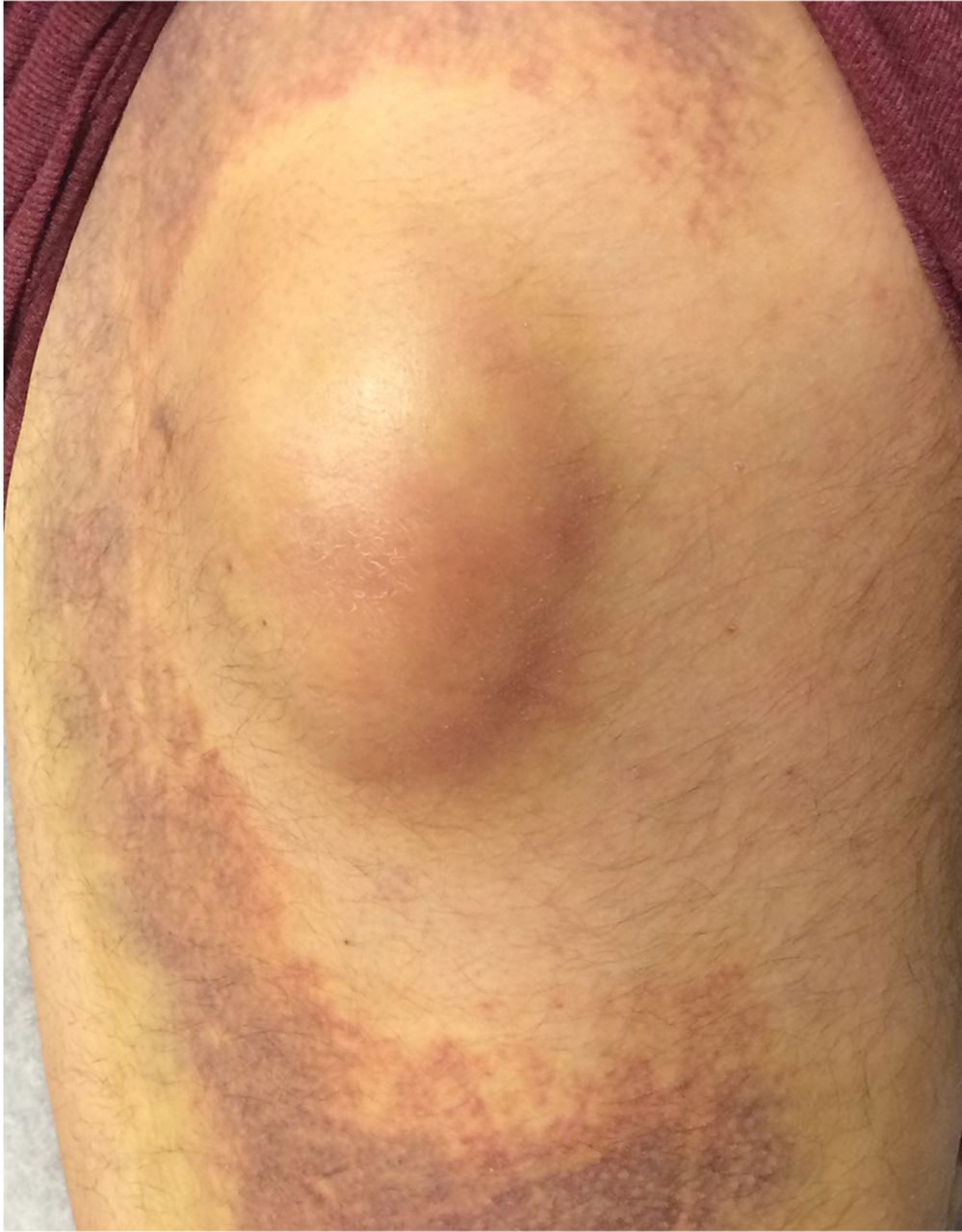
Left lateral hip 2 weeks after a fall.

Ultrasonography to evaluate for fluid collection revealed a subcutaneous heterogeneous and hypoechoic area superficial to the greater trochanter, muscular structure, and the fascia (Figure 2). There was no enlargement of the greater trochanter bursa. Aspiration with an 18-gauge needle under ultrasonographic guidance provided no fluid. Based on the clinical presentation and imaging, the diagnosis of Morel-Lavallée lesion was made.

FIGURE 2
  • Download figure
  • Open in new tab
  • Download powerpoint
FIGURE 2

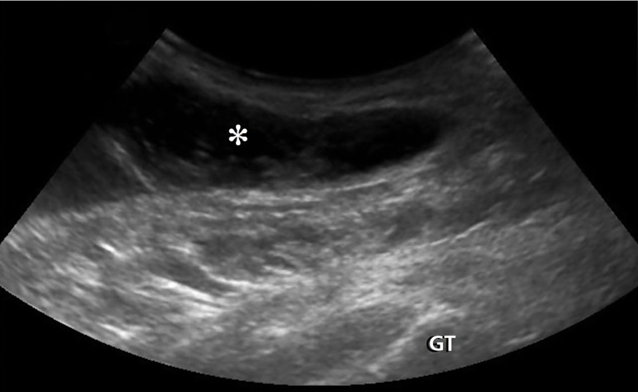
Ultrasonography with a curvilinear 2- to 5-MHz probe and a long-axis view revealed a superficial heterogeneous and hypoechoic area (asterisk) at the greater trochanter (GT).

THE DIFFERENTIAL DIAGNOSIS

Traumatic swelling of the lateral hip is an uncommon condition with few possible causes (Table 1).1–5 A comprehensive evaluation beginning with a history and physical examination is essential.

View this table:
  • View inline
  • View popup
TABLE 1

Differential diagnosis of a traumatic lateral hip lump

Morel-Lavallée lesion is an uncommon condition resulting from shearing trauma and collection of fluid in the space between deep fatty tissue and superficial fascia.6 It is usually the result of severe trauma, as in a motor vehicle accident, but it can also result from sports-related trauma, as in our patient.6–8 Lateral hip, gluteal, and sacral regions are the most common locations for Morel-Lavallée lesions and are often associated with an underlying fracture.6,9

Morel-Lavallée lesions usually develop hours or days after trauma, although they may develop weeks or even months later.2 Symptoms include bulging, pain, and loss of cutaneous sensation over the affected area. Although ultrasonography can be used, magnetic resonance imaging (MRI) is the gold standard for diagnosis and staging.6,10 If there is concern for fracture, plain radiography should be performed.

Mellado and Bencardino classified Morel-Lavallée lesions into 6 types based on their morphology, presence or absence of a capsule, signal behavior on MRI, and enhancement pattern.10 The exact rate of infection in patients with Morel-Lavallée lesions is unknown; however, the risk of infection seems to be highest after surgical intervention or aspiration.5,6

Another potential complication is fluid reaccumulation, which most often occurs with large lesions (> 50 mL) and lesions with a fibrous capsule or pseudocapsule.5 Large lesions can compromise adjacent neurovascular structures, particularly in the extremities.5 Potential consequences include dermal necrosis, compartment syndrome, and tissue necrosis.5

MANAGEMENT APPROACH

Aspiration of a fluid-filled mass is useful in both diagnosis and management of Morel-Lavallée lesions. Treatment includes watchful waiting; compression and pressure wraps; injection of a sclerosing agent (eg, doxycyline, alcohol); needle aspiration; percutaneous drainage with debridement, irrigation, and suction; and incision and evacuation.6

The approach to treatment depends on the stage of the lesion and whether an underlying fracture is present. Depending on the amount of blood and lymphatic products and the acuity of the collected fluid (hours to days post-trauma), aspiration with a large-bore needle (eg, 14 to 22 gauge) may or may not be successful.7 In general, traumatic serosanguinous fluid collections are less painful and resolve faster than well-formed coagulated hematomas.

Patients who have a large lesion, significant pain, or decreased range of motion should be referred to an orthopedic surgeon.

Our patient was managed conservatively, and his symptoms completely resolved in 2 months.

  • Copyright © 2017 The Cleveland Clinic Foundation. All Rights Reserved.

REFERENCES

  1. ↵
    1. Ahmad Z,
    2. Tibrewal S,
    3. Waters G,
    4. Nolan J
    . Solitary amyloidoma related to THA. Orthopedics 2013; 36:e971–e973.
    OpenUrl
  2. ↵
    1. Harris-Spinks C,
    2. Nabhan D,
    3. Khodaee M
    . Noniatrogenic septic olecranon bursitis: report of two cases and review of the literature. Curr Sports Med Rep 2016; 15:33–37.
    OpenUrl
    1. Miller MD,
    2. Sanders TG
    1. Price MD,
    2. Busconi BD,
    3. McMillan S
    . Proximal femur fractures. In: Miller MD, , Sanders TG, eds. Presentation, Imaging and Treatment of Common Musculoskeletal Conditions. Philadelphia, PA: Saunders; 2011:365–376.
    1. Stanton MC,
    2. Maloney MD,
    3. Dehaven KE,
    4. Giordano BD
    . Acute traumatic tear of gluteus medius and minimus tendons in a patient without antecedant peritrochanteric hip pain. Geriatr Orthop Surg Rehabil 2012; 3:84–88.
    OpenUrlCrossRefPubMed
  3. ↵
    1. Khodaee M,
    2. Deu RS,
    3. Mathern S,
    4. Bravman JT
    . Morel-Lavallée lesion in sports. Curr Sports Med Rep 2016; 15:417–422.
    OpenUrlPubMed
  4. ↵
    1. Bonilla-Yoon I,
    2. Masih S,
    3. Patel DB,
    4. et al
    . The Morel-Lavallée lesion: pathophysiology, clinical presentation, imaging features, and treatment options. Emerg Radiol 2014; 21:35–43.
    OpenUrlCrossRefPubMed
  5. ↵
    1. Khodaee M,
    2. Deu RS
    . Ankle Morel-Lavallée lesion in a recreational racquetball player. J Sports Med Phys Fitness 2016. Epub ahead of print.
  6. ↵
    1. Shmerling A,
    2. Bravman JT,
    3. Khodaee M
    . Morel-Lavallée lesion of the knee in a recreational frisbee player. Case Rep Orthop 2016; 2016:8723489.
    OpenUrl
  7. ↵
    1. Miller J,
    2. Daggett J,
    3. Ambay R,
    4. Payne WG
    . Morel-Lavallée lesion. Eplasty 2014; 14:ic12.
    OpenUrl
  8. ↵
    1. Mellado JM,
    2. Bencardino JT
    . Morel-Lavallée lesion: review with emphasis on MR imaging. Magn Reson Imaging Clin N Am 2005; 13:775–782.
    OpenUrlCrossRefPubMed
PreviousNext
Back to top

In this issue

Cleveland Clinic Journal of Medicine: 84 (8)
Cleveland Clinic Journal of Medicine
Vol. 84, Issue 8
1 Aug 2017
  • Table of Contents
  • Table of Contents (PDF)
  • Index by author
Print
Download PDF
Article Alerts
Sign In to Email Alerts with your Email Address
Email Article

Thank you for your interest in spreading the word on Cleveland Clinic Journal of Medicine.

NOTE: We only request your email address so that the person you are recommending the page to knows that you wanted them to see it, and that it is not junk mail. We do not capture any email address.

Enter multiple addresses on separate lines or separate them with commas.
A lump on the hip
(Your Name) has sent you a message from Cleveland Clinic Journal of Medicine
(Your Name) thought you would like to see the Cleveland Clinic Journal of Medicine web site.
CAPTCHA
This question is for testing whether or not you are a human visitor and to prevent automated spam submissions.
Citation Tools
A lump on the hip
Morteza Khodaee, Seth Mathern, Wendy S. Madigosky, Jonathan T. Bravman
Cleveland Clinic Journal of Medicine Aug 2017, 84 (8) 594-596; DOI: 10.3949/ccjm.84a.16065

Citation Manager Formats

  • BibTeX
  • Bookends
  • EasyBib
  • EndNote (tagged)
  • EndNote 8 (xml)
  • Medlars
  • Mendeley
  • Papers
  • RefWorks Tagged
  • Ref Manager
  • RIS
  • Zotero
Share
A lump on the hip
Morteza Khodaee, Seth Mathern, Wendy S. Madigosky, Jonathan T. Bravman
Cleveland Clinic Journal of Medicine Aug 2017, 84 (8) 594-596; DOI: 10.3949/ccjm.84a.16065
Twitter logo Facebook logo Mendeley logo
  • Tweet Widget
  • Facebook Like
  • Linkedin Share Button

Jump to section

  • Article
    • THE DIFFERENTIAL DIAGNOSIS
    • MANAGEMENT APPROACH
    • REFERENCES
  • Figures & Data
  • Info & Metrics
  • PDF

Related Articles

  • No related articles found.
  • PubMed
  • Google Scholar

Cited By...

  • No citing articles found.
  • Google Scholar

More in this TOC Section

  • Severe cutaneous reaction induced by allopurinol
  • Amoxicillin rash in infectious mononucleosis
  • Tinea incognito
Show more The Clinical Picture

Similar Articles

Subjects

  • Emergency Medicine
  • Rheumatology

Navigate

  • Current Issue
  • Past Issues
  • Supplements
  • Article Type
  • Specialty
  • CME/MOC Articles
  • CME/MOC Calendar
  • Media Kit

Authors & Reviewers

  • Manuscript Submission
  • Authors & Reviewers
  • Subscriptions
  • About CCJM
  • Contact Us
  • Cleveland Clinic Center for Continuing Education
  • Consult QD

Share your suggestions!

Copyright © 2025 The Cleveland Clinic Foundation. All rights reserved. The information provided is for educational purposes only. Use of this website is subject to the website terms of use and privacy policy. 

Powered by HighWire