


Article Figures & Data
Figures
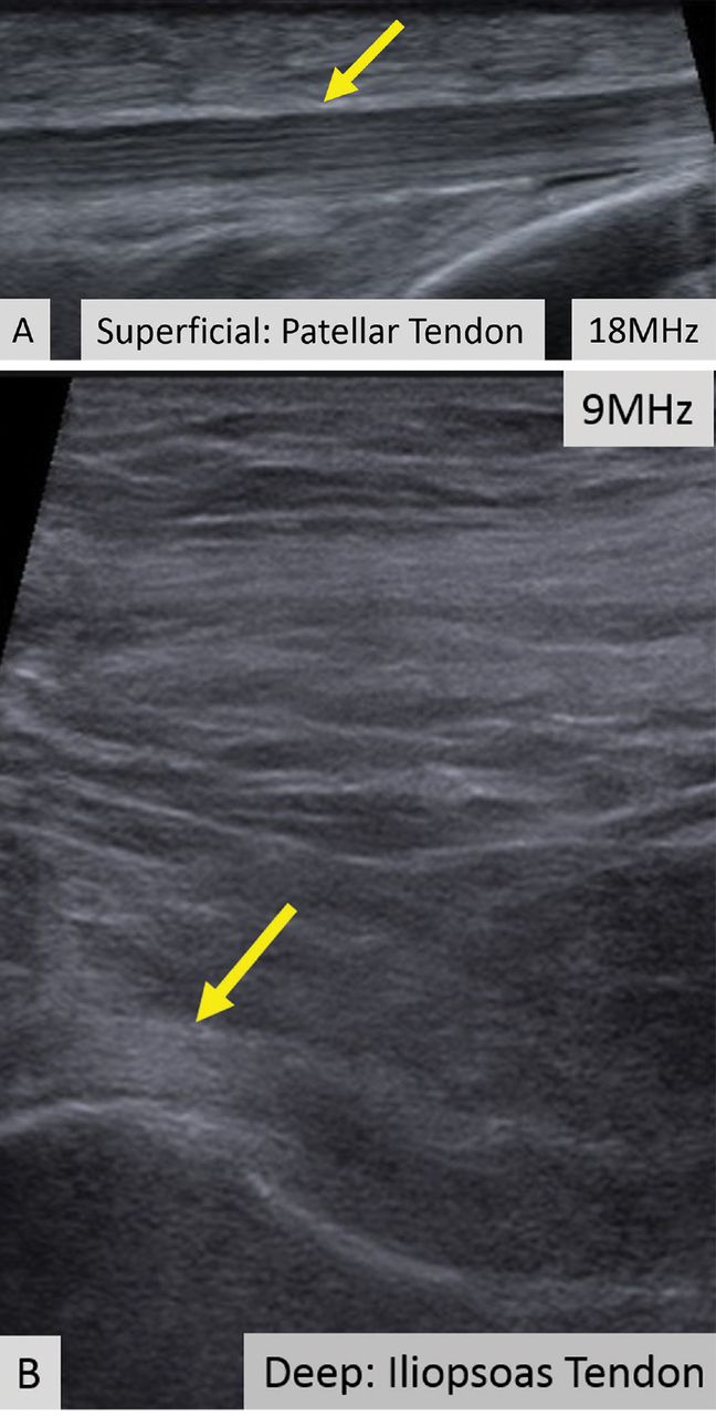
- Figure 1
In ultrasonography, a trade-off exists between image resolution and penetration depth. The superficial patellar tendon (A, arrow) can be seen with high resolution, demonstrating its fine internal structure. The much deeper iliopsoas tendon cannot be seen with the same high resolution because of its deep location (B, arrow).
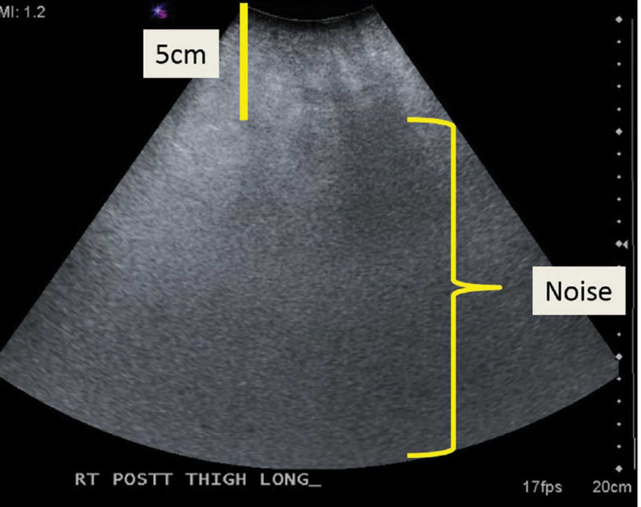
- Figure 2
Ultrasonography of the posterior thigh in a patient with obesity. Because subcutaneous fat attenuates sound waves, examination of soft tissues greater than a few centimeters in thickness is nondiagnostic.
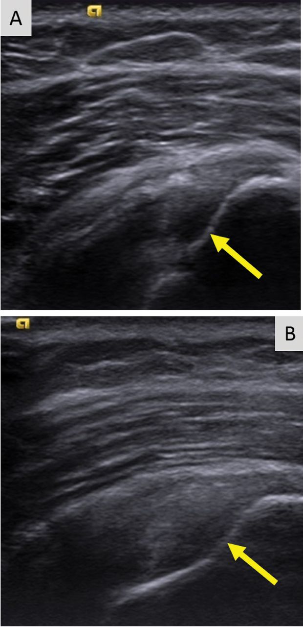
- Figure 3
On ultrasonography, anisotropy causes a hypoechoic defect of the articular supraspinatus tendon fibers (A, arrow). With improved transducer angle, anisotropy is decreased and intact fibers can be seen (B, arrow). Sonographers and interpreting physicians must be careful not to mistake aberrations due to anisotropy for tissue disease.
- Figure 4
Musculoskeletal ultrasonography is inappropriate for evaluating large areas. Here, ultrasonography did not fully demonstrate the extent or nature of the abnormality within the adductor musculature of the patient’s thigh (A, arrow). MRI demonstrated multiple large enhancing metastatic intramuscular masses (B, arrows).
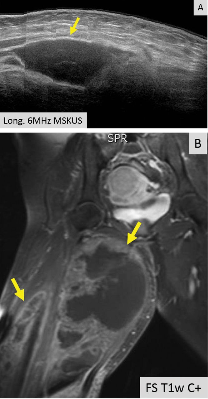
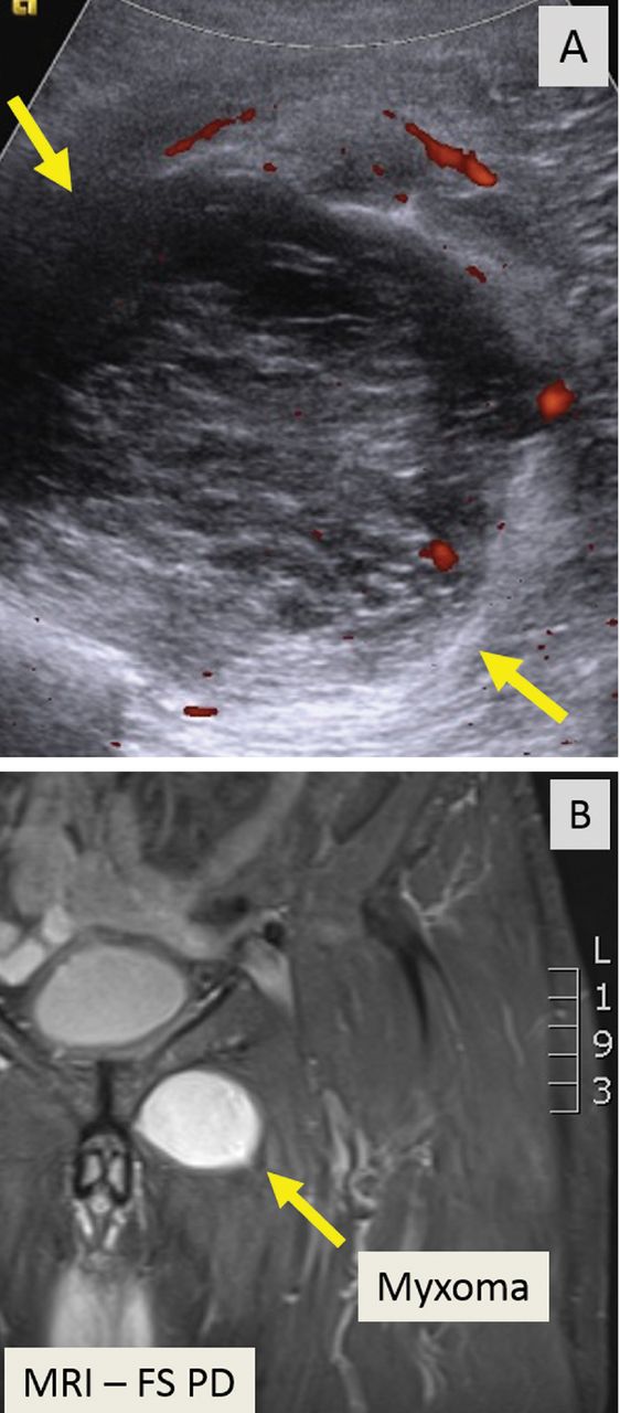
- Figure 5
A deep, complex intramuscular soft-tissue mass seen on ultrasonography (A, arrows) required further evaluation with MRI (B, arrow), which better demonstrated the mass’s margins and its relationship to surrounding structures.


Tables
Condition Characteristics on ultrasonography Power Doppler Normal tendon Hyperechoic, compact internal fibrillar pattern
Anisotropy may be present, mimicking tendinosis or tendon tearingNo signal Tendinosis Hypoechoic, focal or diffuse, abnormally thick-ened, loss of compact fibrillar structure and possibly regions of fiber disruption48,49 With or without increased power Doppler signal Tearing Can differentiate partial- vs full-thickness tear Complex fluid and blood can mimic intact tendon fibers; if tear is present, fluid does not move with joint movement as intact fibers would23 Tenosynovitis or peritendinitis Thickened tendon sheath or peritendon with increased fluid With or without increased power Doppler signal Normal ligament Hyperechoic, compact morphology, less ordered fibrillar pattern than tendon Low-grade ligament injury May be normal or thickened, hypoechoic With or without increased power Doppler signal Intermediate or high- grade ligament injury Fiber disruption, surrounding hematoma or fluid With or without increased power Doppler signal Normal muscle Mostly hypoechoic, interspersed hyperechoic lines and dots (perimysium and epimysium) Normal nerve Less compact-appearing and more varied shape than tendon and ligament
Semicompact bundle of hypoechoic nerve fascicles surrounded by hyperechoic tissueNeuritis (focal or diffuse) Abnormal nerve enlargement, fascicular swelling, blurring of the interstitium, perineural thickening (in the chronic state), possible scarring in entrapment cases20 With or without increased power Doppler signal


