
Main menu

  • Home
  • Content
    • Current Issue
    • Ahead of Print
    • Past Issues
    • Supplements
    • Article Type
  • Specialty
    • Articles by Specialty
  • CME/MOC
    • Articles
    • Calendar
  • Info For
    • Manuscript Submission
    • Authors & Reviewers
    • Subscriptions
    • About CCJM
    • Contact Us
    • Media Kit
  • Conversations with Leaders
  • Conference Coverage
    • Kidney Week 2024
    • CHEST 2024
    • ACR Convergence 2023
    • Kidney Week 2023
    • ObesityWeek 2023
    • IDWeek 2023
    • CHEST 2023
    • MDS 2023
    • IAS 2023
    • ACP 2023
    • AAN 2023
    • ACC / WCC 2023
    • AAAAI Meeting 2023
    • ACR Convergence 2022
    • Kidney Week 2022
    • AIDS 2022
  • Other Publications
    • www.clevelandclinic.org

User menu

  • Register
  • Log in

Search

  • Advanced search
Cleveland Clinic Journal of Medicine
  • Other Publications
    • www.clevelandclinic.org
  • Register
  • Log in
Cleveland Clinic Journal of Medicine

Advanced Search

  • Home
  • Content
    • Current Issue
    • Ahead of Print
    • Past Issues
    • Supplements
    • Article Type
  • Specialty
    • Articles by Specialty
  • CME/MOC
    • Articles
    • Calendar
  • Info For
    • Manuscript Submission
    • Authors & Reviewers
    • Subscriptions
    • About CCJM
    • Contact Us
    • Media Kit
  • Conversations with Leaders
  • Conference Coverage
    • Kidney Week 2024
    • CHEST 2024
    • ACR Convergence 2023
    • Kidney Week 2023
    • ObesityWeek 2023
    • IDWeek 2023
    • CHEST 2023
    • MDS 2023
    • IAS 2023
    • ACP 2023
    • AAN 2023
    • ACC / WCC 2023
    • AAAAI Meeting 2023
    • ACR Convergence 2022
    • Kidney Week 2022
    • AIDS 2022
COVID-19 Curbside Consults

Thoracic imaging in COVID-19

Ruchi Yadav, MD, Debasis Sahoo, MD, FCCP and Ruffin Graham, MD
Cleveland Clinic Journal of Medicine August 2020, 87 (8) 469-476; DOI: https://doi.org/10.3949/ccjm.87a.ccc032
Ruchi Yadav
Departments of Diagnostic Radiology and Nuclear Medicine, Imaging Institute, Cleveland Clinic; Clinical Assistant Professor, Cleveland Clinic Lerner College of Medicine of Case Western Reserve University, Cleveland, OH
  • Find this author on Google Scholar
  • Find this author on PubMed
  • Search for this author on this site
  • For correspondence: [email protected]
Debasis Sahoo
Department of Pulmonary Medicine, Respiratory Institute, Cleveland Clinic; Clinical Assistant Professor, Cleveland Clinic Lerner College of Medicine of Case Western Reserve University, Cleveland, OH
  • Find this author on Google Scholar
  • Find this author on PubMed
  • Search for this author on this site
Ruffin Graham
Section Head, Department of Diagnostic Radiology, Imaging Institute, Cleveland Clinic; Assistant Professor, Cleveland Clinic Lerner College of Medicine of Case Western Reserve University, Cleveland, OH
  • Find this author on Google Scholar
  • Find this author on PubMed
  • Search for this author on this site
  • Article
  • Figures & Data
  • Info & Metrics
  • PDF
Loading

Article Figures & Data

Figures

  • Tables
  • Figure 1
    • Download figure
    • Open in new tab
    • Download powerpoint
    Figure 1

    Portable chest radiographs of a patients with COVID-19 demonstrating classic bilateral, multifocal peripheral airspace opacities in the mid-lower-lung zones.
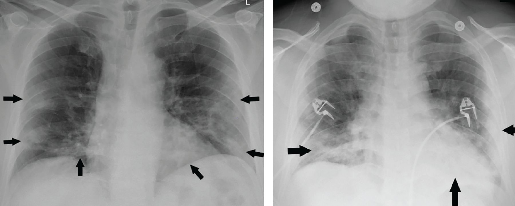
  • Figure 2
    • Download figure
    • Open in new tab
    • Download powerpoint
    Figure 2

    Portable chest radiographs of patients with COVID-19 demonstrating atypical features of diffuse bilateral interstitial changes (A) and unilateral consolidation (B).
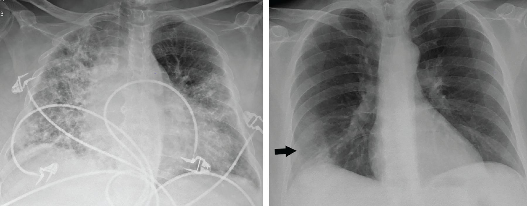
  • Figure 3
    • Download figure
    • Open in new tab
    • Download powerpoint
    Figure 3

    Serial chest radiographs in a 53-year-old woman with COVID-19 demonstrate peripheral opacities on the day of onset of symptoms (A) with rapid worsening by day 3 (B). The patient was subsequently intubated, with the peak severity of parenchymal findings on day 11 (C).

  • Figure 4
    • Download figure
    • Open in new tab
    • Download powerpoint
    Figure 4

    (Left) Setup for obtaining chest radiographs through the glass wall of the room of a patient with suspected or confirmed COVID-19. (Middle) A radiograph obtained in the conventional manner. (Right) A radiograph obtained through the glass.
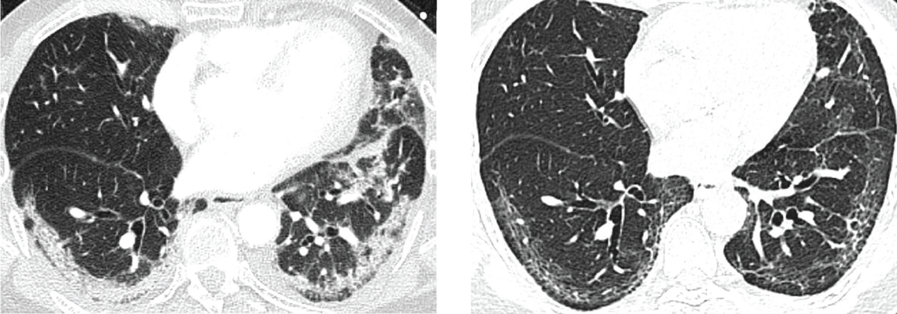
  • Figure 5
    • Download figure
    • Open in new tab
    • Download powerpoint
    Figure 5

    Typical computed tomographic features of COVID-19. Unenhanced axial images of the lungs of 4 different patients with COVID-19 demonstrate bilateral, multifocal, peripheral ground-glass opacities, and consolidation, most with rounded morphology. “Crazy paving” (ground-glass opacities with superimposed interlobular septal thickening and intralobular lines) is seen in (C) (white arrow).

  • Figure 6
    • Download figure
    • Open in new tab
    • Download powerpoint
    Figure 6

    CT scans in a 73-year-old man with COVID-19 demonstrate resolution of subpleural consolidation with residual reticular and fibrotic changes.

  • Figure 7
    • Download figure
    • Open in new tab
    • Download powerpoint
    Figure 7

    CT images of 2 different patients with COVID-19 demonstrating pulmonary embolism.

Tables

  • Figures
    • View popup
    TABLE 1

    Stages of COVID-19 on chest CT

    Early stage (0–2 days)
    Approximately 50% of patients have negative chest CT
    The remaining have ground-glass opacities (44%) and consolidation (17%), more often unilateral
    The less pulmonary consolidation identified on CT, the greater the probability of initial negative reverse transcriptase polymerase chain reaction results11
    Intermediate stage (3–5 days)
    9% of patients have negative chest CT
    88% have ground-glass opacities with or without crazy paving (a sign of progression or peak stage), and 55% have consolidation (bilateral in 76%, peripheral in distribution in 64% with rounded morphology)12
    Late phase (6–12 days)
    Most patients have positive CT findings
    Progressive consolidation, evolving linear consolidation, and organizing pneumonia
    Reverse-halo appearance (a sign of healing or evolving lesion)12
    Ground-glass opacities in 88% with or without crazy paving
    Severe phase
    Massive pulmonary consolidation and “white lungs”
    Recovery phase
    Parenchymal abnormalities resolve with residual linear opacities (Figure 6)
    • Based on information from references 5 and 11–14.

PreviousNext
Back to top

In this issue

Cleveland Clinic Journal of Medicine: 87 (8)
Cleveland Clinic Journal of Medicine
Vol. 87, Issue 8
1 Aug 2020
  • Table of Contents
  • Table of Contents (PDF)
  • Index by author
  • Complete Issue (PDF)
Print
Download PDF
Article Alerts
Sign In to Email Alerts with your Email Address
Email Article

Thank you for your interest in spreading the word on Cleveland Clinic Journal of Medicine.

NOTE: We only request your email address so that the person you are recommending the page to knows that you wanted them to see it, and that it is not junk mail. We do not capture any email address.

Enter multiple addresses on separate lines or separate them with commas.
Thoracic imaging in COVID-19
(Your Name) has sent you a message from Cleveland Clinic Journal of Medicine
(Your Name) thought you would like to see the Cleveland Clinic Journal of Medicine web site.
CAPTCHA
This question is for testing whether or not you are a human visitor and to prevent automated spam submissions.
Citation Tools
Thoracic imaging in COVID-19
Ruchi Yadav, Debasis Sahoo, Ruffin Graham
Cleveland Clinic Journal of Medicine Aug 2020, 87 (8) 469-476; DOI: 10.3949/ccjm.87a.ccc032

Citation Manager Formats

  • BibTeX
  • Bookends
  • EasyBib
  • EndNote (tagged)
  • EndNote 8 (xml)
  • Medlars
  • Mendeley
  • Papers
  • RefWorks Tagged
  • Ref Manager
  • RIS
  • Zotero
Share
Thoracic imaging in COVID-19
Ruchi Yadav, Debasis Sahoo, Ruffin Graham
Cleveland Clinic Journal of Medicine Aug 2020, 87 (8) 469-476; DOI: 10.3949/ccjm.87a.ccc032
Twitter logo Facebook logo Mendeley logo
  • Tweet Widget
  • Facebook Like
  • Linkedin Share Button

Jump to section

  • Article
    • ABSTRACT
    • CHEST RADIOGRAPHY IN COVID-19
    • RADIOGRAPHY ‘THROUGH GLASS’ TO AVOID SPREADING THE VIRUS
    • COMPUTED TOMOGRAPHY IN COVID-19
    • THE RADIOLOGICAL SOCIETY OF NORTH AMERICA CONSENSUS STATEMENT
    • ROLE OF CT IN COVID-19
    • CURRENT RECOMMENDATIONS
    • FLEISCHNER SOCIETY STATEMENT
    • REFERENCES
  • Figures & Data
  • Info & Metrics
  • PDF

Related Articles

  • No related articles found.
  • PubMed
  • Google Scholar

Cited By...

  • Point-of-care lung ultrasound assessment for risk stratification and therapy guiding in COVID-19 patients: a prospective noninterventional study
  • Google Scholar

More in this TOC Section

  • Update to COVID-19 serologic testing : FAQs and caveats
  • Update to post-acute sequelae of SARS-CoV-2 infection: Caring for the 'long-haulers'
  • COVID-19 in older adults
Show more COVID-19 Curbside Consults

Similar Articles

Subjects

  • Covid-19
  • Critical Care
  • Hospital Medicine
  • Imaging
  • Infectious Diseases
  • Pulmonology

Navigate

  • Current Issue
  • Past Issues
  • Supplements
  • Article Type
  • Specialty
  • CME/MOC Articles
  • CME/MOC Calendar
  • Media Kit

Authors & Reviewers

  • Manuscript Submission
  • Authors & Reviewers
  • Subscriptions
  • About CCJM
  • Contact Us
  • Cleveland Clinic Center for Continuing Education
  • Consult QD

Share your suggestions!

Copyright © 2025 The Cleveland Clinic Foundation. All rights reserved. The information provided is for educational purposes only. Use of this website is subject to the website terms of use and privacy policy. 

Powered by HighWire