
Main menu

  • Home
  • Content
    • Current Issue
    • Ahead of Print
    • Past Issues
    • Supplements
    • Article Type
  • Specialty
    • Articles by Specialty
  • CME/MOC
    • Articles
    • Calendar
  • Info For
    • Manuscript Submission
    • Authors & Reviewers
    • Subscriptions
    • About CCJM
    • Contact Us
    • Media Kit
  • Conversations with Leaders
  • Conference Coverage
    • Kidney Week 2024
    • CHEST 2024
    • ACR Convergence 2023
    • Kidney Week 2023
    • ObesityWeek 2023
    • IDWeek 2023
    • CHEST 2023
    • MDS 2023
    • IAS 2023
    • ACP 2023
    • AAN 2023
    • ACC / WCC 2023
    • AAAAI Meeting 2023
    • ACR Convergence 2022
    • Kidney Week 2022
    • AIDS 2022
  • Other Publications
    • www.clevelandclinic.org

User menu

  • Register
  • Log in

Search

  • Advanced search
Cleveland Clinic Journal of Medicine
  • Other Publications
    • www.clevelandclinic.org
  • Register
  • Log in
Cleveland Clinic Journal of Medicine

Advanced Search

  • Home
  • Content
    • Current Issue
    • Ahead of Print
    • Past Issues
    • Supplements
    • Article Type
  • Specialty
    • Articles by Specialty
  • CME/MOC
    • Articles
    • Calendar
  • Info For
    • Manuscript Submission
    • Authors & Reviewers
    • Subscriptions
    • About CCJM
    • Contact Us
    • Media Kit
  • Conversations with Leaders
  • Conference Coverage
    • Kidney Week 2024
    • CHEST 2024
    • ACR Convergence 2023
    • Kidney Week 2023
    • ObesityWeek 2023
    • IDWeek 2023
    • CHEST 2023
    • MDS 2023
    • IAS 2023
    • ACP 2023
    • AAN 2023
    • ACC / WCC 2023
    • AAAAI Meeting 2023
    • ACR Convergence 2022
    • Kidney Week 2022
    • AIDS 2022
Review

Diagnosis and management of pancreatic cystic lesions for the non-gastroenterologist

Arjun Chatterjee, MD, Tyler Stevens, MD, FACG, FASGE and Prabhleen Chahal, MD, FACG, FASGE
Cleveland Clinic Journal of Medicine February 2024, 91 (2) 96-102; DOI: https://doi.org/10.3949/ccjm.91a.23019
Arjun Chatterjee
Department of Internal Medicine, Cleveland Clinic, Cleveland, OH
  • Find this author on Google Scholar
  • Find this author on PubMed
  • Search for this author on this site
Tyler Stevens
Department of Gastroenterology, Hepatology, and Nutrition, Cleveland Clinic, Cleveland, OH; Associate Professor, Cleveland Clinic Lerner College of Medicine of Case Western Reserve University, Cleveland, OH
  • Find this author on Google Scholar
  • Find this author on PubMed
  • Search for this author on this site
  • Find this author on Cleveland Clinic
Prabhleen Chahal
Director, Advanced Endoscopy Training, Department of Gastroenterology, Hepatology, and Nutrition, Cleveland Clinic, Cleveland, OH
  • Find this author on Google Scholar
  • Find this author on PubMed
  • Search for this author on this site
  • For correspondence: [email protected]
  • Find this author on Cleveland Clinic
  • Article
  • Figures & Data
  • Info & Metrics
  • PDF
Loading

ABSTRACT

Although most pancreatic cystic lesions do not progress to cancer, they create concern for patients and their primary care physicians. The lack of consensus guidelines on diagnosis and surveillance of these lesions can lead to a management conundrum. We review current guidelines on diagnosis and management.

KEY POINTS
  • Magnetic resonance cholangiopancreatography with dynamic magnetic resonance imaging is the test of choice for diagnosis and assessment of high-risk or worrisome characteristics in cysts. Pancreatic-protocol computed tomography and endoscopic ultrasonography are suitable options if magnetic resonance imaging is contraindicated.

  • High-risk clinical and laboratory features include obstructive jaundice, recurrent pancreatitis, elevated serum carbohydrate antigen 19-9, presence of cells demonstrating high-grade dysplasia or neoplasia, and new-onset or worsening diabetes.

  • Pancreatic cystic lesions with high-risk features and those with a known high risk of malignancy, such as main duct intraductal papillary mucinous neoplasms and solid pseudopapillary tumors, should be referred for surgical excision.

  • Depending on clinical symptoms, suspected pancreatic cystic lesion type, and the presence of certain high-risk features, the monitoring period might range from 3 months to 2 years.

With the enhanced quality and increased frequency of abdominal cross-sectional imaging, pancreatic cystic lesions (PCLs) are incidentally detected in apparently asymptomatic individuals,1 with a pooled prevalence of up to 8%.2 Although most of these lesions do not progress to cancer, their high prevalence and unclear potential for malignancy raise concern for patients and primary care physicians.3–5 Thus, before making management decisions, it is necessary to describe PCLs by combining clinical and imaging data to determine the risk of malignancy. Several organizations have released guidelines6–10 on the diagnosis and surveillance of PCLs, each with subtle distinctions, and none are aimed specifically at primary care physicians. In this review, we present a summary of current guidelines for diagnosis and management.

EPIDEMIOLOGY

The prevalence of PCLs in the general population has not been thoroughly investigated due to the inherent difficulty of examining a typically asymptomatic condition. Due to the increased use of abdominal imaging and developments in high-resolution cross-sectional imaging,11 the prevalence of PCLs has gradually increased over the past 10 years.12 Depending on the imaging modality used, the proportion of incidentally discovered PCLs ranges from 0.2% to 45.9%, with a pooled prevalence of 8%.2,5,13 The estimated prevalence also varies according to geographical region, with an estimated frequency of 12.6% in the United States and South America, 8.6% in Europe, and 3.1% in Asia.2 In general, the incidence of PCLs normally increases with age. However, certain PCLs demonstrate a higher propensity to develop in either females or males, as well as at specific ages or in particular locations within the pancreas.2,14

PCL CLASSIFICATION

PCLs are categorized as benign or neoplastic. Benign PCLs include simple cysts, lymphoepithelial cysts, and retention cysts. Neoplastic PCLs include serous cystic neoplasms, solid pseudopapillary tumors, mucinous cystic neoplasms, intraductal papillary mucinous neoplasms, cystic pancreatic endocrine neoplasms, and pancreatic adenocarcinomas with a cystic component.2,14–18

Benign PCLs

Simple cysts (also known as true epithelial cysts) are unilocular, lined by a single epithelial layer, have no communication with the pancreatic ductal system, and have no malignant potential.16

Lymphoepithelial cysts are more common in males in their 50s and are observed throughout the pancreas. They are sometimes mistaken for pseudocysts and have a mean size of 5 cm, and around half of them are multilocular.17,18

Retention cysts are dilated side branches of the pancreatic duct produced by blockage (eg, calculi, mucin), and they may have mucinous mucosal lining and can be difficult to differentiate from intraductal papillary mucinous neoplasms, PCLs with malignant potential.19

In some classification schemes, acute pancreatic fluid collection, pseudocyst, acute necrotic collection, and walled-off necrosis are considered benign inflammatory fluid collections, but these are not true PCLs because the contents are not lined by epithelial cells and the lesions are not always found in the pancreas.

Neoplastic PCLs

Table 16–10,19 lists the key epidemiologic, clinical, and imaging characteristics of neoplastic PCLs.

View this table:
  • View inline
  • View popup
TABLE 1

Characteristics of neoplastic pancreatic cystic lesions

Mucinous cysts are lined with a mucin-producing epithelium and include intraductal papillary mucinous neoplasms and mucinous cystic neoplasms.6 The difference between mucinous cystic neoplasms and intraductal papillary mucinous neoplasms is the presence of a connection to the pancreatic ductal system. Mucinous cystic neoplasms do not communicate with the ductal system, whereas intraductal papillary mucinous neoplasms originate from the ductal system.9,20

Mucinous cystic neoplasms are found in the body and tail of the pancreas and are almost exclusively found in women ages 40 to 60 (with a peak incidence at age 40 to 50). These neoplasms have a characteristic ovarian-type stroma and have been found to be malignant in 0% to 34% of cases.20,21

Intraductal papillary mucinous neoplasms are divided into 3 types: main duct type, branch duct type, and mixed type. The mixed type involves the main duct and the branch duct, based on imaging studies with or without histology. Main duct intraductal papillary mucinous neoplasm causes dilation of the main pancreatic duct of more than 5 mm without other identified causes. Branch duct intraductal papillary mucinous neoplasms have a lower risk of malignancy, ranging between 12% and 47%, while main duct intraductal papillary mucinous neoplasms and mixed type have a higher risk of being malignant, ranging from 38% to 68%.22

Serous cystic neoplasms are most frequent in women between the ages of 50 and 60, exhibit a honeycomb appearance on imaging (many tiny cysts surrounding a central stellate scar with calcification), and have a negligible chance of becoming malignant.22,23 There are 4 distinct morphological patterns: microcystic, macrocystic, mixed microcystic and macrocystic, and solid.23,24

Solid pseudopapillary tumors are large solid, cystic, or mixed solid-cystic tumors that primarily affect young women. Although their malignant potential has not been well investigated, these are certainly malignant tumors with both local and metastatic potential, and surgical excision is recommended.25

Other lesions with a cystic appearance include pancreatic adenocarcinomas with a cystic component and cystic pancreatic endocrine neoplasm.7,26

PCL CANCER RISK

Determining the cancer risk of PCLs should be approached in 2 steps. First, determine whether the cyst is neoplastic. Next, look for clinical and imaging signs that have been linked to an elevated risk of cancer and are described as “high-risk” or “worrisome” characteristics.6

High-risk clinical features include obstructive jaundice without other explanation, recurrent pancreatitis due to a PCL, a significantly elevated serum carbohydrate antigen 19-9 level, or, if cytology is obtained, the presence of cells demonstrating high-grade dysplasia or neoplasia, and new-onset or worsening diabetes. Worrisome characteristics include main pancreatic duct dilation greater than or equal to 5 mm, cyst size greater than or equal to 3 cm, and the presence of a solid component or mural nodule in the PCL.6,7,9,10 Of the PCLs with malignant potential, mucinous cystic neoplasms and intraductal papillary mucinous neoplasms are the most commonly observed in clinical practice.

Table 26,7 lists the high-risk and worrisome traits for presumed intraductal papillary mucinous neoplasms. When 1 or more of these characteristics are present, the patient should be referred to a center of excellence for additional examination and treatment by a multidisciplinary expert group.

View this table:
  • View inline
  • View popup
TABLE 2

High-risk and worrisome features in intraductal papillary mucinous neoplasms

DIAGNOSIS

PCLs are frequently seen on cross-sectional imaging of the abdomen in asymptomatic patients. If a PCL is an incidental finding, dedicated magnetic resonance cholangio pancreatography with dynamic magnetic resonance imaging is the test of choice and should be performed to identify the cyst characteristics and any high-risk or worrisome characteristics.6–8

In patients who are unable to undergo magnetic resonance imaging, pancreatic-protocol computed tomography and endoscopic ultrasonography are suitable options.7 If the diagnosis is ambiguous, or if the PCL has clinical or radiologic worrisome features, endoscopic ultrasonography can give further diagnostic information.3 Fine-needle aspiration for cystic fluid cytology and biomarker analysis can provide information on amylase concentration, intracystic glucose level, carcinoembryonic antigen levels, or molecular markers.7,27,28 Notably, while certain PCLs can be accurately diagnosed using cross-sectional imaging with or without endoscopic ultrasonography with fine-needle aspiration for cytology, surgical pathology is required for definitive histologic classification.29

Lately, remarkable progress has been made in the identification and validation of molecular cyst fluid biomarkers such as KRAS, GNAS, SPINK1, interleukin-1-beta, cancer antigen 72-4, vascular endothelial growth factor-A, vascular endothelial growth factor receptor 2, prostaglandin E2, and methylated DNA biomarkers.30–35 These biomarkers play a pivotal role in aiding the diagnostic process, contributing to improved accuracy in assessing PCLs.

MANAGEMENT AND PROGNOSIS

PCLs that have the potential to become malignant are managed by active monitoring or surgical excision. PCLs with high-risk characteristics, and those with a known high risk of malignancy, such as main duct intraductal papillary mucinous neoplasms and solid pseudopapillary tumors, should be referred for surgical excision. Patients with PCLs who have symptoms such as pancreatitis, nausea and vomiting caused by intestinal obstruction, or abdominal discomfort should undergo surgical evaluation regardless of cancer risk.

Patients with asymptomatic cysts and those without high-risk characteristics can undergo active surveillance, as the likelihood of advanced neoplasia is low. Surveillance is not advised for people older than 85 or people with too many medical comorbidities to undergo surgery.6,7,9,10 Simple cysts and asymptomatic pseudocysts don’t require monitoring.7,9 Depending on clinical symptoms, suspected PCL type, and the existence of high-risk traits, the monitoring period might range from 3 months to 2 years.6,7,9,10

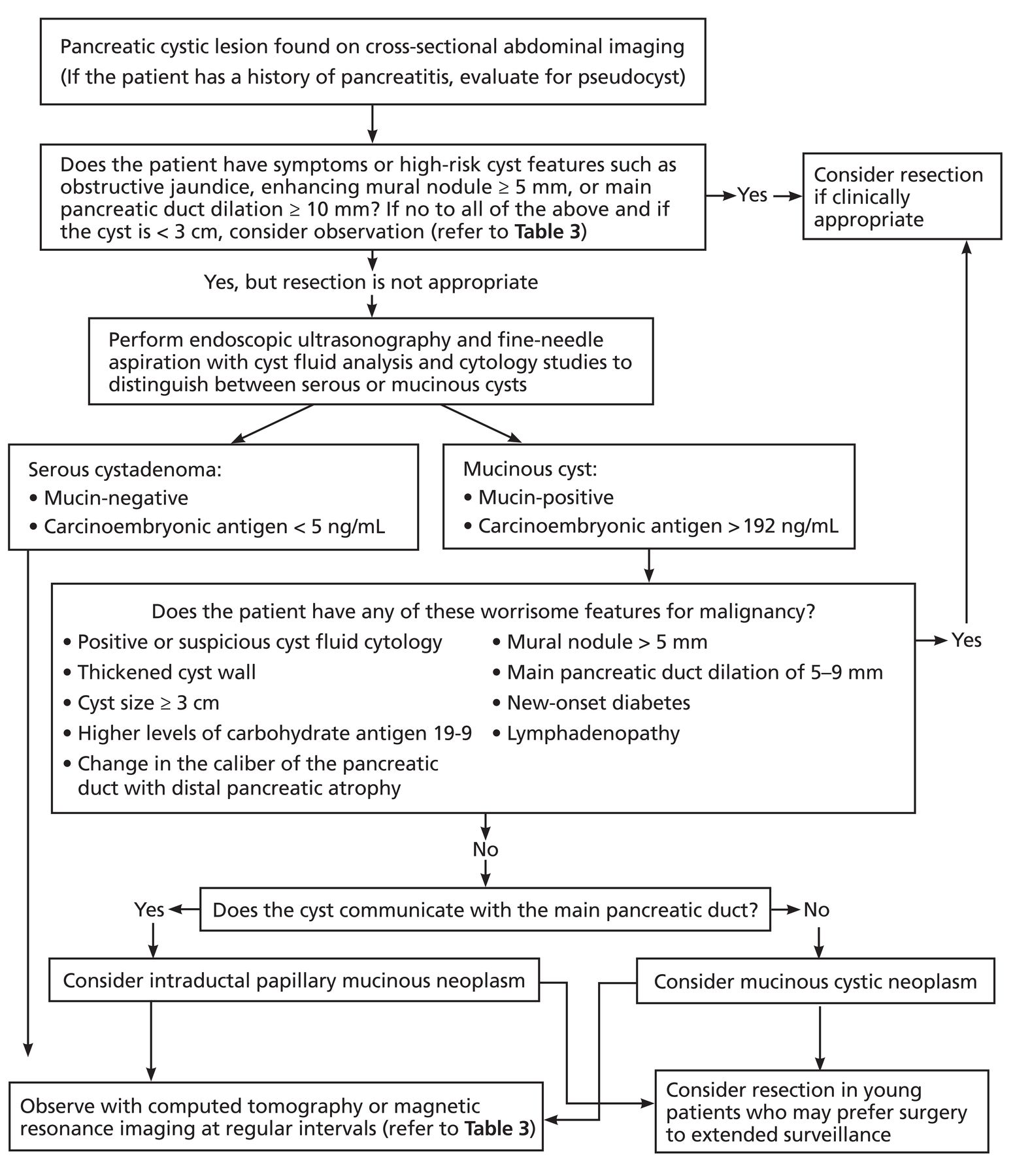
Due to its exceptional resolution and ability to discern the main pancreatic duct effectively, magnetic resonance imaging and magnetic resonance cholangiopancreatography are used for surveillance. On the other hand, endoscopic ultrasonography is reserved for patients displaying concerning characteristics, in addition to fine-needle aspiration of cyst fluid for a precise diagnosis through biomarker analysis. However, for lesions with no worrisome features, a combination of history, examination, and radiologic characteristics may commonly define the type of PCL and assess the risk of malignant degeneration.26 Figure 1 outlines a strategy to evaluate and manage PCLs.

Figure 1
  • Download figure
  • Open in new tab
  • Download powerpoint
Figure 1

Strategy to evaluate and manage pancreatic cystic lesions.

The duration of PCL surveillance is debatable, with most current guidelines recommending the surveillance interval based on radiologic PCL appearance and changes over time compared with previous imaging,6,7,10 while some advocate stopping after 5 years if the PCL is stable and has not progressed.9 If the patient is unwilling to undergo pancreatic surgery or is unfit for surgery, then asymptomatic PCL surveillance may be stopped as it is unlikely to impact clinical management or survival.6,7,10

Experts advocate maintaining surveillance till age 75 and individualizing follow-up between ages 76 and 85 (Table 3).6–10 It is also advised to inform patients that they may require continued surveillance even after undergoing partial pancreatic resection, as recurrence may occur in the remnant pancreas.6,7,35,36 Although it is difficult to find strong prospective evidence that surveillance reduces mortality, studies have shown that PCLs with the potential to become malignant can take years to develop, and that pancreatic cancers detected through surveillance were more frequently at an earlier stage in patients with intraductal papillary mucinous neoplasms.7,36

View this table:
  • View inline
  • View popup
TABLE 3

Approach to surveillance of pancreatic cystic neoplasms based on the different society guidelines

CONCLUSION

Pancreatic cysts are frequently found incidentally on cross-sectional imaging. The possibility of malignancy varies depending on the type of PCL. Magnetic resonance cholangiopancreatography with dynamic magnetic resonance imaging is the preferred test to identify cyst characteristics and high-risk or worrisome features. PCLs with malignant potential are treated by close surveillance or surgical excision. A multidisciplinary team should assess PCLs with high-risk characteristics and those with a known high risk of malignancy, such as main duct intraductal papillary mucinous neoplasms, mucinous cystic neoplasms, and solid pseudopapillary tumors.

Because advanced neoplasia is unlikely, active surveillance is appropriate for asymptomatic cysts and those that do not have any high-risk characteristics. Surgery should be performed to remove high-risk PCLs or those that progress while under surveillance. The overall prognosis is favorable, with early detection and active surveillance serving as the cornerstones of management.

DISCLOSURES

Dr. Stevens has disclosed teaching and speaking for Abbvie Pharmaceuticals. The other authors report no relevant financial relationships which, in the context of their contributions, could be perceived as a potential conflict of interest.

  • Copyright © 2024 The Cleveland Clinic Foundation. All Rights Reserved.

REFERENCES

  1. ↵
    1. Morris-Stiff G,
    2. Falk GA,
    3. Chalikonda S,
    4. Walsh RM
    . Natural history of asymptomatic pancreatic cystic neoplasms. HPB (Oxford) 2013; 15(3):175–181. doi:10.1111/j.1477-2574.2012.00522.x
    OpenUrlCrossRef
  2. ↵
    1. Zerboni G,
    2. Signoretti M,
    3. Crippa S,
    4. Falconi M,
    5. Arcidiacono PG,
    6. Capurso G
    . Systematic review and meta-analysis: prevalence of incidentally detected pancreatic cystic lesions in asymptomatic individuals. Pancreatology 2019; 19(1):2–9. doi:10.1016/j.pan.2018.11.014
    OpenUrlCrossRef
  3. ↵
    1. Perri G,
    2. Marchegiani G,
    3. Frigerio I, et al
    . Management of pancreatic cystic lesions. Dig Surg 2020; 37(1):1–9. doi:10.1159/000496509
    OpenUrlCrossRef
    1. Correa-Gallego C,
    2. Ferrone CR,
    3. Thayer SP,
    4. Wargo JA,
    5. Warshaw AL,
    6. Fernández-Del Castillo C
    . Incidental pancreatic cysts: do we really know what we are watching? Pancreatology 2010; 10(2–3):144–150. doi:10.1159/000243733
    OpenUrlCrossRefPubMed
  4. ↵
    1. de la Fuente J,
    2. Chatterjee A,
    3. Lui J, et al
    . Long-term outcomes and risk of pancreatic cancer in intraductal papillary mucinous neoplasms. JAMA Netw Open 2023;6(10):e2337799. doi:10.1001/jamanetworkopen.2023.37799
    OpenUrlCrossRef
  5. ↵
    1. Ohtsuka T,
    2. Fernandez-Del Castillo C,
    3. Furukawa T, et al
    . International evidence-based Kyoto guidelines for the management of intra-ductal papillary mucinous neoplasm of the pancreas. Pancreatology 2023; 28:S1424–3903(23)01883–5. doi:10.1016/j.pan.2023.12.009
    OpenUrlCrossRef
  6. ↵
    1. Elta GH,
    2. Enestvedt BK,
    3. Sauer BG,
    4. Lennon AM
    . ACG clinical guideline: diagnosis and management of pancreatic cysts. Am J Gastroenterol 2018; 113(4):464–479. doi:10.1038/ajg.2018.14
    OpenUrlCrossRefPubMed
  7. ↵
    1. Megibow AJ,
    2. Baker ME,
    3. Morgan DE, et al
    . Management of incidental pancreatic cysts: a white paper of the ACR Incidental Findings Committee. J Am Coll Radiol 2017; 14(7):911–923. doi:10.1016/j.jacr.2017.03.010
    OpenUrlCrossRefPubMed
  8. ↵
    1. Vege SS,
    2. Ziring B,
    3. Jain R,
    4. Moayyedi P; Clinical Guidelines Committee; American Gastroenterology Association
    . American Gastroenterological Association Institute guideline on the diagnosis and management of asymptomatic neoplastic pancreatic cysts. Gastroenterology 2015; 148(4):819–822. doi:10.1053/j.gastro.2015.01.015
    OpenUrlCrossRefPubMed
  9. ↵
    1. European Study Group on Cystic Tumours of the Pancreas
    . European evidence-based guidelines on pancreatic cystic neoplasms. Gut 2018; 67(5):789–804. doi:10.1136/gutjnl-2018-316027
    OpenUrlAbstract/FREE Full Text
  10. ↵
    1. Smith-Bindman R,
    2. Miglioretti DL,
    3. Larson EB
    . Rising use of diagnostic medical imaging in a large integrated health system. Health Aff (Millwood) 2008; 27(6):1491–1502. doi:10.1377/hlthaff.27.6.1491
    OpenUrlAbstract/FREE Full Text
  11. ↵
    1. Moris M,
    2. Bridges MD,
    3. Pooley RA, et al
    . Association between advances in high-resolution cross-section imaging technologies and increase in prevalence of pancreatic cysts from 2005 to 2014. Clin Gastroenterol Hepatol 2016; 14(4):585–593.e3. doi:10.1016/j.cgh.2015.08.038
    OpenUrlCrossRefPubMed
  12. ↵
    1. Zhu S,
    2. Wang WT,
    3. Shang XS, et al
    . Difference analysis in prevalence of incidental pancreatic cystic lesions between computed tomography and magnetic resonance imaging. BMC Med Imaging 2019; 19(1):43. doi:10.1186/s12880-019-0341-5
    OpenUrlCrossRef
  13. ↵
    1. Kromrey ML,
    2. Bülow R,
    3. Hübner J, et al
    . Prospective study on the incidence, prevalence and 5-year pancreatic-related mortality of pancreatic cysts in a population-based study. Gut 2018; 67(1): 138–145. doi:10.1136/gutjnl-2016-313127
    OpenUrlAbstract/FREE Full Text
    1. Banks PA,
    2. Bollen TL,
    3. Dervenis C, et al
    . Classification of acute pancreatitis—2012: revision of the Atlanta classification and definitions by international consensus. Gut 2013; 62(1):102–111. doi:10.1136/gutjnl-2012-302779
    OpenUrlAbstract/FREE Full Text
  14. ↵
    1. Bergin D,
    2. Ho LM,
    3. Jowell PS,
    4. Pappas TN,
    5. Paulson EK
    . Simple pancreatic cysts: CT and endosonographic appearances. AJR Am J Roentgenol 2002; 178(4):837–840. doi:10.2214/ajr.178.4.1780837
    OpenUrlCrossRefPubMed
  15. ↵
    1. Adsay NV,
    2. Hasteh F,
    3. Cheng JD, et al
    . Lymphoepithelial cysts of the pancreas: a report of 12 cases and a review of the literature. Mod Pathol 2002; 15(5):492–501. doi:10.1038/modpathol.3880553
    OpenUrlCrossRefPubMed
  16. ↵
    1. Nasr J,
    2. Sanders M,
    3. Fasanella K,
    4. Khalid A,
    5. McGrath K
    . Lymphoepithelial cysts of the pancreas: an EUS case series. Gastrointest Endosc 2008; 68(1):170–173. doi:10.1016/j.gie.2008.02.044
    OpenUrlCrossRefPubMed
  17. ↵
    1. Assifi MM,
    2. Nguyen PD,
    3. Agrawal N, et al
    . Non-neoplastic epithelial cysts of the pancreas: a rare, benign entity. J Gastrointest Surg 2014; 18(3):523–531. doi:10.1007/s11605-014-2459-7
    OpenUrlCrossRef
  18. ↵
    1. Ayoub F,
    2. Davis AM,
    3. Chapman CG
    . Pancreatic cysts-an overview and summary of society guidelines, 2021. JAMA 2021; 325(4):391–392. doi:10.1001/jama.2020.18678
    OpenUrlCrossRef
  19. ↵
    1. Nilsson LN,
    2. Keane MG,
    3. Shamali A, et al
    . Nature and management of pancreatic mucinous cystic neoplasm (MCN): a systematic review of the literature. Pancreatology 2016; 16(6):1028–1036. doi:10.1016/j.pan.2016.09.011
    OpenUrlCrossRef
  20. ↵
    1. Stark A,
    2. Donahue TR,
    3. Reber HA,
    4. Hines OJ
    . Pancreatic cyst disease: a review. JAMA 2016; 315(17):1882–1893. doi:10.1001/jama.2016.4690
    OpenUrlCrossRef
  21. ↵
    1. Jais B,
    2. Rebours V,
    3. Malleo G, et al
    . Serous cystic neoplasm of the pancreas: a multinational study of 2622 patients under the auspices of the International Association of Pancreatology and European Pancreatic Club (European Study Group on Cystic Tumors of the Pancreas). Gut 2016; 65(2):305–312. doi:10.1136/gutjnl-2015-309638
    OpenUrlAbstract/FREE Full Text
  22. ↵
    1. Kimura W,
    2. Moriya T,
    3. Hirai I, et al
    . Multicenter study of serous cystic neoplasm of the Japan pancreas society [published correction appears in Pancreas 2013; 42(1):186]. Pancreas 2012; 41(3):380–387. doi:10.1097/MPA.0b013e31822a27db
    OpenUrlCrossRefPubMed
  23. ↵
    1. Law JK,
    2. Ahmed A,
    3. Singh VK, et al
    . A systematic review of solid-pseudopapillary neoplasms: are these rare lesions? Pancreas 2014; 43(3):331–337. doi:10.1097/MPA.0000000000000061
    OpenUrlCrossRefPubMed
  24. ↵
    1. Smith ZL,
    2. Satyavada S,
    3. Simons-Linares R, et al
    . Intracystic glucose and carcinoembryonic antigen in differentiating histologically confirmed pancreatic mucinous neoplastic cysts. Am J Gastroenterol 2022; 117(3):478–485. doi:10.14309/ajg.0000000000001623
    OpenUrlCrossRef
  25. ↵
    1. Raman A,
    2. Lennon AM
    . Cyst fluid biomarkers—diagnosis and prediction of malignancy for cystic lesions of the pancreas. Visc Med 2018; 34(3):178–181. doi:10.1159/000490137
    OpenUrlCrossRef
  26. ↵
    1. Del Chiaro M,
    2. Segersvärd R,
    3. Pozzi Mucelli R, et al
    . Comparison of preoperative conference-based diagnosis with histology of cystic tumors of the pancreas. Ann Surg Oncol 2014; 21(5):1539–1544. doi:10.1245/s10434-013-3465-9
    OpenUrlCrossRefPubMed
  27. ↵
    1. Das KK
    . The “next generation” of pancreatic cyst fluid biomarkers? Gastroenterology 2023; 164(1):21–23. doi:10.1053/j.gastro.2022.10.023
    OpenUrlCrossRef
  28. ↵
    1. Singhi AD,
    2. McGrath K,
    3. Brand RE, et al
    . Preoperative next-generation sequencing of pancreatic cyst fluid is highly accurate in cyst classification and detection of advanced neoplasia. Gut 2018; 67(12):2131–2141. doi:10.1136/gutjnl-2016-313586
    OpenUrlAbstract/FREE Full Text
    1. Majumder S,
    2. Chatterjee A,
    3. Taylor WR, et al
    . Sa301: Feasibility of detecting cancer in intraductal papillary mucinous neoplasms using plasma methylated DNA markers. Gastroenterology 2021; 160(6):S–476. doi:10.1016/s0016-5085(21)01844-8
    OpenUrlCrossRef
    1. Majumder S,
    2. Taylor WR,
    3. Yab TC, et al
    . Novel methylated DNA markers discriminate advanced neoplasia in pancreatic cysts: marker discovery, tissue validation, and cyst fluid testing. Am J Gastroenterol 2019; 114(9):1539–1549. doi:10.14309/ajg.0000000000000284
    OpenUrlCrossRef
    1. Alles AJ,
    2. Warshaw AL,
    3. Southern JF,
    4. Compton CC,
    5. Lewandrowski KB
    . Expression of CA 72-4 (TAG-72) in the fluid contents of pancreatic cysts. a new marker to distinguish malignant pancreatic cystic tumors from benign neoplasms and pseudocysts. Ann Surg 1994; 219(2):131–134. doi:10.1097/00000658-199402000-00004
    OpenUrlCrossRefPubMed
    1. Okasha HH,
    2. Abdellatef A,
    3. Elkholy S, et al
    . Role of endoscopic ultrasound and cyst fluid tumor markers in diagnosis of pancreatic cystic lesions. World J Gastrointest Endosc 2022; 14(6):402–415. doi:10.4253/wjge.v14.i6.402
    OpenUrlCrossRef
  29. ↵
    1. Majumder S,
    2. Philip NA,
    3. Singh Nagpal SJ, et al
    . High-grade dysplasia in resected main-duct intraductal papillary mucinous neoplasm (MD-IPMN) is associated with an increased risk of subsequent pancreatic cancer. Am J Gastroenterol 2019; 114(3):524–529. doi:10.1038/s41395-018-0403-2
    OpenUrlCrossRef
  30. ↵
    1. de la Fuente J,
    2. Lui J,
    3. Lennon RJ, et al
    . Pancreatic cancer is more frequently early stage at diagnosis in surgically resected intraductal papillary mucinous neoplasms with preoperative surveillance. Gastro Hep Adv 2022; 1(6):1099–1107. doi:10.1016/j.gastha.2022.07.004
    OpenUrlCrossRef
PreviousNext
Back to top

In this issue

Cleveland Clinic Journal of Medicine: 91 (2)
Cleveland Clinic Journal of Medicine
Vol. 91, Issue 2
1 Feb 2024
  • Table of Contents
  • Table of Contents (PDF)
  • Index by author
  • Complete Issue (PDF)
Print
Download PDF
Article Alerts
Sign In to Email Alerts with your Email Address
Email Article

Thank you for your interest in spreading the word on Cleveland Clinic Journal of Medicine.

NOTE: We only request your email address so that the person you are recommending the page to knows that you wanted them to see it, and that it is not junk mail. We do not capture any email address.

Enter multiple addresses on separate lines or separate them with commas.
Diagnosis and management of pancreatic cystic lesions for the non-gastroenterologist
(Your Name) has sent you a message from Cleveland Clinic Journal of Medicine
(Your Name) thought you would like to see the Cleveland Clinic Journal of Medicine web site.
CAPTCHA
This question is for testing whether or not you are a human visitor and to prevent automated spam submissions.
Citation Tools
Diagnosis and management of pancreatic cystic lesions for the non-gastroenterologist
Arjun Chatterjee, Tyler Stevens, Prabhleen Chahal
Cleveland Clinic Journal of Medicine Feb 2024, 91 (2) 96-102; DOI: 10.3949/ccjm.91a.23019

Citation Manager Formats

  • BibTeX
  • Bookends
  • EasyBib
  • EndNote (tagged)
  • EndNote 8 (xml)
  • Medlars
  • Mendeley
  • Papers
  • RefWorks Tagged
  • Ref Manager
  • RIS
  • Zotero
Share
Diagnosis and management of pancreatic cystic lesions for the non-gastroenterologist
Arjun Chatterjee, Tyler Stevens, Prabhleen Chahal
Cleveland Clinic Journal of Medicine Feb 2024, 91 (2) 96-102; DOI: 10.3949/ccjm.91a.23019
Twitter logo Facebook logo Mendeley logo
  • Tweet Widget
  • Facebook Like
  • Linkedin Share Button

Jump to section

  • Article
    • ABSTRACT
    • EPIDEMIOLOGY
    • PCL CLASSIFICATION
    • PCL CANCER RISK
    • DIAGNOSIS
    • MANAGEMENT AND PROGNOSIS
    • CONCLUSION
    • DISCLOSURES
    • REFERENCES
  • Figures & Data
  • Info & Metrics
  • PDF

Related Articles

  • No related articles found.
  • PubMed
  • Google Scholar

Cited By...

  • No citing articles found.
  • Google Scholar

More in this TOC Section

  • Alpha-gal syndrome: Recognizing and managing a tick-bite–related meat allergy
  • Managing right ventricular failure in the setting of pulmonary embolism
  • Psychedelics, spirituality, and existential distress in patients at the end of life
Show more Review

Similar Articles

Subjects

  • Gastroenterology
  • Imaging

Navigate

  • Current Issue
  • Past Issues
  • Supplements
  • Article Type
  • Specialty
  • CME/MOC Articles
  • CME/MOC Calendar
  • Media Kit

Authors & Reviewers

  • Manuscript Submission
  • Authors & Reviewers
  • Subscriptions
  • About CCJM
  • Contact Us
  • Cleveland Clinic Center for Continuing Education
  • Consult QD

Share your suggestions!

Copyright © 2025 The Cleveland Clinic Foundation. All rights reserved. The information provided is for educational purposes only. Use of this website is subject to the website terms of use and privacy policy. 

Powered by HighWire