
Main menu

  • Home
  • Content
    • Current Issue
    • Ahead of Print
    • Past Issues
    • Supplements
    • Article Type
  • Specialty
    • Articles by Specialty
  • CME/MOC
    • Articles
    • Calendar
  • Info For
    • Manuscript Submission
    • Authors & Reviewers
    • Subscriptions
    • About CCJM
    • Contact Us
    • Media Kit
  • Conversations with Leaders
  • Conference Coverage
    • Kidney Week 2024
    • CHEST 2024
    • ACR Convergence 2023
    • Kidney Week 2023
    • ObesityWeek 2023
    • IDWeek 2023
    • CHEST 2023
    • MDS 2023
    • IAS 2023
    • ACP 2023
    • AAN 2023
    • ACC / WCC 2023
    • AAAAI Meeting 2023
    • ACR Convergence 2022
    • Kidney Week 2022
    • AIDS 2022
  • Other Publications
    • www.clevelandclinic.org

User menu

  • Register
  • Log in

Search

  • Advanced search
Cleveland Clinic Journal of Medicine
  • Other Publications
    • www.clevelandclinic.org
  • Register
  • Log in
Cleveland Clinic Journal of Medicine

Advanced Search

  • Home
  • Content
    • Current Issue
    • Ahead of Print
    • Past Issues
    • Supplements
    • Article Type
  • Specialty
    • Articles by Specialty
  • CME/MOC
    • Articles
    • Calendar
  • Info For
    • Manuscript Submission
    • Authors & Reviewers
    • Subscriptions
    • About CCJM
    • Contact Us
    • Media Kit
  • Conversations with Leaders
  • Conference Coverage
    • Kidney Week 2024
    • CHEST 2024
    • ACR Convergence 2023
    • Kidney Week 2023
    • ObesityWeek 2023
    • IDWeek 2023
    • CHEST 2023
    • MDS 2023
    • IAS 2023
    • ACP 2023
    • AAN 2023
    • ACC / WCC 2023
    • AAAAI Meeting 2023
    • ACR Convergence 2022
    • Kidney Week 2022
    • AIDS 2022
The Clinical Picture

Page kidney after a renal biopsy

Hiroyuki Teruya, MD, Hiroyuki Yano, MD, Hitoshi Miyasato, MD and Mitsuyo Kinjo, MD, MPH
Cleveland Clinic Journal of Medicine May 2020, 87 (5) 271-272; DOI: https://doi.org/10.3949/ccjm.87a.19130
Hiroyuki Teruya
Department of Internal Medicine, Okinawa Miyako Hospital, Okinawa, Japan
  • Find this author on Google Scholar
  • Find this author on PubMed
  • Search for this author on this site
Hiroyuki Yano
Department of Internal Medicine, Okinawa Chubu Hospital, Okinawa, Japan
  • Find this author on Google Scholar
  • Find this author on PubMed
  • Search for this author on this site
Hitoshi Miyasato
Department of Internal Medicine, Okinawa Chubu Hospital, Okinawa, Japan
  • Find this author on Google Scholar
  • Find this author on PubMed
  • Search for this author on this site
Mitsuyo Kinjo
Department of Internal Medicine, Okinawa Chubu Hospital, Okinawa, Japan
  • Find this author on Google Scholar
  • Find this author on PubMed
  • Search for this author on this site
  • For correspondence: [email protected]
  • Article
  • Figures & Data
  • Info & Metrics
  • PDF
Loading

A 25-year-old man presented with sudden onset of left flank pain upon standing up from a chair 3 weeks after undergoing renal biopsy. The procedure had been an ultrasonographically guided percutaneous renal biopsy of the lower pole of the left kidney, with 3 passes, to evaluate hematuria and nephritic-range proteinuria and had resulted in the diagnosis of immunoglobulin A (IgA) nephropathy.

The patient had a history of hypertension, for which he was taking nifedipine 40 mg daily, which had been keeping his blood pressure below 130/80 mm Hg. As an infant he had had congenital hydrocephalus, for which a ventriculoperitoneal shunt had been placed.

On examination, his blood pressure was elevated at 160/80 mm Hg; other vital signs were stable. Abdominal examination revealed left flank tenderness and left costovertebral angle tenderness. The rest of the physical examination was unremarkable.

His blood urea nitrogen level was 13 mg/dL (reference range 8–20), and his serum creatinine was 1.40 mg/dL, up from 1.20 mg/dL before the biopsy (reference range 0.65–1.07). His electrolyte levels, liver function test results, and complete blood cell counts were normal.

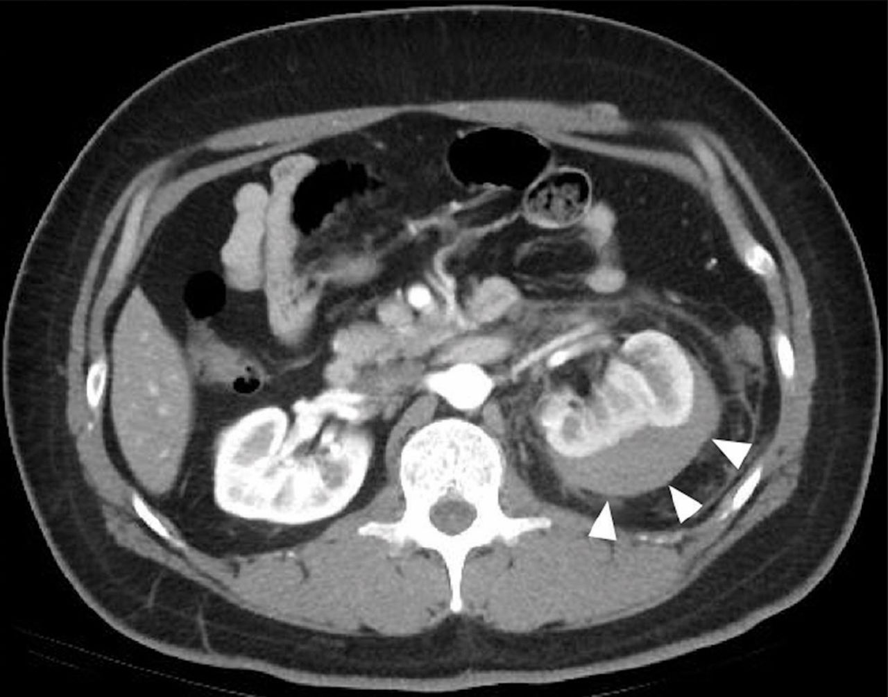
Computed tomography (CT) with contrast revealed a large pericapsular collection with high attenuation (70 Hounsfield units [HU]) in the left kidney, compressing the renal parenchyma (Figures 1 and 2).

Figure 1
  • Download figure
  • Open in new tab
  • Download powerpoint
Figure 1

Axial computed tomography with contrast of the mid-kidney shows that the renal parenchyma is compressed by a contained, high-attenuation (70 HU), pericapsular collection (arrowheads).

Figure 2
  • Download figure
  • Open in new tab
  • Download powerpoint
Figure 2

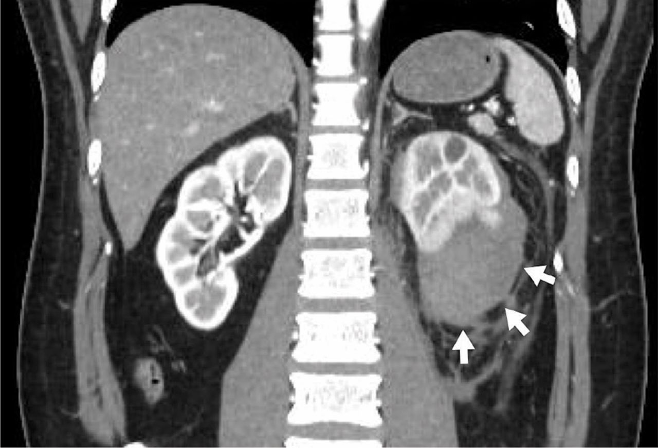
Coronal computed tomography with contrast shows that the lower pole is compressed by a contained, high-attenuation, pericapsular collection (arrows).

In view of his worsening hypertension and the pericapsular hematoma in his left kidney, we suspected Page kidney as a complication of renal biopsy.

Two days after admission, his creatinine level had increased to 2.75 mg/dL. Four days after admission, his plasma renin activity was elevated to 11 μg/L/hour (reference range 0.3–2.9) and his serum aldosterone level was 337 pg/mL (reference range 29.9–159), establishing the diagnosis of Page kidney.

Intravenous nicardipine and fentanyl were given. Although his pain subsided significantly, his blood pressure remained high. Renin-angiotensin-aldosterone system (RAAS) inhibitors were initially deferred in view of his acute kidney injury, and nicardipine was continued.

The patient subsequently underwent laparoscopic evacuation of the hematoma. Afterward, CT confirmed that the hematoma was gone, but his kidney function did not improve. Therefore, 6 days after admission, enalapril 2.5 mg daily was added to his regimen, and his blood pressure returned to normal over the next several days. One month later, his serum creatinine level was down to 1.28 mg/dL, his plasma renin activity was 0.3 μg/L/hour, and his aldosterone level was 59.7 pg/mL.

PAGE KIDNEY

Page kidney, a condition in which an affected kidney is compressed by external force, is an uncommon cause of secondary hypertension and renal insufficiency. Other possible causes of secondary hypertension include renal artery stenosis, juxtaglomerular cell tumor, and malignant hypertension.

Page kidney was first reported in 1939 by Irvine Page,1 who induced it in a dog by wrapping the animal’s kidney in cellophane. The presumed mechanism is that direct external compression of the kidney causes decreased renal perfusion and increased renin secretion, resulting in activation of the RAAS system and secondary hypertension.2

The most common cause of constrictive pressure on the kidney is hematoma due to abdominal trauma, surgery, or percutaneous interventions. Page kidney has often been reported after traumatic biopsy of kidney allografts, but more rarely after native kidney biopsy.

Ultrasonography and CT are useful for detecting hematoma.

As activation of the RAAS is the central mechanism of hypertension in Page kidney, we deemed it suitable to give enalapril, a drug that blocks the RAAS, and nicardipine, a calcium channel blocker, to control hypertension and prevent further kidney damage. However, few studies have investigated optimal antihypertensive therapy in Page kidney. Percutaneous or open drainage of the hematoma may be needed for patients with uncontrolled hypertension or worsening renal function. Surgical nephrectomy is occasionally required to control hypertension.3

Page kidney should be considered in a patient with new-onset hypertension and flank pain after native kidney biopsy. Early recognition can allow for conservative treatment, which can improve this condition and preserve kidney function.4

Acknowledgment

The authors thank Dr. Rita McGill for editing this article.

  • Copyright © 2020 The Cleveland Clinic Foundation. All Rights Reserved.

REFERENCES

  1. ↵
    1. Page IH
    . The production of persistent arterial hypertension by cellophane perinephritis. JAMA 1939; 113(23):2046–2048. doi:10.1001/jama.1939.02800480032008
    OpenUrlCrossRef
  2. ↵
    1. Wahdat R,
    2. Schwartz C,
    3. Espinosa J,
    4. Lucerna A
    . Page kidney: taking a page from history. Am J Emerg Med 2017; 35(1):193.e1–193.e2. doi:10.1016/j.ajem.2016.06.095
    OpenUrlCrossRef
  3. ↵
    1. Kiczek M,
    2. Udayasankar U
    . Page kidney. J Urol 2015; 194(4):1109–1110. doi:10.1016/j.juro.2015.07.035
    OpenUrlCrossRef
  4. ↵
    1. Sokhal AK,
    2. Prakash G,
    3. Saini DK,
    4. Singh K,
    5. Sankhwar S,
    6. Singh BP
    . Page kidney: a rare but surgically treatable cause of hypertension. Saudi J Kidney Dis Transpl 2018; 29(1):193–197. doi:10.4103/1319-2442.225183
    OpenUrlCrossRef
PreviousNext
Back to top

In this issue

Cleveland Clinic Journal of Medicine: 87 (5)
Cleveland Clinic Journal of Medicine
Vol. 87, Issue 5
1 May 2020
  • Table of Contents
  • Table of Contents (PDF)
  • Index by author
  • Complete Issue (PDF)
Print
Download PDF
Article Alerts
Sign In to Email Alerts with your Email Address
Email Article

Thank you for your interest in spreading the word on Cleveland Clinic Journal of Medicine.

NOTE: We only request your email address so that the person you are recommending the page to knows that you wanted them to see it, and that it is not junk mail. We do not capture any email address.

Enter multiple addresses on separate lines or separate them with commas.
Page kidney after a renal biopsy
(Your Name) has sent you a message from Cleveland Clinic Journal of Medicine
(Your Name) thought you would like to see the Cleveland Clinic Journal of Medicine web site.
CAPTCHA
This question is for testing whether or not you are a human visitor and to prevent automated spam submissions.
Citation Tools
Page kidney after a renal biopsy
Hiroyuki Teruya, Hiroyuki Yano, Hitoshi Miyasato, Mitsuyo Kinjo
Cleveland Clinic Journal of Medicine May 2020, 87 (5) 271-272; DOI: 10.3949/ccjm.87a.19130

Citation Manager Formats

  • BibTeX
  • Bookends
  • EasyBib
  • EndNote (tagged)
  • EndNote 8 (xml)
  • Medlars
  • Mendeley
  • Papers
  • RefWorks Tagged
  • Ref Manager
  • RIS
  • Zotero
Share
Page kidney after a renal biopsy
Hiroyuki Teruya, Hiroyuki Yano, Hitoshi Miyasato, Mitsuyo Kinjo
Cleveland Clinic Journal of Medicine May 2020, 87 (5) 271-272; DOI: 10.3949/ccjm.87a.19130
Twitter logo Facebook logo Mendeley logo
  • Tweet Widget
  • Facebook Like
  • Linkedin Share Button

Jump to section

  • Article
    • PAGE KIDNEY
    • Acknowledgment
    • REFERENCES
  • Figures & Data
  • Info & Metrics
  • PDF

Related Articles

  • No related articles found.
  • PubMed
  • Google Scholar

Cited By...

  • No citing articles found.
  • Google Scholar

More in this TOC Section

  • Tinea incognito
  • Prolonged venous filling time and dependent rubor in a patient with peripheral artery disease
  • Sarcoidosis with diffuse purplish erythematous plaques on the hands
Show more The Clinical Picture

Similar Articles

Subjects

  • Imaging
  • Nephrology

Navigate

  • Current Issue
  • Past Issues
  • Supplements
  • Article Type
  • Specialty
  • CME/MOC Articles
  • CME/MOC Calendar
  • Media Kit

Authors & Reviewers

  • Manuscript Submission
  • Authors & Reviewers
  • Subscriptions
  • About CCJM
  • Contact Us
  • Cleveland Clinic Center for Continuing Education
  • Consult QD

Share your suggestions!

Copyright © 2025 The Cleveland Clinic Foundation. All rights reserved. The information provided is for educational purposes only. Use of this website is subject to the website terms of use and privacy policy. 

Powered by HighWire