


Article Figures & Data
Figures
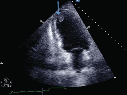
- FIGURE 1
Transthoracic echocardiography, apical four-chamber view, shows thrombus in the left ventricular apical cavity. The blue arrow points to the well-demarcated thrombus adhering to the endocardium.
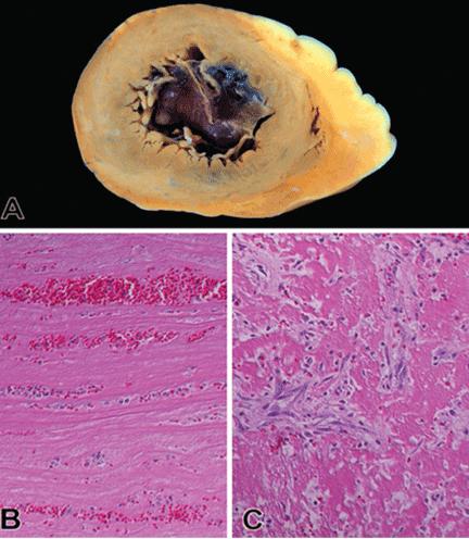
- FIGURE 2
(A) A cross section of the apical segment of the left ventricle shows a mildly dilated cavity filled with mural thrombus. (B) Photomicrograph of an acute thrombus shows alternating layers of fibrin and platelet with red and white blood cells (hematoxylin and eosin, original magnification × 200). (C) Organization of a thrombus is characterized by infiltration of fibroblasts and newly formed capillaries (hematoxylin and eosin, original magnification × 200).
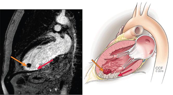
- FIGURE 3
Cardiac magnetic resonance imaging with a delayed-enhancement phase-sensitive inversion recovery image, vertical long-axis view. The red arrow points to dense subendocardial delayed enhancement in the apex extending into the mid-inferior wall, consistent with scar in the distal left anterior descending artery territory. The orange arrow shows a nonenhancing mass in the apex, consistent with thrombus.

Tables
- TABLE 1
Incidence of left ventricular thrombosis after ST-segment elevation myocardial infarction
Authors No. of patientsa Incidence of left ventricular thrombosis Predictors of left ventricular thrombosis Kalra and Jang,4
200071 4% Anterior infarction Nayak et al,5
2004200 11% Anterior infarction Rehan et al,6
200692 4% Anterior infarction Zielinska et al,7
20082,911 2.5% Anterior infarction
Left ventricular ejection fraction < 40%
HypertensionOsherov et al,8
2009642 6% Severe mitral valve regurgitation
Low left ventricular ejection fractionSolheim et al,9
2010100 15% Higher peak creatine kinase level
Larger infarcts
Lower left ventricular ejection fractionShacham et al,10
2013207 5% Higher C-reactive protein levels
Higher fibrinogen levelsGianstefani et al,11
20141,059 4% Low left ventricular ejection fraction
Anterior infarction
Use of glycoprotein IIb/IIIa inhibitors↵a Most patients underwent percutaneous coronary intervention.
- TABLE 2
Bleeding risk after percutaneous coronary intervention: Triple thrombotic therapy vs dual antiplatelet therapy
Authors No. of patients Absolute difference in major bleeding with warfarin Comments Khurram et al,49 2006 107 6.6% higher Bleeding defined as requiring > 2 units of packed red blood cells, or intraocular or disabling bleeding DeEugenio et al,52 2007 194 11% higher Hazard ratio 5.0 (95% confidence interval [CI] 1.4–17.8) with warfarin Karjalainen et al,40 2007 478 5.6% higher Odds ratio 3.4 (95% CI 1.2–9.3) with warfarin
Increased stent thrombosis with warfarin-aspirin combinationRuiz-Nodar et al,53 2008 426 5.9% higher All patients had atrial fibrillation
Mortality rate was higher without anticoagulationSarafoff et al,54 2008 515 1.7% lower (not statistically significant) Both dual antiplatelet therapy and triple therapy had favorable efficacy and safety Rossini et al,55 2008b 204 0.9% higher (not statistically significant) Bleeding rate was lower if the international normalized ratio was kept between 2 and 2.5: 4.9% vs 33% at 3 months ↵a This was a prospective study. The other studies in this table were retrospective.


Jump to section
- Article
- THE INCIDENCE OF LEFT VENTRICULAR THROMBOSIS IN ACUTE MI
- WHAT IS THE PATHOGENESIS OF LEFT VENTRICULAR THROMBOSIS?
- HOW IS LEFT VENTRICULAR THROMBOSIS DIAGNOSED?
- WHAT COMPLICATIONS ARISE FROM LEFT VENTRICULAR THROMBOSIS?
- ANTICOAGULATION TREATMENT
- BLEEDING COMPLICATIONS WITH TRIPLE ANTITHROMBOTIC THERAPY
- CASE FOLLOW-UP
- TAKE-HOME POINTS
- REFERENCES
- Figures & Data
- Info & Metrics
Related Articles
Cited By...
- No citing articles found.