
Main menu

  • Home
  • Content
    • Current Issue
    • Ahead of Print
    • Past Issues
    • Supplements
    • Article Type
  • Specialty
    • Articles by Specialty
  • CME/MOC
    • Articles
    • Calendar
  • Info For
    • Manuscript Submission
    • Authors & Reviewers
    • Subscriptions
    • About CCJM
    • Contact Us
    • Media Kit
  • Conversations with Leaders
  • Conference Coverage
    • ACC / WCC 2023
    • AAAAI Meeting 2023
    • ACR Convergence 2022
    • Kidney Week 2022
    • AIDS 2022
    • CHEST 2021
    • IDWeek 2021
    • IAS 2021
    • ADA 2021
    • ATS 2021
    • ACC 2021
    • ACP 2021
    • AAN 2021
  • Other Publications
    • www.clevelandclinic.org

User menu

  • Register
  • Log in

Search

  • Advanced search
Cleveland Clinic Journal of Medicine
  • Other Publications
    • www.clevelandclinic.org
  • Register
  • Log in
Cleveland Clinic Journal of Medicine

Advanced Search

  • Home
  • Content
    • Current Issue
    • Ahead of Print
    • Past Issues
    • Supplements
    • Article Type
  • Specialty
    • Articles by Specialty
  • CME/MOC
    • Articles
    • Calendar
  • Info For
    • Manuscript Submission
    • Authors & Reviewers
    • Subscriptions
    • About CCJM
    • Contact Us
    • Media Kit
  • Conversations with Leaders
  • Conference Coverage
    • ACC / WCC 2023
    • AAAAI Meeting 2023
    • ACR Convergence 2022
    • Kidney Week 2022
    • AIDS 2022
    • CHEST 2021
    • IDWeek 2021
    • IAS 2021
    • ADA 2021
    • ATS 2021
    • ACC 2021
    • ACP 2021
    • AAN 2021
The Clinical Picture

Imaging suggestive, but symptoms atypical

Mohammad Afzalimoghaddam, MD, Maryam Edalatifard, MD, Shervin Farahmand, MD, Shahram Bagheri-Hariri, MD and Hadi Mirfazaelian, MD
Cleveland Clinic Journal of Medicine January 2017, 84 (1) 25-26; DOI: https://doi.org/10.3949/ccjm.84a.15144
Mohammad Afzalimoghaddam
Associate Professor of Emergency Medicine, Department of Emergency Medicine, Tehran University of Medical Sciences, Tehran, Iran
  • Find this author on Google Scholar
  • Find this author on PubMed
  • Search for this author on this site
  • For correspondence: mafzalimoghadam@yahoo.com
Maryam Edalatifard
Assistant Professor of Emergency Medicine, Department of Emergency Medicine, Tehran University of Medical Sciences, Tehran, Iran
  • Find this author on Google Scholar
  • Find this author on PubMed
  • Search for this author on this site
Shervin Farahmand
Associate Professor of Emergency Medicine, Department of Emergency Medicine, Tehran University of Medical Sciences, Tehran, Iran
  • Find this author on Google Scholar
  • Find this author on PubMed
  • Search for this author on this site
Shahram Bagheri-Hariri
Assistant Professor of Emergency Medicine, Department of Emergency Medicine, Tehran University of Medical Sciences, Tehran, Iran
  • Find this author on Google Scholar
  • Find this author on PubMed
  • Search for this author on this site
Hadi Mirfazaelian
Assistant Professor of Emergency Medicine, Department of Emergency Medicine, Tehran University of Medical Sciences, Tehran, Iran
  • Find this author on Google Scholar
  • Find this author on PubMed
  • Search for this author on this site
  • Article
  • Figures & Data
  • Info & Metrics
  • PDF
Loading

A 66-year-old man with chronic obstructive pulmonary disease (COPD) was brought to the emergency department with a 2-week history of progressive dyspnea followed by altered mental status. He had no history of diabetes mellitus, hypertension, or drug abuse.

On physical examination, he was stuporous. He had no fever or hypotension, but his pulse and breathing were rapid, and he had central cyanosis, bilateral conjunctival congestion, a puffy face, generalized wheezing, basilar crackles in both lungs, and leg edema.

Laboratory testing showed hypoxia and severe hypercarbia. His hematocrit was 65% (reference range 39–51) and his hemoglobin level was 21.5 g/dL (13–17).

The patient was diagnosed with an exacerbation of COPD. He was intubated, placed on mechanical ventilation, and admitted to the intensive care unit.

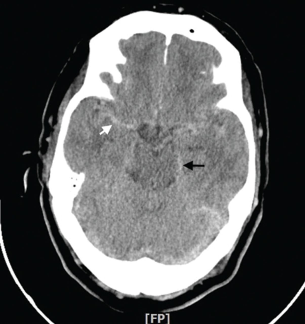
Computed tomography (CT) performed because of his decreased level of consciousness (Figure 1) showed increased attenuation in the ambient cistern and the lateral aspect of the lateral cerebral fissure, suggesting subarachnoid hemorrhage. The attenuation value in these areas was 89 Hounsfield units (typical values for brain tissue are in the 20s to 30s, and for blood in the 30s to 40s). To further evaluate for subarachnoid hemorrhage, lumbar puncture was performed, but analysis of the fluid sample showed normal protein and glucose levels and no cells.

FIGURE 1
  • Download figure
  • Open in new tab
  • Download powerpoint
FIGURE 1

Computed tomography showed increased attenuation in the ambient cistern (black arrow) and cortical sulci (white arrow).

Based on the results of cerebrospinal fluid evaluation and on the CT attenuation value, a diagnosis of pseudosubarachnoid hemorrhage due to polycythemia was made.

SUBARACHNOID VS PSEUDOSUBARACHNOID HEMORRHAGE

Subarachnoid hemorrhage typically begins with a “thunder-clap” headache (beginning suddenly and described by patients as “the worst headache ever.”) While not all patients have this presentation, if imaging suggests subarachnoid hemorrhage but the patient has atypical signs and symptoms (eg, other than headache), then pseudo-subarachnoid hemorrhage should be considered.

Brain CT is one of the most reliable tools for diagnosing subarachnoid hemorrhage in the emergency department. Done within 6 hours of symptom onset, it has a sensitivity of 98.7% and a specificity of 99.9%.1 Magnetic resonance imaging can also visualize subarachnoid hemorrhage within the first 12 hours, typically as a hyperintensity in the subarachnoid space on fluid-attenuated inversion-recovery sequences2 and on susceptibility-weighted sequences.

Lumbar puncture is also an important diagnostic tool but carries a risk of brain herniation in patients with brain edema.

Pseudosubarachnoid hemorrhage is an artifact of CT imaging. It is rare, and its prevalence is unknown.3 However, it may be seen in up to 20% of patients after resuscitation, as a result of diffuse cerebral edema that lowers the attenuation of brain tissue on CT, making the vessels relatively conspicuous. It can also be seen in purulent meningitis (due to proteinaceous influx after blood-brain barrier disruption),4 in meningeal leukemia (due to increased cellularity in the leptomeninges), and in severe polycythemia (from a higher concentration of blood and hemoglobin in the vessels).3,5–7

Although the level of attenuation on CT may help distinguish subarachnoid from pseu-dosubarachnoid hemorrhage, its accuracy has not been defined. Inspecting the CT images may clarify whether areas with high attenuation look like blood vessels vs subarachnoid hemorrhage.

Our patient recovered and had an uneventful follow-up. The cause of his elevated hematocrit was likely chronic hypoxia from COPD.

ACKNOWLEDGMENT

We thank Dr. Saeide Khanbagi and Dr. Azade Nasr-lari for their cooperation.

  • Copyright © 2017 The Cleveland Clinic Foundation. All Rights Reserved.

REFERENCES

  1. ↵
    1. Dubosh NM,
    2. Bellolio MF,
    3. Rabinstein AA,
    4. Edlow JA
    . Sensitivity of early brain computed tomography to exclude aneurysmal subarachnoid hemorrhage: a systematic review and meta-analysis. Stroke 2016; 47:750–755.
    OpenUrlAbstract/FREE Full Text
  2. ↵
    1. Sohn CH,
    2. Baik SK,
    3. Lee HJ,
    4. et al
    . MR imaging of hyperacute subarachnoid and intraventricular hemorrhage at 3T: a preliminary report of gradient echo T2*-weighted sequences. AJNR Am J Neuroradiol 2005; 26:662–665.
    OpenUrlAbstract/FREE Full Text
  3. ↵
    1. Yuzawa H,
    2. Higano S,
    3. Mugikura S,
    4. et al
    . Pseudo-subarachnoid hemorrhage found in patients with postresuscitation encephalopathy: characteristics of CT findings and clinical importance. AJNR Am J Neuroradiol 2008; 29:1544–1549.
    OpenUrlAbstract/FREE Full Text
  4. ↵
    1. Given CA 2nd.,
    2. Burdette JH,
    3. Elster AD,
    4. Williams DW 3rd.
    Pseudo-subarachnoid hemorrhage: a potential imaging pitfall associated with diffuse cerebral edema. AJNR Am J Neuroradiol 2003; 24:254–256.
    OpenUrlAbstract/FREE Full Text
  5. ↵
    1. Avrahami E,
    2. Katz R,
    3. Rabin A,
    4. Friedman V
    . CT diagnosis of non-traumatic subarachnoid haemorrhage in patients with brain edema. Eur J Radiol 1998; 28:222–225.
    OpenUrlCrossRefPubMed
    1. Ben Salem D,
    2. Osseby GV,
    3. Rezaizadeh-Bourdariat K,
    4. et al
    . Spontaneous hyperdense intracranial vessels seen on CT scan in polycythemia cases. J Radiol 2003; 84:605–608. French.
    OpenUrlPubMed
  6. ↵
    1. Hsieh SW,
    2. Khor GT,
    3. Chen CN,
    4. Huang P
    . Pseudo subarachnoid hemorrhage in meningeal leukemia. J Emerg Med 2012; 42:e109–e111.
    OpenUrlPubMed
PreviousNext
Back to top

In this issue

Cleveland Clinic Journal of Medicine: 84 (1)
Cleveland Clinic Journal of Medicine
Vol. 84, Issue 1
1 Jan 2017
  • Table of Contents
  • Table of Contents (PDF)
  • Index by author
Print
Download PDF
Article Alerts
Sign In to Email Alerts with your Email Address
Email Article

Thank you for your interest in spreading the word on Cleveland Clinic Journal of Medicine.

NOTE: We only request your email address so that the person you are recommending the page to knows that you wanted them to see it, and that it is not junk mail. We do not capture any email address.

Enter multiple addresses on separate lines or separate them with commas.
Imaging suggestive, but symptoms atypical
(Your Name) has sent you a message from Cleveland Clinic Journal of Medicine
(Your Name) thought you would like to see the Cleveland Clinic Journal of Medicine web site.
CAPTCHA
This question is for testing whether or not you are a human visitor and to prevent automated spam submissions.
Citation Tools
Imaging suggestive, but symptoms atypical
Mohammad Afzalimoghaddam, Maryam Edalatifard, Shervin Farahmand, Shahram Bagheri-Hariri, Hadi Mirfazaelian
Cleveland Clinic Journal of Medicine Jan 2017, 84 (1) 25-26; DOI: 10.3949/ccjm.84a.15144

Citation Manager Formats

  • BibTeX
  • Bookends
  • EasyBib
  • EndNote (tagged)
  • EndNote 8 (xml)
  • Medlars
  • Mendeley
  • Papers
  • RefWorks Tagged
  • Ref Manager
  • RIS
  • Zotero
Share
Imaging suggestive, but symptoms atypical
Mohammad Afzalimoghaddam, Maryam Edalatifard, Shervin Farahmand, Shahram Bagheri-Hariri, Hadi Mirfazaelian
Cleveland Clinic Journal of Medicine Jan 2017, 84 (1) 25-26; DOI: 10.3949/ccjm.84a.15144
Reddit logo Twitter logo Facebook logo Mendeley logo
  • Tweet Widget
  • Facebook Like
  • Linkedin Share Button

Jump to section

  • Article
    • SUBARACHNOID VS PSEUDOSUBARACHNOID HEMORRHAGE
    • ACKNOWLEDGMENT
    • REFERENCES
  • Figures & Data
  • Info & Metrics
  • PDF

Related Articles

  • No related articles found.
  • PubMed
  • Google Scholar

Cited By...

  • No citing articles found.
  • Google Scholar

More in this TOC Section

  • Mass under the lip
  • Oral leukoplakia and oral cancer
  • Median rhomboid glossitis caused by tongue-brushing
Show more The Clinical Picture

Similar Articles

Subjects

  • Neurology
  • Vascular Medicine
  • Imaging
  • Emergency Medicine

Navigate

  • Current Issue
  • Past Issues
  • Supplements
  • Article Type
  • Specialty
  • CME/MOC Articles
  • CME/MOC Calendar
  • Media Kit

Authors & Reviewers

  • Manuscript Submission
  • Authors & Reviewers
  • Subscriptions
  • About CCJM
  • Contact Us
  • Cleveland Clinic Center for Continuing Education
  • Consult QD

Share your suggestions!

Copyright © 2023 The Cleveland Clinic Foundation. All rights reserved. The information provided is for educational purposes only. Use of this website is subject to the website terms of use and privacy policy. 

Powered by HighWire