
Main menu

  • Home
  • Content
    • Current Issue
    • Ahead of Print
    • Past Issues
    • Supplements
    • Article Type
  • Specialty
    • Articles by Specialty
  • CME/MOC
    • Articles
    • Calendar
  • Info For
    • Manuscript Submission
    • Authors & Reviewers
    • Subscriptions
    • About CCJM
    • Contact Us
    • Media Kit
  • Conversations with Leaders
  • Conference Coverage
    • Kidney Week 2024
    • CHEST 2024
    • ACR Convergence 2023
    • Kidney Week 2023
    • ObesityWeek 2023
    • IDWeek 2023
    • CHEST 2023
    • MDS 2023
    • IAS 2023
    • ACP 2023
    • AAN 2023
    • ACC / WCC 2023
    • AAAAI Meeting 2023
    • ACR Convergence 2022
    • Kidney Week 2022
    • AIDS 2022
  • Other Publications
    • www.clevelandclinic.org

User menu

  • Register
  • Log in

Search

  • Advanced search
Cleveland Clinic Journal of Medicine
  • Other Publications
    • www.clevelandclinic.org
  • Register
  • Log in
Cleveland Clinic Journal of Medicine

Advanced Search

  • Home
  • Content
    • Current Issue
    • Ahead of Print
    • Past Issues
    • Supplements
    • Article Type
  • Specialty
    • Articles by Specialty
  • CME/MOC
    • Articles
    • Calendar
  • Info For
    • Manuscript Submission
    • Authors & Reviewers
    • Subscriptions
    • About CCJM
    • Contact Us
    • Media Kit
  • Conversations with Leaders
  • Conference Coverage
    • Kidney Week 2024
    • CHEST 2024
    • ACR Convergence 2023
    • Kidney Week 2023
    • ObesityWeek 2023
    • IDWeek 2023
    • CHEST 2023
    • MDS 2023
    • IAS 2023
    • ACP 2023
    • AAN 2023
    • ACC / WCC 2023
    • AAAAI Meeting 2023
    • ACR Convergence 2022
    • Kidney Week 2022
    • AIDS 2022
Review

Detecting and managing device leads inadvertently placed in the left ventricle

Richard G. Trohman, MD, MBA and Parikshit S. Sharma, MD, MPH
Cleveland Clinic Journal of Medicine January 2018, 85 (1) 69-75; DOI: https://doi.org/10.3949/ccjm.85a.17012
Richard G. Trohman
Electrophysiology, Arrhythmia, and Pacemaker Section, Division of Cardiology, Department of Internal Medicine, Rush University Medical Center, Chicago, IL
  • Find this author on Google Scholar
  • Find this author on PubMed
  • Search for this author on this site
  • For correspondence: [email protected]
Parikshit S. Sharma
Electrophysiology, Arrhythmia, and Pacemaker Section, Division of Cardiology, Department of Internal Medicine, Rush University Medical Center, Chicago, IL
  • Find this author on Google Scholar
  • Find this author on PubMed
  • Search for this author on this site
  • Article
  • Figures & Data
  • Info & Metrics
  • PDF
Loading

Article Figures & Data

Figures

  • Tables
  • Figure 1
    • Download figure
    • Open in new tab
    • Download powerpoint
    Figure 1

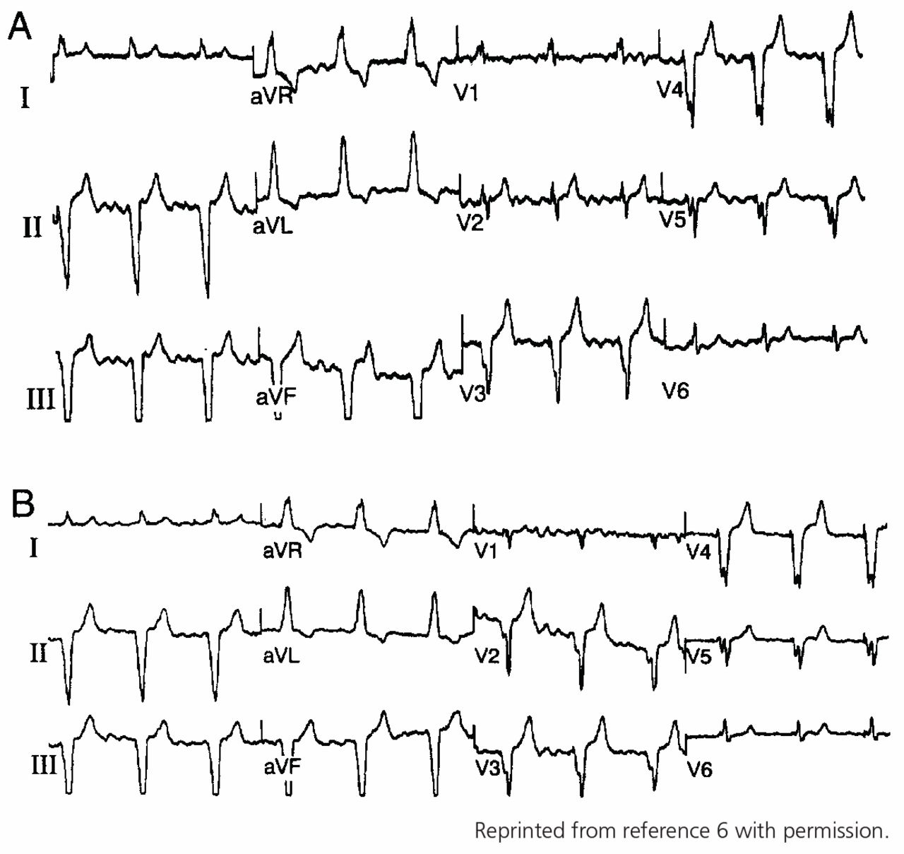
    Typical 12-lead electrocardiogram showing right bundle branch block morphology from the right ventricular apex with (A) standard V1 and V2 lead positions and (B) return to left bundle branch block morphology after V1 and V2 are moved 1 interspace lower than standard.

  • Figure 2
    • Download figure
    • Open in new tab
    • Download powerpoint
    Figure 2

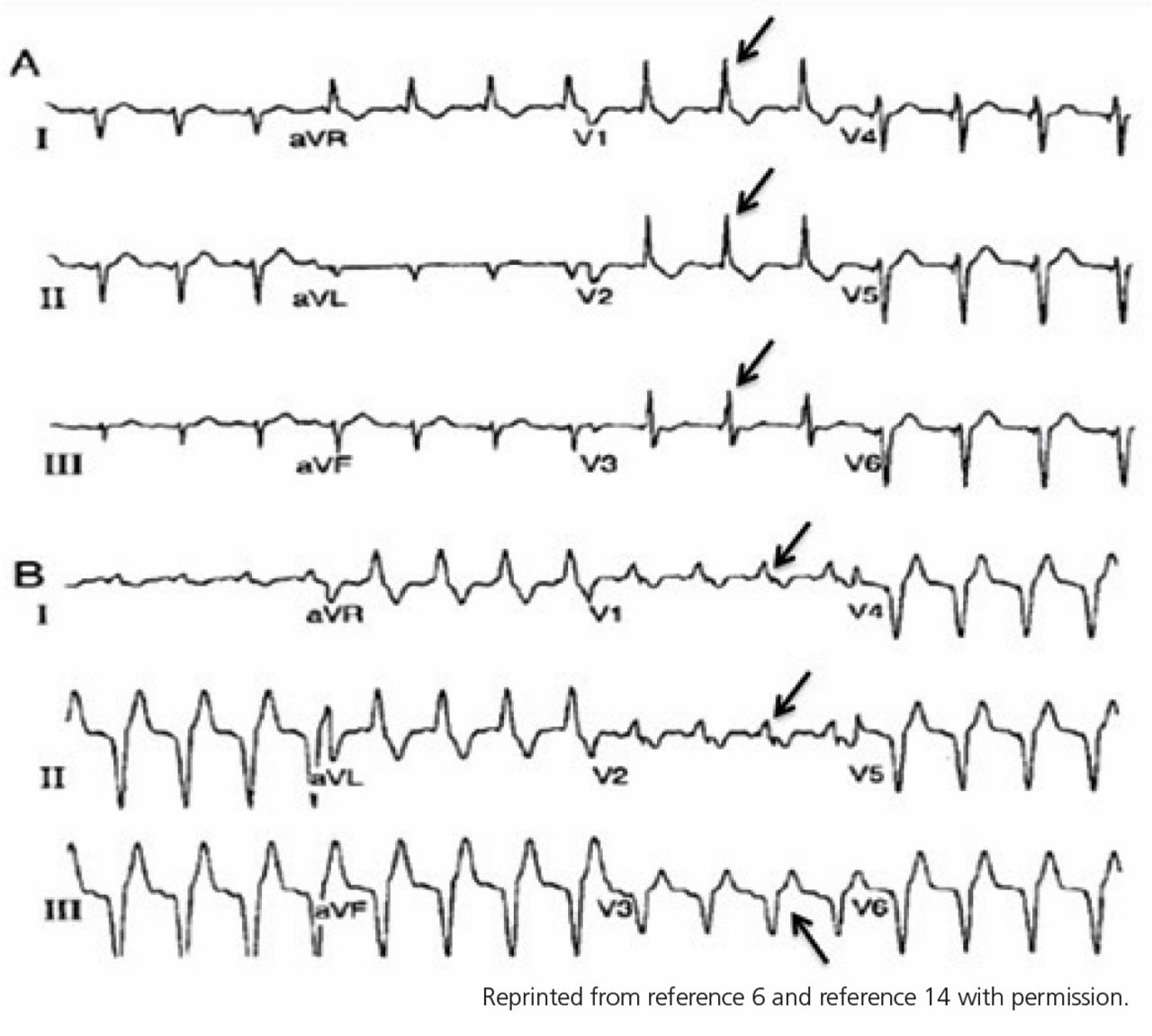
    (A) Electrocardiogram from a patient with known left ventricular lead position through a patent foramen ovale. Arrows point to dominant R waves in leads V1, V2, and V3, compatible with left ventricular pacing. (B) The same patient after revision and placement in the right ventricle. Arrows point to dominant R waves in leads V1 and V2, with a precordial transition to a dominant S wave occurring at lead V3.

  • Figure 3
    • Download figure
    • Open in new tab
    • Download powerpoint
    Figure 3

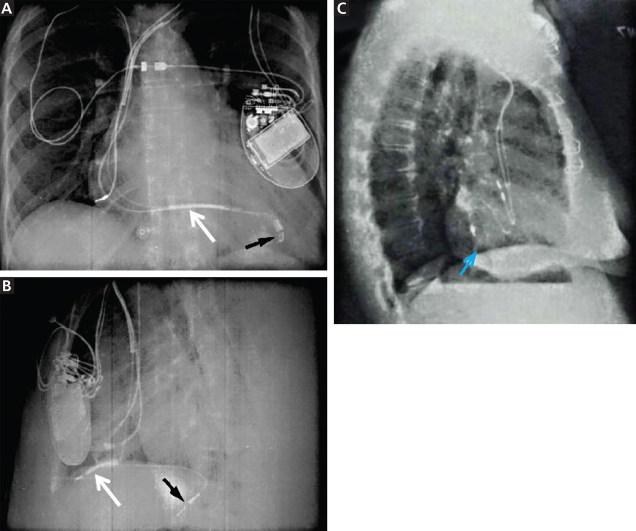
    (A) Normal lead positions in a cardiac resynchronization therapy defibrillator device on a posteroanterior chest radiograph. An adapter has been added to a pre-existing right-sided atrial lead that has been tunneled to the left-sided pocket. Note that the right ventricular (white arrow) and left ventricular (black arrow) leads appear to overlap. (B) On the lateral chest radiograph, the left ventricular lead is correctly positioned posteriorly (black arrow) and the right ventricular lead is positioned anteriorly (white arrow). (C) In this graphically enhanced image, a ventricular lead has passed through a patent foramen ovale and is positioned posteriorly in the left ventricle endocardium (blue arrow).

    Adapted with permission from references 14 and 15.

  • Figure 4
    • Download figure
    • Open in new tab
    • Download powerpoint
    Figure 4

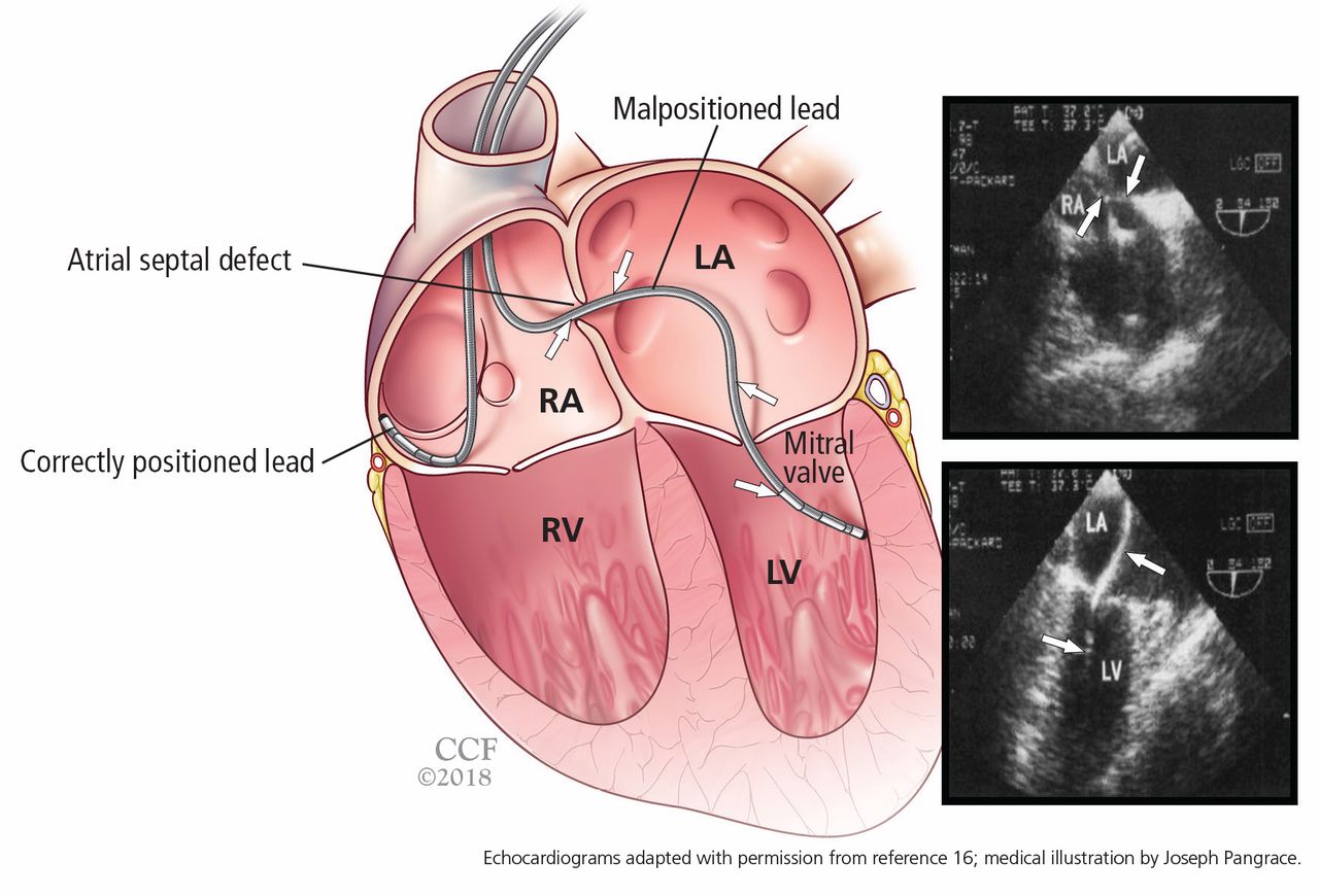
    Left, correctly positioned and malpositioned leads. As shown in the transesophageal echocardiogram (right), the malpositioned lead passed through an atrial septal defect (top) through the mitral valve into the left ventricle (bottom).

Tables

  • Figures
    • View popup
    TABLE 1

    Electrocardiographic localization of leads that exhibit right bundle branch block morphologies during pacing

    Frontal axisPrecordial transitionLocationSensitivity (%)Specificity (%)Positive predictive value (%)
    0° to –90°By V3RV septum or apex869995
    By V4RV septum or apex1009264
    By V4Posterior LV or coronary vein268336
    After V4Posterior LV or coronary vein72100100
    −90° to –180°By V3LV apex and distal anterior LV85100100
    90° to 180°Proximal anterior and anterolateral LV1009790
    • LV = left ventricular; RV = right ventricular

    • Reprinted from reference 6 with permission.

PreviousNext
Back to top

In this issue

Cleveland Clinic Journal of Medicine: 85 (1)
Cleveland Clinic Journal of Medicine
Vol. 85, Issue 1
1 Jan 2018
  • Table of Contents
  • Table of Contents (PDF)
  • Index by author
Print
Download PDF
Article Alerts
Sign In to Email Alerts with your Email Address
Email Article

Thank you for your interest in spreading the word on Cleveland Clinic Journal of Medicine.

NOTE: We only request your email address so that the person you are recommending the page to knows that you wanted them to see it, and that it is not junk mail. We do not capture any email address.

Enter multiple addresses on separate lines or separate them with commas.
Detecting and managing device leads inadvertently placed in the left ventricle
(Your Name) has sent you a message from Cleveland Clinic Journal of Medicine
(Your Name) thought you would like to see the Cleveland Clinic Journal of Medicine web site.
CAPTCHA
This question is for testing whether or not you are a human visitor and to prevent automated spam submissions.
Citation Tools
Detecting and managing device leads inadvertently placed in the left ventricle
Richard G. Trohman, Parikshit S. Sharma
Cleveland Clinic Journal of Medicine Jan 2018, 85 (1) 69-75; DOI: 10.3949/ccjm.85a.17012

Citation Manager Formats

  • BibTeX
  • Bookends
  • EasyBib
  • EndNote (tagged)
  • EndNote 8 (xml)
  • Medlars
  • Mendeley
  • Papers
  • RefWorks Tagged
  • Ref Manager
  • RIS
  • Zotero
Share
Detecting and managing device leads inadvertently placed in the left ventricle
Richard G. Trohman, Parikshit S. Sharma
Cleveland Clinic Journal of Medicine Jan 2018, 85 (1) 69-75; DOI: 10.3949/ccjm.85a.17012
Twitter logo Facebook logo Mendeley logo
  • Tweet Widget
  • Facebook Like
  • Linkedin Share Button

Jump to section

  • Article
    • ABSTRACT
    • RARE, BUT LIKELY UNDERREPORTED
    • HOW LEADS CAN END UP IN THE WRONG PLACE
    • PREVENTION IS THE BEST MANAGEMENT
    • POSTOPERATIVE DETECTION BY ECG
    • POSTOPERATIVE DETECTION BY CHEST RADIOGRAPHY
    • ECHOCARDIOGRAPHY TO CONFIRM
    • CT AND MRI: LIMITED ROLES
    • MANAGING MALPOSITIONED LEADS
    • Footnotes
    • REFERENCES
  • Figures & Data
  • Info & Metrics
  • PDF

Related Articles

  • No related articles found.
  • PubMed
  • Google Scholar

Cited By...

  • No citing articles found.
  • Google Scholar

More in this TOC Section

  • Alpha-gal syndrome: Recognizing and managing a tick-bite–related meat allergy
  • Managing right ventricular failure in the setting of pulmonary embolism
  • Psychedelics, spirituality, and existential distress in patients at the end of life
Show more Review

Similar Articles

Subjects

  • Cardiology
  • Imaging

Navigate

  • Current Issue
  • Past Issues
  • Supplements
  • Article Type
  • Specialty
  • CME/MOC Articles
  • CME/MOC Calendar
  • Media Kit

Authors & Reviewers

  • Manuscript Submission
  • Authors & Reviewers
  • Subscriptions
  • About CCJM
  • Contact Us
  • Cleveland Clinic Center for Continuing Education
  • Consult QD

Share your suggestions!

Copyright © 2025 The Cleveland Clinic Foundation. All rights reserved. The information provided is for educational purposes only. Use of this website is subject to the website terms of use and privacy policy. 

Powered by HighWire