
Main menu

  • Home
  • Content
    • Current Issue
    • Ahead of Print
    • Past Issues
    • Supplements
    • Article Type
  • Specialty
    • Articles by Specialty
  • CME/MOC
    • Articles
    • Calendar
  • Info For
    • Manuscript Submission
    • Authors & Reviewers
    • Subscriptions
    • About CCJM
    • Contact Us
    • Media Kit
  • Conversations with Leaders
  • Conference Coverage
    • Kidney Week 2024
    • CHEST 2024
    • ACR Convergence 2023
    • Kidney Week 2023
    • ObesityWeek 2023
    • IDWeek 2023
    • CHEST 2023
    • MDS 2023
    • IAS 2023
    • ACP 2023
    • AAN 2023
    • ACC / WCC 2023
    • AAAAI Meeting 2023
    • ACR Convergence 2022
    • Kidney Week 2022
    • AIDS 2022
  • Other Publications
    • www.clevelandclinic.org

User menu

  • Register
  • Log in

Search

  • Advanced search
Cleveland Clinic Journal of Medicine
  • Other Publications
    • www.clevelandclinic.org
  • Register
  • Log in
Cleveland Clinic Journal of Medicine

Advanced Search

  • Home
  • Content
    • Current Issue
    • Ahead of Print
    • Past Issues
    • Supplements
    • Article Type
  • Specialty
    • Articles by Specialty
  • CME/MOC
    • Articles
    • Calendar
  • Info For
    • Manuscript Submission
    • Authors & Reviewers
    • Subscriptions
    • About CCJM
    • Contact Us
    • Media Kit
  • Conversations with Leaders
  • Conference Coverage
    • Kidney Week 2024
    • CHEST 2024
    • ACR Convergence 2023
    • Kidney Week 2023
    • ObesityWeek 2023
    • IDWeek 2023
    • CHEST 2023
    • MDS 2023
    • IAS 2023
    • ACP 2023
    • AAN 2023
    • ACC / WCC 2023
    • AAAAI Meeting 2023
    • ACR Convergence 2022
    • Kidney Week 2022
    • AIDS 2022
The Clinical Picture

Acute necrotizing esophagitis

Takeshi Kondo, MD
Cleveland Clinic Journal of Medicine December 2018, 85 (12) 912-913; DOI: https://doi.org/10.3949/ccjm.85a.18054
Takeshi Kondo
Department of General Medicine, Chiba University Hospital, Chiba, Japan
  • Find this author on Google Scholar
  • Find this author on PubMed
  • Search for this author on this site
  • For correspondence: [email protected]
  • Article
  • Figures & Data
  • Info & Metrics
  • PDF
Loading

An 82-year-old man with poorly controlled diabetes mellitus presented to our emergency department with a 1-day history of confusion and coffee-ground emesis.

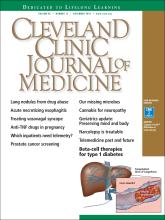
Blood test results suggested diabetic ketoacidosis. Upper endoscopy showed diffuse black discoloration of the esophageal mucosa that affected the distal esophagus and stopped abruptly at the gastroesophageal junction (Figure 1), with normal gastric and duodenal mucosa.

Figure 1
  • Download figure
  • Open in new tab
  • Download powerpoint
Figure 1

Upper endoscopy on the day of admission showed diffuse black discoloration of the esophageal mucosa affecting the distal esophagus and stopping abruptly at the gastroesophageal junction.

Biopsy study revealed necrosis of the esophageal mucosa. A diagnosis of acute necrotizing esophagitis was made.

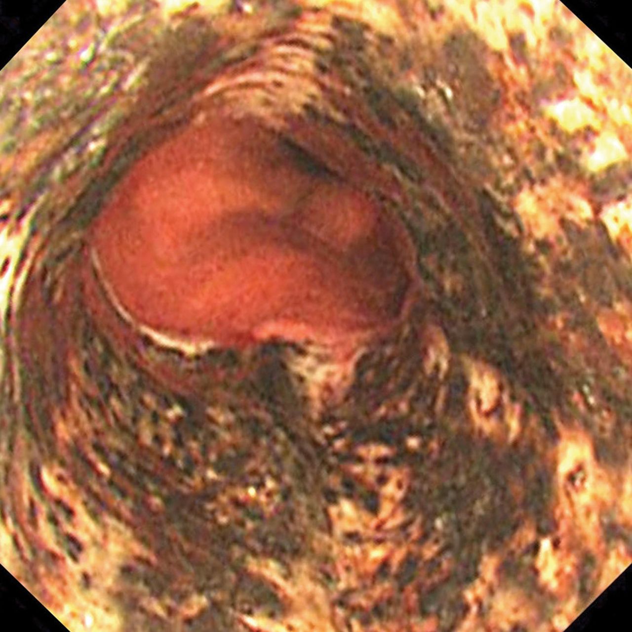
The patient received nothing by mouth, and he was treated with intravenous fluids, insulin, and a proton pump inhibitor. His symptoms re solved, and upper endoscopy repeated on hospital day 14 demonstrated improvement of the mucosal lesions and esophagitis (Figure 2).

Figure 2
  • Download figure
  • Open in new tab
  • Download powerpoint
Figure 2

Upper endoscopy repeated 14 days after admission showed improvement of the mucosal lesions and esophagitis.

ACUTE NECROTIZING ESOPHAGITIS

Acute necrotizing esophagitis is thought to arise from a combination of an ischemic insult to the esophagus, an impaired mucosal barrier system, and a backflow injury from chemical contents of gastric secretions.1 The tissue hypoperfusion derives from vasculopathy, hypotension, or malnutrition. It is associated with diabetes mellitus, diabetic ketoacidosis, lactic acidosis, alcohol abuse, cirrhosis, renal insufficiency, malignancy, antibiotic use, esophageal infections, and aortic dissection.

The esophagus has a diverse blood supply. The upper esophagus is supplied by the inferior thyroid arteries, the mid-esophagus by the bronchial, proper esophageal, and intercostal arteries, and the distal esophagus by the left gastric and left inferior phrenic arteries.1

KEY FEATURES AND DIAGNOSTIC CLUES

The necrotic changes are prominent in the distal esophagus, which is more susceptible to ischemia and mucosal injury. The characteristic endoscopic finding is a diffuse black esophagus with a sharp transition to normal mucosa at the gastroesophageal junction.

The differential diagnosis includes melanosis, pseudomelanosis, malignant melanoma, acanthosis nigricans, coal dust deposition, caustic ingestion, radiation esophagitis, and infectious esophagitis caused by cytomegalovirus, herpes simplex virus, Candida albicans, or Klebsiella pneumoniae.2–4

TREATMENT AND OUTCOME

Avoidance of oral intake and gastric acid suppression with intravenous proton pump inhibitors are recommended to prevent additional injury of the esophageal mucosa.

The condition generally resolves with restored blood flow and treatment of any coexisting illness. However, it may be complicated by perforation (6.8%), mediastinitis (5.7%), or subsequent development of esophageal stricture (10.2%).5 Patients with esophageal stricture require endoscopic dilation after mucosal recovery.

The overall risk of death in acute necrotizing esophagitis is high (31.8%) and most often due to the underlying disease, such as sepsis, malignancy, cardiogenic shock, or hypovolemic shock.5 The mortality rate directly attributed to complications of acute necrotizing esophagitis is much lower (5.7%).5

  • Copyright © 2018 The Cleveland Clinic Foundation. All Rights Reserved.

REFERENCES

  1. ↵
    1. Gurvits GE
    . Black esophagus: acute esophageal necrosis syndrome. World J Gastroenterol 2010; 16(26):3219–3225. pmid:20614476
    OpenUrlCrossRefPubMed
  2. ↵
    1. Khan HA
    . Coal dust deposition—rare cause of “black esophagus.” Am J Gastroenterol 1996; 91(10):2256. pmid:8855776
    OpenUrlPubMed
    1. Ertekin C,
    2. Alimoglu O,
    3. Akyildiz H,
    4. Guloglu R,
    5. Taviloglu K
    . The results of caustic ingestions. Hepatogastroenterology 2004; 51(59):1397–1400. pmid:15362762
    OpenUrlPubMed
  3. ↵
    1. Kozlowski LM,
    2. Nigra TP
    . Esophageal acanthosis nigricans in association with adenocarcinoma from an unknown primary site. J Am Acad Dermatol 1992; 26(2 pt 2):348–351. pmid:1569256
    OpenUrlCrossRefPubMed
  4. ↵
    1. Gurvits GE,
    2. Shapsis A,
    3. Lau N,
    4. Gualtieri N,
    5. Robilotti JG
    . Acute esophageal necrosis: a rare syndrome. J Gastroenterol 2007; 42(1):29–38. doi:10.1007/s00535-006-1974-z
    OpenUrlCrossRefPubMed
PreviousNext
Back to top

In this issue

Cleveland Clinic Journal of Medicine: 85 (12)
Cleveland Clinic Journal of Medicine
Vol. 85, Issue 12
1 Dec 2018
  • Table of Contents
  • Table of Contents (PDF)
  • Index by author
Print
Download PDF
Article Alerts
Sign In to Email Alerts with your Email Address
Email Article

Thank you for your interest in spreading the word on Cleveland Clinic Journal of Medicine.

NOTE: We only request your email address so that the person you are recommending the page to knows that you wanted them to see it, and that it is not junk mail. We do not capture any email address.

Enter multiple addresses on separate lines or separate them with commas.
Acute necrotizing esophagitis
(Your Name) has sent you a message from Cleveland Clinic Journal of Medicine
(Your Name) thought you would like to see the Cleveland Clinic Journal of Medicine web site.
CAPTCHA
This question is for testing whether or not you are a human visitor and to prevent automated spam submissions.
Citation Tools
Acute necrotizing esophagitis
Takeshi Kondo
Cleveland Clinic Journal of Medicine Dec 2018, 85 (12) 912-913; DOI: 10.3949/ccjm.85a.18054

Citation Manager Formats

  • BibTeX
  • Bookends
  • EasyBib
  • EndNote (tagged)
  • EndNote 8 (xml)
  • Medlars
  • Mendeley
  • Papers
  • RefWorks Tagged
  • Ref Manager
  • RIS
  • Zotero
Share
Acute necrotizing esophagitis
Takeshi Kondo
Cleveland Clinic Journal of Medicine Dec 2018, 85 (12) 912-913; DOI: 10.3949/ccjm.85a.18054
Twitter logo Facebook logo Mendeley logo
  • Tweet Widget
  • Facebook Like
  • Linkedin Share Button

Jump to section

  • Article
    • ACUTE NECROTIZING ESOPHAGITIS
    • KEY FEATURES AND DIAGNOSTIC CLUES
    • TREATMENT AND OUTCOME
    • REFERENCES
  • Figures & Data
  • Info & Metrics
  • PDF

Related Articles

  • No related articles found.
  • PubMed
  • Google Scholar

Cited By...

  • No citing articles found.
  • Google Scholar

More in this TOC Section

  • Tinea incognito
  • Prolonged venous filling time and dependent rubor in a patient with peripheral artery disease
  • Sarcoidosis with diffuse purplish erythematous plaques on the hands
Show more The Clinical Picture

Similar Articles

Subjects

  • Diabetes
  • Gastroenterology
  • Geriatrics

Navigate

  • Current Issue
  • Past Issues
  • Supplements
  • Article Type
  • Specialty
  • CME/MOC Articles
  • CME/MOC Calendar
  • Media Kit

Authors & Reviewers

  • Manuscript Submission
  • Authors & Reviewers
  • Subscriptions
  • About CCJM
  • Contact Us
  • Cleveland Clinic Center for Continuing Education
  • Consult QD

Share your suggestions!

Copyright © 2025 The Cleveland Clinic Foundation. All rights reserved. The information provided is for educational purposes only. Use of this website is subject to the website terms of use and privacy policy. 

Powered by HighWire