
Main menu

  • Home
  • Content
    • Current Issue
    • Ahead of Print
    • Past Issues
    • Supplements
    • Article Type
  • Specialty
    • Articles by Specialty
  • CME/MOC
    • Articles
    • Calendar
  • Info For
    • Manuscript Submission
    • Authors & Reviewers
    • Subscriptions
    • About CCJM
    • Contact Us
    • Media Kit
  • Conversations with Leaders
  • Conference Coverage
    • Kidney Week 2024
    • CHEST 2024
    • ACR Convergence 2023
    • Kidney Week 2023
    • ObesityWeek 2023
    • IDWeek 2023
    • CHEST 2023
    • MDS 2023
    • IAS 2023
    • ACP 2023
    • AAN 2023
    • ACC / WCC 2023
    • AAAAI Meeting 2023
    • ACR Convergence 2022
    • Kidney Week 2022
    • AIDS 2022
  • Other Publications
    • www.clevelandclinic.org

User menu

  • Register
  • Log in

Search

  • Advanced search
Cleveland Clinic Journal of Medicine
  • Other Publications
    • www.clevelandclinic.org
  • Register
  • Log in
Cleveland Clinic Journal of Medicine

Advanced Search

  • Home
  • Content
    • Current Issue
    • Ahead of Print
    • Past Issues
    • Supplements
    • Article Type
  • Specialty
    • Articles by Specialty
  • CME/MOC
    • Articles
    • Calendar
  • Info For
    • Manuscript Submission
    • Authors & Reviewers
    • Subscriptions
    • About CCJM
    • Contact Us
    • Media Kit
  • Conversations with Leaders
  • Conference Coverage
    • Kidney Week 2024
    • CHEST 2024
    • ACR Convergence 2023
    • Kidney Week 2023
    • ObesityWeek 2023
    • IDWeek 2023
    • CHEST 2023
    • MDS 2023
    • IAS 2023
    • ACP 2023
    • AAN 2023
    • ACC / WCC 2023
    • AAAAI Meeting 2023
    • ACR Convergence 2022
    • Kidney Week 2022
    • AIDS 2022
The Clinical Picture

Wolff-Parkinson-White pattern unmasked by severe musculoskeletal pain

Rishi Raj, MD, Leon O’Neill, MD and Nasreen Shaikh, MD
Cleveland Clinic Journal of Medicine July 2018, 85 (7) 507-509; DOI: https://doi.org/10.3949/ccjm.85a.17131
Rishi Raj
Chief Resident, Department of Internal Medicine, Monmouth Medical Center, Long Branch, NJ
  • Find this author on Google Scholar
  • Find this author on PubMed
  • Search for this author on this site
  • For correspondence: [email protected]
Leon O’Neill IV
Monmouth Medical Center, Long Branch, NJ
  • Find this author on Google Scholar
  • Find this author on PubMed
  • Search for this author on this site
Nasreen Shaikh
Monmouth Medical Center, Long Branch, NJ
  • Find this author on Google Scholar
  • Find this author on PubMed
  • Search for this author on this site
  • Article
  • Figures & Data
  • Info & Metrics
  • PDF
Loading

A 55-year-old man with no significant medical history presented to the emergency department with left-sided flank pain that had begun 3 days earlier. He described the pain as continuous, sharp, and aggravated by movement. He worked in construction, and before the pain started he had moved 8 sheets of drywall and lifted 5-gallon buckets of spackling compound. He denied any associated chest pain, palpitations, dyspnea, cough, or light-headedness. His family history included sudden cardiac death in 2 second-degree relatives.

On arrival in the emergency department, his vital signs were normal, as were the rest of the findings on physical examination except for reproducible point tenderness below the left scapula.

Laboratory workup revealed normal blood cell counts, liver enzymes, and kidney function. His initial troponin test was negative.

A routine electrocardiogram (Figure 1) showed normal sinus rhythm with a rate of 65 beats per minute, delta waves (most pronounced in V2), and Q waves in leads II, III, and aVF: the Wolff-Parkinson-White (WPW) pattern. Three subsequent electrocardiograms showed consistent findings.

Figure 1
  • Download figure
  • Open in new tab
  • Download powerpoint
Figure 1

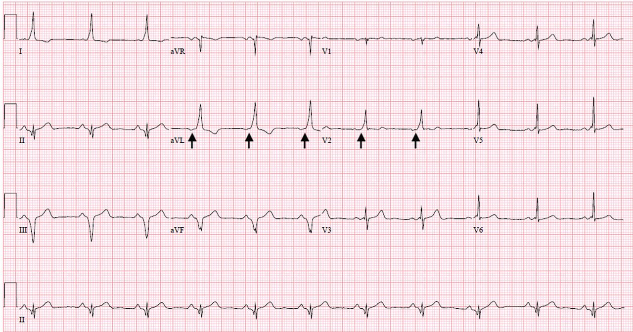
Electrocardiogram showing normal sinus rhythm with delta waves, most pronounced in lead V2 (arrows).

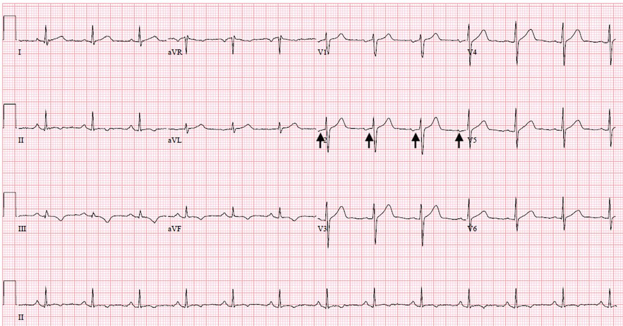
He was admitted to the hospital and was conservatively treated with nonsteroidal anti-inflammatory drugs for his musculoskeletal back pain. A follow-up electrocardiogram 24 hours later no longer showed delta waves (Figure 2). Echocardiography showed a normal ejection fraction with no valvular disease. An exercise stress test was negative for reversible ischemia.

Figure 2
  • Download figure
  • Open in new tab
  • Download powerpoint
Figure 2

Electrocardiography 24 hours after presentation and after the patient’s musculoskeletal pain had been brought under control showed resolution of the delta waves (arrows) and normal PR intervals.

The patient was referred to an electrophysiologist for further evaluation, but he returned to his home country (Haiti) after discharge and was lost to follow-up.

WOLFF-PARKINSON-WHITE PATTERN VS SYNDROME

WPW syndrome is a disorder of the conduction system leading to preexcitation of the ventricles by an accessory pathway between the atria and ventricles. It is characterized by preexcitation manifested on electrocardiography and by symptomatic arrhythmias.

In contrast, the WPW pattern is defined only by preexcitation findings on electrocardiography without symptomatic arrhythmias. Patients with WPW syndrome can present with palpitation, dizziness, and syncope resulting from underlying arrhythmia.1 This is not seen in patients with the WPW pattern.

A short PR interval with or without delta waves can also be seen in the absence of an accessory pathway, eg, in hypoplastic left heart syndrome, atrioventricular canal defect, and Ebstein anomaly. These conditions are termed pseudopreexcitation syndrome.2

Our patient presented with severe musculoskeletal pain that precipitated the electrocardiographic changes of the WPW pattern and resolved with adequate pain control. The WPW pattern can be unmasked under different scenarios, including anesthesia, sympathomimetic drugs, and postoperatively.3–5

Catecholamine challenge has been used to unmask high-risk features in WPW syndrome.3 Our patient may have had a transient spike in catecholamine levels because of severe musculoskeletal pain, leading to unmasking of accessory pathways and resulting in the WPW pattern on electrocardiography.

Most patients with the WPW pattern experience no symptoms, but a small percentage develop arrhythmias.

In rare cases, sudden cardiac death can be the presenting feature of WPW syndrome. The estimated risk of sudden cardiac death in patients with the WPW pattern is 1.25 per 1,000 person-years; ventricular fibrillation is the underlying mechanism.6 As our patient had a family history of sudden cardiac death, he was considered at high risk and was therefore referred to an electrophysiologist.

  • Copyright © 2018 The Cleveland Clinic Foundation. All Rights Reserved.

REFERENCES

  1. ↵
    1. Munger TM,
    2. Packer DL,
    3. Hammill SC,
    4. et al
    . A population study of the natural history of Wolff-Parkinson-White syndrome in Olmsted County, Minnesota, 1953–1989. Circulation 1993; 87(3):866–873. pmid:8443907
    OpenUrlAbstract/FREE Full Text
  2. ↵
    1. Carlson AM,
    2. Turek JW,
    3. Law IH,
    4. Von Bergen NH
    . Pseudo-preexcitation is prevalent among patients with repaired complex congenital heart disease. Pediatr Cardiol. 2015; 36(1):8–13. doi:10.1007/s00246-014-0955-x
    OpenUrlCrossRef
  3. ↵
    1. Aleong RG,
    2. Singh SM,
    3. Levinson JR,
    4. Milan DJ
    . Catecholamine challenge unmasking high-risk features in the Wolff-Parkinson-White syndrome. Europace 2009; 11(10):1396–1398. doi:10.1093/europace/eup211
    OpenUrlCrossRefPubMed
    1. Sahu S,
    2. Karna ST,
    3. Karna A,
    4. Lata I,
    5. Kapoor D
    . Anaesthetic management of Wolff-Parkinson-White syndrome for hysterectomy. Indian J Anaesth 2011; 55(4):378–380. doi:10.4103/0019-5049.84866
    OpenUrlCrossRefPubMed
  4. ↵
    1. Tseng ZH,
    2. Yadav AV,
    3. Scheinman MM
    . Catecholamine dependent accessory pathway automaticity. Pacing Clin Electrophysiol 2004; 27(7):1005–1007. doi:10.1111/j.1540-8159.2004.00574.x
    OpenUrlCrossRefPubMed
  5. ↵
    1. Obeyesekere MN,
    2. Leong-Sit P,
    3. Massel D,
    4. et al
    . Risk of arrhythmia and sudden death in patients with asymptomatic preexcitation: a meta-analysis. Circulation 2012; 125:(19)2308–2315. doi:10.1161/CIRCULATIONAHA.111.055350
    OpenUrlAbstract/FREE Full Text
PreviousNext
Back to top

In this issue

Cleveland Clinic Journal of Medicine: 85 (7)
Cleveland Clinic Journal of Medicine
Vol. 85, Issue 7
1 Jul 2018
  • Table of Contents
  • Table of Contents (PDF)
  • Index by author
Print
Download PDF
Article Alerts
Sign In to Email Alerts with your Email Address
Email Article

Thank you for your interest in spreading the word on Cleveland Clinic Journal of Medicine.

NOTE: We only request your email address so that the person you are recommending the page to knows that you wanted them to see it, and that it is not junk mail. We do not capture any email address.

Enter multiple addresses on separate lines or separate them with commas.
Wolff-Parkinson-White pattern unmasked by severe musculoskeletal pain
(Your Name) has sent you a message from Cleveland Clinic Journal of Medicine
(Your Name) thought you would like to see the Cleveland Clinic Journal of Medicine web site.
CAPTCHA
This question is for testing whether or not you are a human visitor and to prevent automated spam submissions.
Citation Tools
Wolff-Parkinson-White pattern unmasked by severe musculoskeletal pain
Rishi Raj, Leon O’Neill, Nasreen Shaikh
Cleveland Clinic Journal of Medicine Jul 2018, 85 (7) 507-509; DOI: 10.3949/ccjm.85a.17131

Citation Manager Formats

  • BibTeX
  • Bookends
  • EasyBib
  • EndNote (tagged)
  • EndNote 8 (xml)
  • Medlars
  • Mendeley
  • Papers
  • RefWorks Tagged
  • Ref Manager
  • RIS
  • Zotero
Share
Wolff-Parkinson-White pattern unmasked by severe musculoskeletal pain
Rishi Raj, Leon O’Neill, Nasreen Shaikh
Cleveland Clinic Journal of Medicine Jul 2018, 85 (7) 507-509; DOI: 10.3949/ccjm.85a.17131
Twitter logo Facebook logo Mendeley logo
  • Tweet Widget
  • Facebook Like
  • Linkedin Share Button

Jump to section

  • Article
    • WOLFF-PARKINSON-WHITE PATTERN VS SYNDROME
    • REFERENCES
  • Figures & Data
  • Info & Metrics
  • PDF

Related Articles

  • No related articles found.
  • PubMed
  • Google Scholar

Cited By...

  • No citing articles found.
  • Google Scholar

More in this TOC Section

  • Tinea incognito
  • Prolonged venous filling time and dependent rubor in a patient with peripheral artery disease
  • Sarcoidosis with diffuse purplish erythematous plaques on the hands
Show more The Clinical Picture

Similar Articles

Subjects

  • Cardiology
  • Emergency Medicine

Navigate

  • Current Issue
  • Past Issues
  • Supplements
  • Article Type
  • Specialty
  • CME/MOC Articles
  • CME/MOC Calendar
  • Media Kit

Authors & Reviewers

  • Manuscript Submission
  • Authors & Reviewers
  • Subscriptions
  • About CCJM
  • Contact Us
  • Cleveland Clinic Center for Continuing Education
  • Consult QD

Share your suggestions!

Copyright © 2025 The Cleveland Clinic Foundation. All rights reserved. The information provided is for educational purposes only. Use of this website is subject to the website terms of use and privacy policy. 

Powered by HighWire