
Main menu

  • Home
  • Content
    • Current Issue
    • Ahead of Print
    • Past Issues
    • Supplements
    • Article Type
  • Specialty
    • Articles by Specialty
  • CME/MOC
    • Articles
    • Calendar
  • Info For
    • Manuscript Submission
    • Authors & Reviewers
    • Subscriptions
    • About CCJM
    • Contact Us
    • Media Kit
  • Conversations with Leaders
  • Conference Coverage
    • Kidney Week 2024
    • CHEST 2024
    • ACR Convergence 2023
    • Kidney Week 2023
    • ObesityWeek 2023
    • IDWeek 2023
    • CHEST 2023
    • MDS 2023
    • IAS 2023
    • ACP 2023
    • AAN 2023
    • ACC / WCC 2023
    • AAAAI Meeting 2023
    • ACR Convergence 2022
    • Kidney Week 2022
    • AIDS 2022
  • Other Publications
    • www.clevelandclinic.org

User menu

  • Register
  • Log in

Search

  • Advanced search
Cleveland Clinic Journal of Medicine
  • Other Publications
    • www.clevelandclinic.org
  • Register
  • Log in
Cleveland Clinic Journal of Medicine

Advanced Search

  • Home
  • Content
    • Current Issue
    • Ahead of Print
    • Past Issues
    • Supplements
    • Article Type
  • Specialty
    • Articles by Specialty
  • CME/MOC
    • Articles
    • Calendar
  • Info For
    • Manuscript Submission
    • Authors & Reviewers
    • Subscriptions
    • About CCJM
    • Contact Us
    • Media Kit
  • Conversations with Leaders
  • Conference Coverage
    • Kidney Week 2024
    • CHEST 2024
    • ACR Convergence 2023
    • Kidney Week 2023
    • ObesityWeek 2023
    • IDWeek 2023
    • CHEST 2023
    • MDS 2023
    • IAS 2023
    • ACP 2023
    • AAN 2023
    • ACC / WCC 2023
    • AAAAI Meeting 2023
    • ACR Convergence 2022
    • Kidney Week 2022
    • AIDS 2022
The Clinical Picture

Emphysematous cystitis

Waiel Abusnina, MD, Mena Shehata, MD, Sammy Nassri, MD and Fuad Zeid, MD
Cleveland Clinic Journal of Medicine January 2019, 86 (1) 10-11; DOI: https://doi.org/10.3949/ccjm.86a.18063
Waiel Abusnina
Department of Internal Medicine, Joan C. Edwards School of Medicine at Marshall University, Huntington, WV
  • Find this author on Google Scholar
  • Find this author on PubMed
  • Search for this author on this site
  • For correspondence: [email protected]
Mena Shehata
Department of Internal Medicine, Joan C. Edwards School of Medicine at Marshall University, Huntington, WV
  • Find this author on Google Scholar
  • Find this author on PubMed
  • Search for this author on this site
Sammy Nassri
Department of Internal Medicine, Joan C. Edwards School of Medicine at Marshall University, Huntington, WV
  • Find this author on Google Scholar
  • Find this author on PubMed
  • Search for this author on this site
Fuad Zeid
Department of Pulmonary Medicine, Joan C. Edwards School of Medicine at Marshall University, Huntington, WV
  • Find this author on Google Scholar
  • Find this author on PubMed
  • Search for this author on this site
  • Article
  • Figures & Data
  • Info & Metrics
  • PDF
Loading

A 59-year-old woman with a history of chronic kidney disease and atonic bladder was brought to the hospital by emergency medical services. She had fallen in her home 2 days earlier and remained on the floor until neighbors eventually heard her cries and called 911. She complained of abdominal pain and distention along with emesis.

On presentation, she had tachycardia and tachypnea. The examination was notable for pronounced abdominal distention, diminished bowel sounds, and costovertebral angle tenderness.

The emergency department physician started empiric treatment for abdominal sepsis, including fluid resuscitation and broad-spectrum antibiotics. Initial imaging studies included abdominal radiography, which revealed a nonobstructive bowel gas pattern but raised suspicion of gas in the bladder (Figure 1). Arterial blood gas analysis showed lactic acidosis.

Figure 1
  • Download figure
  • Open in new tab
  • Download powerpoint
Figure 1

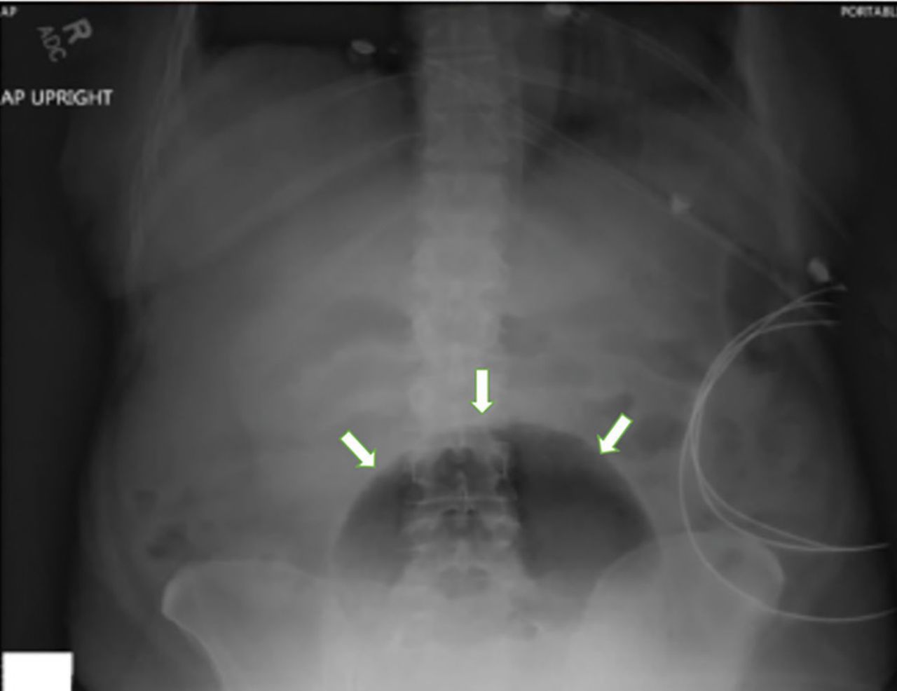
Plain abdominal radiography showed bladder distention with gas (arrows).

While laboratory work was being done, the patient’s tachypnea progressed to respiratory distress, and she ultimately required intubation. Vasopressors were started, as the patient was hemodynamically unstable. A Foley catheter was placed, which yielded about 1,100 mL of purulent urine.

Laboratory workup showed:

  • Procalcitonin 189 ng/mL (reference range < 2.0 ng/mL)

  • White blood cell count 10.7 × 10⁹/L (4.5–10.0)

  • Myoglobin 20,000 ng/mL (< 71)

  • Serum creatinine 4.8 mg/dL (0.06–1.10).

Urinalysis was positive for infection; blood and urine cultures later were positive for Escherichia coli.

Computed tomography of the abdomen and pelvis showed diffuse bladder dilation with urine and gas. It also revealed gas within the bladder wall and moderate hydroureter and hydronephrosis (Figure 2).

Figure 2
  • Download figure
  • Open in new tab
  • Download powerpoint
Figure 2

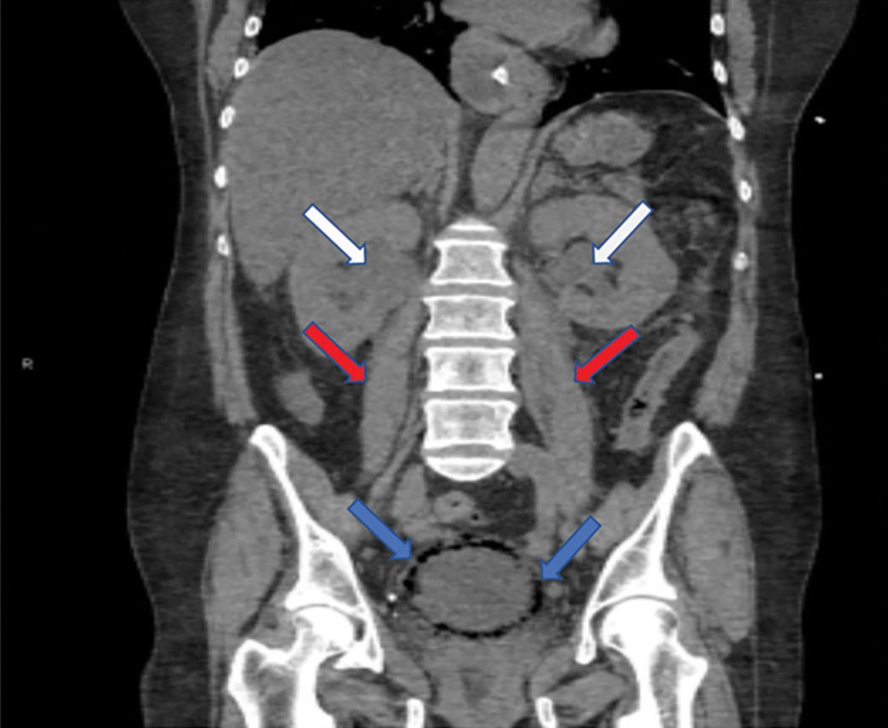
Coronal computed tomography of the abdomen and pelvis revealed a diffuse collection of gas within the bladder wall (blue arrows), bilateral hydronephrosis (white arrows), and bilateral hydroureter (red arrows).

The patient went into shock that was refractory to pressors, culminating in cardiac arrest despite resuscitative measures.

EMPHYSEMATOUS CYSTITIS, A FORM OF URINARY TRACT INFECTION

Emphysematous cystitis is a rare form of complicated urinary tract infection characterized by gas inside the bladder and in the bladder wall. While the exact mechanisms underlying gas formation are not clear, gas-producing pathogens are clearly implicated in severe infection. E coli and Klebsiella pneumoniae are the most common organisms associated with emphysematous cystitis; others include Proteus mirabilis, and Enterobacter and Streptococcus species.1,2

More than 50% of patients with emphysematous cystitis have diabetes mellitus. Other risk factors include bladder outlet obstruction, neurogenic bladder, and female sex.3 The severity of disease ranges from asymptomatic pneumaturia (up to 7% of cases)2 to fulminant emphysematous cystitis, as in our patient.

The clinical presentation of emphysematous cystitis is nonspecific and can range from minimally symptomatic urinary tract infection to acute abdomen and septic shock.4

Some patients present with pneumaturia (the passing of gas through the urethra with micturition). Pneumaturia arises from 3 discrete causes: urologic instrumentation, fistula between the bladder and large or small bowel, and gas-producing bacteria in the bladder (emphysematous cystitis).5 Pneumaturia should always raise the suspicion of emphysematous cystitis.

The diagnosis can be made with either radiographic or computed tomographic evidence of gas within the bladder and bladder wall, in the absence of both bladder fistula and history of iatrogenic pneumaturia. Emphysematous cystitis should prompt urine and blood cultures to direct antimicrobial therapy, as 50% of patients with emphysematous cystitis have concomitant bacteremia.6

Our patient had an elevated serum level of procalcitonin, a marker of bacterial infection. Procalcitonin is a more specific biomarker of bacterial infection than acute-phase reactants such as the erythrocyte sedimentation rate or the C-reactive protein level. Measuring procalcitonin may help physicians make the diagnosis earlier, differentiate infectious from sterile causes of severe systemic inflammation, assess the severity of systemic inflammation caused by bacterial infections, and decide whether to start or discontinue antibiotic therapy.7

Most cases of emphysematous cystitis can be treated with antibiotics, though early diagnosis is crucial to a favorable outcome. Delay in diagnosis may contribute to the 20% mortality rate associated with this condition.6

  • Copyright © 2019 The Cleveland Clinic Foundation. All Rights Reserved.

REFERENCES

  1. ↵
    1. Stein JP,
    2. Spitz A,
    3. Elmajian DA,
    4. et al
    . Bilateral emphyse-matous pyelonephritis: a case report and review of the literature. Urology 1996; 47(1):129–134. pmid:8560648
    OpenUrlPubMed
  2. ↵
    1. Amano M,
    2. Shimizu T
    . Emphysematous cystitis: a review of the literature. Intern Med 2014; 53(2):79–82. pmid:24429444
    OpenUrlCrossRefPubMed
  3. ↵
    1. Wang JH
    . Emphysematous cystitis. Urol Sci 2010; 21(4):185–186. doi:10.1016/S1879-5226(10)60041-3
    OpenUrlCrossRef
  4. ↵
    1. Thomas AA,
    2. Lane BR,
    3. Thomas AZ,
    4. Remer EM,
    5. Campbell SC,
    6. Shoskes DA
    . Emphysematous cystitis: a review of 135 cases. BJU Int 2007; 100(1):17–20. doi:10.1111/j.1464-410X.2007.06930.x
    OpenUrlCrossRefPubMed
  5. ↵
    1. Arthur LM,
    2. Johnson HW
    . Pneumaturia: a case report and review of the literature. J Urol 1948; 60(4):659–665. pmid:18885959
    OpenUrlPubMed
  6. ↵
    1. Grupper M,
    2. Kravtsov A,
    3. Potasman I
    . Emphysematous cystitis: illustrative case report and review of the literature. Medicine (Baltimore) 2007; 86(1):47–53. doi:10.1097/MD.0b013e3180307c3a
    OpenUrlCrossRefPubMed
  7. ↵
    1. Lee H
    . Procalcitonin as a biomarker of infectious diseases. Korean J Intern Med 2013; 28(3):285–291. doi:10.3904/kjim.2013.28.3.285
    OpenUrlCrossRef
PreviousNext
Back to top

In this issue

Cleveland Clinic Journal of Medicine: 86 (1)
Cleveland Clinic Journal of Medicine
Vol. 86, Issue 1
1 Jan 2019
  • Table of Contents
  • Table of Contents (PDF)
  • Index by author
Print
Download PDF
Article Alerts
Sign In to Email Alerts with your Email Address
Email Article

Thank you for your interest in spreading the word on Cleveland Clinic Journal of Medicine.

NOTE: We only request your email address so that the person you are recommending the page to knows that you wanted them to see it, and that it is not junk mail. We do not capture any email address.

Enter multiple addresses on separate lines or separate them with commas.
Emphysematous cystitis
(Your Name) has sent you a message from Cleveland Clinic Journal of Medicine
(Your Name) thought you would like to see the Cleveland Clinic Journal of Medicine web site.
CAPTCHA
This question is for testing whether or not you are a human visitor and to prevent automated spam submissions.
Citation Tools
Emphysematous cystitis
Waiel Abusnina, Mena Shehata, Sammy Nassri, Fuad Zeid
Cleveland Clinic Journal of Medicine Jan 2019, 86 (1) 10-11; DOI: 10.3949/ccjm.86a.18063

Citation Manager Formats

  • BibTeX
  • Bookends
  • EasyBib
  • EndNote (tagged)
  • EndNote 8 (xml)
  • Medlars
  • Mendeley
  • Papers
  • RefWorks Tagged
  • Ref Manager
  • RIS
  • Zotero
Share
Emphysematous cystitis
Waiel Abusnina, Mena Shehata, Sammy Nassri, Fuad Zeid
Cleveland Clinic Journal of Medicine Jan 2019, 86 (1) 10-11; DOI: 10.3949/ccjm.86a.18063
Twitter logo Facebook logo Mendeley logo
  • Tweet Widget
  • Facebook Like
  • Linkedin Share Button

Jump to section

  • Article
    • EMPHYSEMATOUS CYSTITIS, A FORM OF URINARY TRACT INFECTION
    • REFERENCES
  • Figures & Data
  • Info & Metrics
  • PDF

Related Articles

  • No related articles found.
  • PubMed
  • Google Scholar

Cited By...

  • No citing articles found.
  • Google Scholar

More in this TOC Section

  • Tinea incognito
  • Prolonged venous filling time and dependent rubor in a patient with peripheral artery disease
  • Sarcoidosis with diffuse purplish erythematous plaques on the hands
Show more The Clinical Picture

Similar Articles

Subjects

  • Emergency Medicine
  • Imaging
  • Infectious Diseases
  • Nephrology

Navigate

  • Current Issue
  • Past Issues
  • Supplements
  • Article Type
  • Specialty
  • CME/MOC Articles
  • CME/MOC Calendar
  • Media Kit

Authors & Reviewers

  • Manuscript Submission
  • Authors & Reviewers
  • Subscriptions
  • About CCJM
  • Contact Us
  • Cleveland Clinic Center for Continuing Education
  • Consult QD

Share your suggestions!

Copyright © 2025 The Cleveland Clinic Foundation. All rights reserved. The information provided is for educational purposes only. Use of this website is subject to the website terms of use and privacy policy. 

Powered by HighWire