
Main menu

  • Home
  • Content
    • Current Issue
    • Ahead of Print
    • Past Issues
    • Supplements
    • Article Type
  • Specialty
    • Articles by Specialty
  • CME/MOC
    • Articles
    • Calendar
  • Info For
    • Manuscript Submission
    • Authors & Reviewers
    • Subscriptions
    • About CCJM
    • Contact Us
    • Media Kit
  • Conversations with Leaders
  • Conference Coverage
    • Kidney Week 2024
    • CHEST 2024
    • ACR Convergence 2023
    • Kidney Week 2023
    • ObesityWeek 2023
    • IDWeek 2023
    • CHEST 2023
    • MDS 2023
    • IAS 2023
    • ACP 2023
    • AAN 2023
    • ACC / WCC 2023
    • AAAAI Meeting 2023
    • ACR Convergence 2022
    • Kidney Week 2022
    • AIDS 2022
  • Other Publications
    • www.clevelandclinic.org

User menu

  • Register
  • Log in

Search

  • Advanced search
Cleveland Clinic Journal of Medicine
  • Other Publications
    • www.clevelandclinic.org
  • Register
  • Log in
Cleveland Clinic Journal of Medicine

Advanced Search

  • Home
  • Content
    • Current Issue
    • Ahead of Print
    • Past Issues
    • Supplements
    • Article Type
  • Specialty
    • Articles by Specialty
  • CME/MOC
    • Articles
    • Calendar
  • Info For
    • Manuscript Submission
    • Authors & Reviewers
    • Subscriptions
    • About CCJM
    • Contact Us
    • Media Kit
  • Conversations with Leaders
  • Conference Coverage
    • Kidney Week 2024
    • CHEST 2024
    • ACR Convergence 2023
    • Kidney Week 2023
    • ObesityWeek 2023
    • IDWeek 2023
    • CHEST 2023
    • MDS 2023
    • IAS 2023
    • ACP 2023
    • AAN 2023
    • ACC / WCC 2023
    • AAAAI Meeting 2023
    • ACR Convergence 2022
    • Kidney Week 2022
    • AIDS 2022
The Clinical Picture

A right atrial mass

Zeel Shah, MD, M. Chadi Alraies, MD, Ameer Harp, MD, Salam Khalil, MD, Amir Kaki, MD and Mahir Elder, MD
Cleveland Clinic Journal of Medicine July 2019, 86 (7) 445-447; DOI: https://doi.org/10.3949/ccjm.86a.18131
Zeel Shah
Wayne State University School of Medicine, Detroit, MI
  • Find this author on Google Scholar
  • Find this author on PubMed
  • Search for this author on this site
M. Chadi Alraies
Department of Cardiovascular Medicine, Wayne State University School of Medicine/Detroit Medical Center, Detroit, MI
  • Find this author on Google Scholar
  • Find this author on PubMed
  • Search for this author on this site
  • For correspondence: [email protected]
Ameer Harp
Wayne State University School of Medicine/Detroit Medical Center, Detroit, MI
  • Find this author on Google Scholar
  • Find this author on PubMed
  • Search for this author on this site
Salam Khalil
Wayne State University School of Medicine/Detroit Medical Center, Detroit, MI
  • Find this author on Google Scholar
  • Find this author on PubMed
  • Search for this author on this site
Amir Kaki
Wayne State University School of Medicine and Ascension St. John Hospital, Detroit, MI
  • Find this author on Google Scholar
  • Find this author on PubMed
  • Search for this author on this site
Mahir Elder
Wayne State University School of Medicine and Beaumont Hospital, Detroit, MI
  • Find this author on Google Scholar
  • Find this author on PubMed
  • Search for this author on this site
  • Article
  • Figures & Data
  • Info & Metrics
  • PDF
Loading

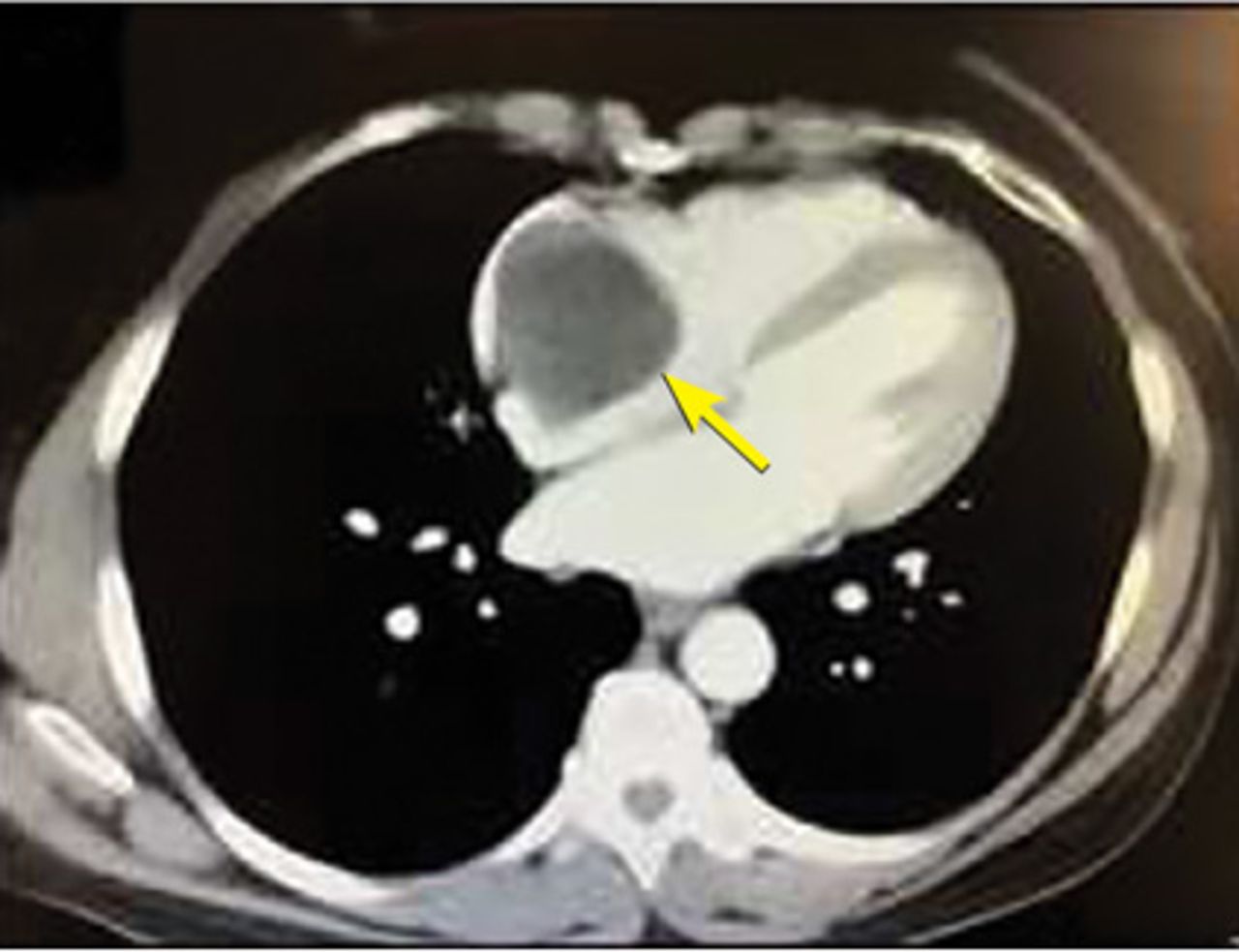
A 55-year-old woman with hypertension, hyperthyroidism, and endometrial adenocarcinoma treated 5 years earlier presented with occasional shortness of breath and dizziness. To rule out pulmonary embolism, computed tomography of the chest was done and showed a high-density lesion in the right atrium of the heart, measuring up to 4.5 cm (Figure 1).

Figure 1
  • Download figure
  • Open in new tab
  • Download powerpoint
Figure 1

Computed tomography of the chest. An arrow points to the mass in the right atrium.

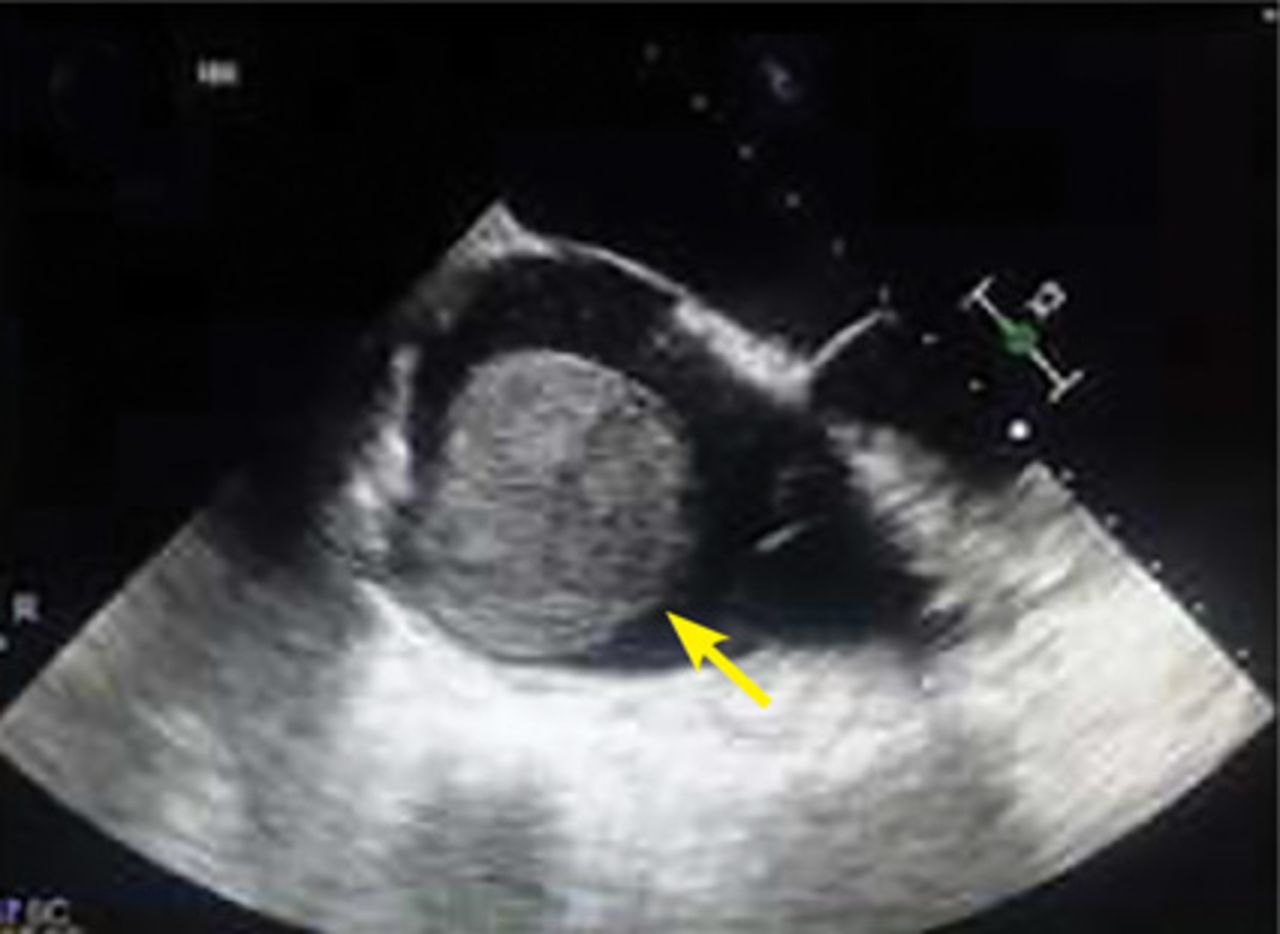
Transthoracic echocardiography confirmed a large round echogenic right atrial mass measuring 4.5 by 4.3 cm (Figure 2). The mass appeared to encroach into the tricuspid valve orifice, but there was no evidence of functional obstruction or stenosis.

Figure 2
  • Download figure
  • Open in new tab
  • Download powerpoint
Figure 2

Transthoracic echocardiography.

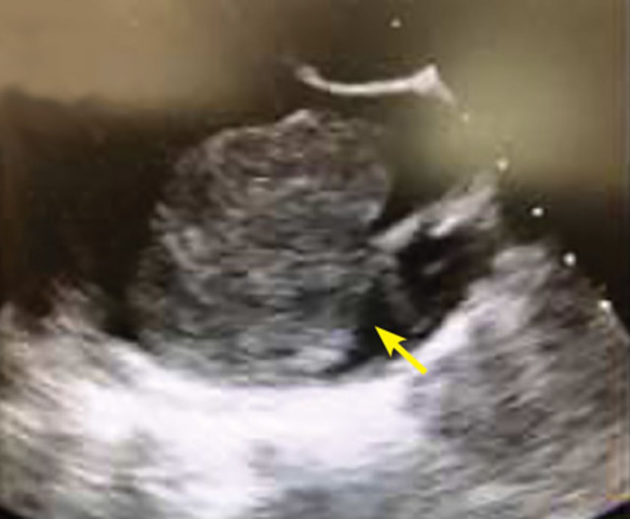
Transesophageal echocardiography showed a large globular echogenic mass measuring 5.5 by 4.6 cm with a stalk originating from the right atrial appendage (Figures 3 and 4). The mass was minimally mobile and did not obstruct the superior vena cava or the tricuspid orifice.

Figure 3
  • Download figure
  • Open in new tab
  • Download powerpoint
Figure 3

Transesophageal echocardiography.

Figure 4
  • Download figure
  • Open in new tab
  • Download powerpoint
Figure 4

Transesophageal echocardiography.

Given her history, the intracardiac mass strongly suggested metastatic adenocarcinoma. Other possibilities in the differential diagnosis were thrombus, myxoma, lipoma, angiosarcoma, and lymphoma.1,2

The cardiovascular team was consulted, and they recommended proceeding with open heart surgery and resection of the mass. In preparation for the surgery, cardiac catheterization was performed, and did not show any significant coronary artery disease. However, using a pigtail catheter, right atrial angiography demonstrated a large, round, mobile mass attached to the wall of the right atrium (Figure 5).

Figure 5
  • Download figure
  • Open in new tab
  • Download powerpoint
Figure 5

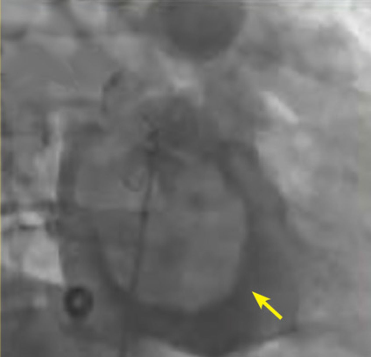
Angiography with pigtail catheter.

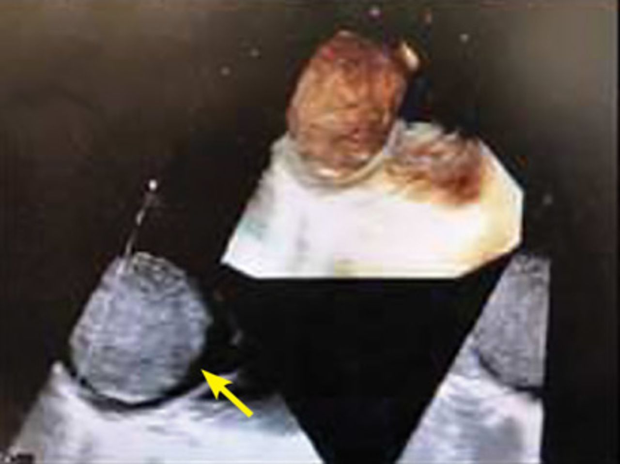
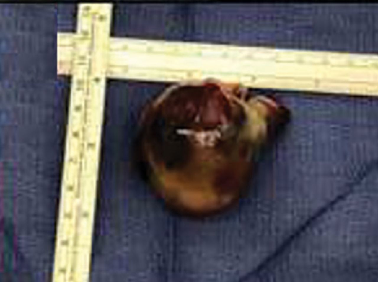
The patient underwent surgery, and the tumor was resected (Figures 6 and 7). Pathologic study confirmed the diagnosis of cardiac benign myxoma. The patient recovered and was eventually discharged to her home.

Figure 6
  • Download figure
  • Open in new tab
  • Download powerpoint
Figure 6

Operative photograph.

Figure 7
  • Download figure
  • Open in new tab
  • Download powerpoint
Figure 7

Gross section.

PRIMARY HEART TUMORS ARE RARE

Metastatic cardiac tumors are more common than primary cardiac tumors. On autopsy, between 0.01% and 0.1% of people in the general population have primary cardiac tumors, while 0.7% to 3.5% have metastatic tumors.3 About 75% of primary cardiac tumors are benign. In autopsies of patients with malignant neoplasms, cardiac metastasis has been found in 6% to 20%.4 Further, right-sided masses typically raise the concern of metastatic disease, whereas benign masses are more commonly found on the left side.

The most common neoplasms that metastasize to the heart are malignant melanoma, lymphoma, leukemia, breast, and lung cancers. The layers of the heart affected by malignant neoplasms in order of frequency from highest to lowest are the pericardium, epicardium, myocardium, and endocardium.3

MYXOMA: A PRIMARY CARDIAC TUMOR

The most common type of primary cardiac tumor is myxoma. Most—75% to 80%—occur in the left atrium, while 15% to 20% occur in the right atrium.5 Right atrial myxomas are usually found in the intraatrial septum at the border of the fossa ovalis.6 Myxomas can occur at any age, but are most common in women between the third and sixth decades.2

The cause of atrial myxomas is currently unknown. Most cases are sporadic. However, 10% are familial, with an autosomal-dominant pattern.7

The clinical symptoms of right atrial myxoma depend on the tumor’s size, location, and mobility and on the patient’s physical activity and body position.4 Common presenting symptoms include shortness of breath, pulmonary edema, cough, hemoptysis, and fatigue. Thirty percent of patients present with constitutional symptoms.4

Auscultation may reveal a characteristic “tumor plop” early in diastole.4,7 About 35% of patients have laboratory abnormalities such as elevations in erythrocyte sedimentation rate, C-reactive protein, and globulin levels and anemia. Our patient did not.

Embolization occurs in about 10% of cases of right-sided myxoma and can result in pulmonary artery embolism or a stroke. Pulmonary artery embolism occurs with myxoma embolization to the lungs. Strokes can occur in patients who have a patent foramen ovale or atrial septal defect, through which embolism to the systemic arterial circulation can occur.

The primary treatment for myxoma is complete resection of the tumor and its base with wide safety margins. This is particularly important to prevent recurrence of the myxoma and need for repeat operations, with their risk of surgical complications.9

  • Copyright © 2019 The Cleveland Clinic Foundation. All Rights Reserved.

REFERENCES

  1. ↵
    1. Dujardin KS,
    2. Click RL,
    3. Oh JK
    . The role of intraoperative transesophageal echocardiography in patients undergoing cardiac mass removal. J Am Soc Echocardiogr 2000; 13(12):1080–1083. pmid:11119275
    OpenUrlCrossRefPubMed
  2. ↵
    1. Jang KH,
    2. Shin DH,
    3. Lee C,
    4. Jang JK,
    5. Cheong S,
    6. Yoo SY
    . Left atrial mass with stalk: thrombus or myxoma? J Cardiovasc Ultrasound 2010; 18(4):154–156. doi:10.4250/jcu.2010.18.4.154
    OpenUrlCrossRefPubMed
  3. ↵
    1. Goldberg AD,
    2. Blankstein R,
    3. Padera RF
    . Tumors metastatic to the heart. Circulation 2013; 128(16):1790–1794. doi:10.1161/CIRCULATIONAHA.112.000790
    OpenUrlFREE Full Text
  4. ↵
    1. Aggarwal SK,
    2. Barik R,
    3. Sarma TC,
    4. et al
    . Clinical presentation and investigation findings in cardiac myxomas: new insights from the developing world. Am Heart J 2007; 154(6):1102–1107. doi:10.1016/j.ahj.2007.07.032
    OpenUrlCrossRefPubMed
  5. ↵
    1. Diaz A,
    2. Di Salvo C,
    3. Lawrence D,
    4. Hayward M
    . Left atrial and right ventricular myxoma: an uncommon presentation of a rare tumour. Interact Cardiovasc Thorac Surg 2011; 12(4):622–623. doi:10.1510/icvts.2010.255661
    OpenUrlCrossRefPubMed
  6. ↵
    1. Reynen K
    . Cardiac myxomas. N Engl J Med 1995; 333(24):1610–1617. doi:10.1056/NEJM199512143332407
    OpenUrlCrossRefPubMed
  7. ↵
    1. Kolluru A,
    2. Desai D,
    3. Cohen GI
    . The etiology of atrial myxoma tumor plop. J Am Coll Cardiol 2011; 57(21):e371. doi:10.1016/j.jacc.2010.09.085
    OpenUrlFREE Full Text
    1. Kassab R,
    2. Wehbe L,
    3. Badaoui G,
    4. el Asmar B,
    5. Jebara V,
    6. Ashoush R
    . Recurrent cerebrovascular accident: unusual and isolated manifestation of myxoma of the left atrium. J Med Liban 1999; 47(4):246–250. French. pmid:10641454
    OpenUrlPubMed
  8. ↵
    1. Guhathakurta S,
    2. Riordan JP
    . Surgical treatment of right atrial myxoma. Tex Heart Inst J 2000; 27(1):61–63. pmid:10830633
    OpenUrlPubMed
PreviousNext
Back to top

In this issue

Cleveland Clinic Journal of Medicine: 86 (7)
Cleveland Clinic Journal of Medicine
Vol. 86, Issue 7
1 Jul 2019
  • Table of Contents
  • Table of Contents (PDF)
  • Index by author
Print
Download PDF
Article Alerts
Sign In to Email Alerts with your Email Address
Email Article

Thank you for your interest in spreading the word on Cleveland Clinic Journal of Medicine.

NOTE: We only request your email address so that the person you are recommending the page to knows that you wanted them to see it, and that it is not junk mail. We do not capture any email address.

Enter multiple addresses on separate lines or separate them with commas.
A right atrial mass
(Your Name) has sent you a message from Cleveland Clinic Journal of Medicine
(Your Name) thought you would like to see the Cleveland Clinic Journal of Medicine web site.
CAPTCHA
This question is for testing whether or not you are a human visitor and to prevent automated spam submissions.
Citation Tools
A right atrial mass
Zeel Shah, M. Chadi Alraies, Ameer Harp, Salam Khalil, Amir Kaki, Mahir Elder
Cleveland Clinic Journal of Medicine Jul 2019, 86 (7) 445-447; DOI: 10.3949/ccjm.86a.18131

Citation Manager Formats

  • BibTeX
  • Bookends
  • EasyBib
  • EndNote (tagged)
  • EndNote 8 (xml)
  • Medlars
  • Mendeley
  • Papers
  • RefWorks Tagged
  • Ref Manager
  • RIS
  • Zotero
Share
A right atrial mass
Zeel Shah, M. Chadi Alraies, Ameer Harp, Salam Khalil, Amir Kaki, Mahir Elder
Cleveland Clinic Journal of Medicine Jul 2019, 86 (7) 445-447; DOI: 10.3949/ccjm.86a.18131
Twitter logo Facebook logo Mendeley logo
  • Tweet Widget
  • Facebook Like
  • Linkedin Share Button

Jump to section

  • Article
    • PRIMARY HEART TUMORS ARE RARE
    • MYXOMA: A PRIMARY CARDIAC TUMOR
    • REFERENCES
  • Figures & Data
  • Info & Metrics
  • PDF

Related Articles

  • No related articles found.
  • PubMed
  • Google Scholar

Cited By...

  • No citing articles found.
  • Google Scholar

More in this TOC Section

  • Tinea incognito
  • Prolonged venous filling time and dependent rubor in a patient with peripheral artery disease
  • Sarcoidosis with diffuse purplish erythematous plaques on the hands
Show more The Clinical Picture

Similar Articles

Subjects

  • Cardiology
  • Imaging
  • Oncology

Navigate

  • Current Issue
  • Past Issues
  • Supplements
  • Article Type
  • Specialty
  • CME/MOC Articles
  • CME/MOC Calendar
  • Media Kit

Authors & Reviewers

  • Manuscript Submission
  • Authors & Reviewers
  • Subscriptions
  • About CCJM
  • Contact Us
  • Cleveland Clinic Center for Continuing Education
  • Consult QD

Share your suggestions!

Copyright © 2025 The Cleveland Clinic Foundation. All rights reserved. The information provided is for educational purposes only. Use of this website is subject to the website terms of use and privacy policy. 

Powered by HighWire