
Main menu

  • Home
  • Content
    • Current Issue
    • Ahead of Print
    • Past Issues
    • Supplements
    • Article Type
  • Specialty
    • Articles by Specialty
  • CME/MOC
    • Articles
    • Calendar
  • Info For
    • Manuscript Submission
    • Authors & Reviewers
    • Subscriptions
    • About CCJM
    • Contact Us
    • Media Kit
  • Conversations with Leaders
  • Conference Coverage
    • Kidney Week 2024
    • CHEST 2024
    • ACR Convergence 2023
    • Kidney Week 2023
    • ObesityWeek 2023
    • IDWeek 2023
    • CHEST 2023
    • MDS 2023
    • IAS 2023
    • ACP 2023
    • AAN 2023
    • ACC / WCC 2023
    • AAAAI Meeting 2023
    • ACR Convergence 2022
    • Kidney Week 2022
    • AIDS 2022
  • Other Publications
    • www.clevelandclinic.org

User menu

  • Register
  • Log in

Search

  • Advanced search
Cleveland Clinic Journal of Medicine
  • Other Publications
    • www.clevelandclinic.org
  • Register
  • Log in
Cleveland Clinic Journal of Medicine

Advanced Search

  • Home
  • Content
    • Current Issue
    • Ahead of Print
    • Past Issues
    • Supplements
    • Article Type
  • Specialty
    • Articles by Specialty
  • CME/MOC
    • Articles
    • Calendar
  • Info For
    • Manuscript Submission
    • Authors & Reviewers
    • Subscriptions
    • About CCJM
    • Contact Us
    • Media Kit
  • Conversations with Leaders
  • Conference Coverage
    • Kidney Week 2024
    • CHEST 2024
    • ACR Convergence 2023
    • Kidney Week 2023
    • ObesityWeek 2023
    • IDWeek 2023
    • CHEST 2023
    • MDS 2023
    • IAS 2023
    • ACP 2023
    • AAN 2023
    • ACC / WCC 2023
    • AAAAI Meeting 2023
    • ACR Convergence 2022
    • Kidney Week 2022
    • AIDS 2022
The Clinical Picture

An uncommon cause of chest pain

Salman Salahuddin, MD, DM, MRCP (UK)
Cleveland Clinic Journal of Medicine September 2021, 88 (9) 487-488; DOI: https://doi.org/10.3949/ccjm.88a.20160
Salman Salahuddin
Department of Cardiology, Aster Malabar Institute of Medical Sciences, Ltd, Calicut, Kerala, India
  • Find this author on Google Scholar
  • Find this author on PubMed
  • Search for this author on this site
  • For correspondence: [email protected]
  • Article
  • Figures & Data
  • Info & Metrics
  • PDF
Loading

A 20-year-old man was referred to the chest pain unit with acute-onset right-sided chest pain. A chest radiograph taken at another facility (Figure 1) had been reported as normal, and electrocardiography had revealed “T inversion in lead III.” The patient was referred because of this finding.

Figure 1
  • Download figure
  • Open in new tab
  • Download powerpoint
Figure 1

The chest radiograph taken at another facility was ruled to be normal.

The patient had no history of significant medical illness. He was lean and thin-built. Clinical examination in the emergency department revealed no significant findings, with normal oxygen saturation on room air. Echocardiography showed no regional wall-motion abnormality, and acute coronary syndrome and aortic dissection were ruled out.

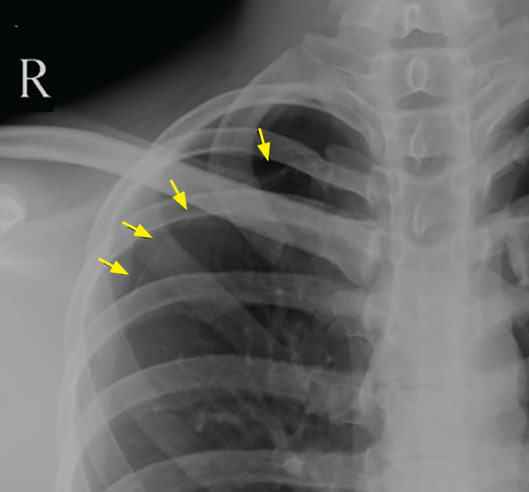
However, re-review of the chest radiograph from the previous facility noted a mild displacement of the pleural line, with absence of lung markings in the right upper lung zone, suggestive of a small apical pneumothorax (Figure 2). Based on this finding, on the absence of a precipitating cause or other significant finding, and on his chest pain atypical for angina, a diagnosis of small spontaneous primary pneumothorax was made.

Figure 2
  • Download figure
  • Open in new tab
  • Download powerpoint
Figure 2

On re-review of the chest radiograph from another facility, magnification showed a thin visceral pleural marking (arrows) displaced from the chest wall at the right apex, indicating a small right apical pneumothorax.

The patient was treated conservatively with high-flow oxygen for several hours, his cardiorespiratory status stabilized, and he was discharged.

SPONTANEOUS PNEUMOTHORAX: RISK FACTORS AND DIAGNOSIS

A small pneumothorax can be easily missed on a chest radiograph unless there is a strong index of clinical suspicion.

Pneumothorax is considered spontaneous if there is no apparent precipitating cause. Spontaneous pneumothorax is considered “primary” if it occurs in individuals with no underlying lung disease. Primary spontaneous pneumothorax typically occurs in tall, thin individuals. Other risk factors are male sex and cigarette smoking. Spontaneous rupture of a subpleural bleb or bulla is usually the cause.

Patients typically present with an abrupt onset of chest pain that is usually pleuritic, with or without breathlessness. Some patients may experience shoulder-tip pain.

Posteroanterior chest radiography is the standard view in patients with suspected primary spontaneous pneumothorax and may be combined with lateral radiographs in difficult cases. Expiratory films, though traditionally believed to enhance diagnostic sensitivity, may not be routinely necessary.1,2 The radiographic hallmark is displacement of the pleural line and the absence of lung markings between the edge of the pleura and chest wall. If these features are difficult to see on the posteroanterior radiograph, a lateral decubitus view with the affected side up would clearly show the lung “falling away” from the chest wall, demonstrating the pneumothorax.

Computed tomography is more sensitive in the detection of pneumothorax but is not usually required. Bedside lung ultrasonography has emerged as a new sensitive tool to diagnose pneumothorax in the emergency department, especially in trauma and critically ill patients.1

TREATMENT

Conservative management with observation for 6 hours is recommended if the patient is clinically stable and no enlargement of the pneumothorax is noted on repeat radiographs. An outpatient follow-up visit 2 days after discharge is desirable.3,4

High-flow oxygen may be administered to hasten absorption of intrapleural air.4 Additional inpatient care and pleural drainage may be necessary for patients with larger pneumothoraces (distance from lung apex to visceral pleura ≥ 3 cm, or ≥ 15% of thoracic volume)3–5 or patients who are clinically unstable due to hypotension, respiratory distress, or significant oxygen desaturation (< 92%).

Primary spontaneous pneumothorax typically occurs at rest. Therefore, there is no need to recommend avoiding exercise to prevent recurrences.

The acute nature of the presentation can mimic an acute coronary syndrome or an acute aortic syndrome and therefore requires a strong index of suspicion for the diagnosis. A small pneumothorax can be easily missed on chest radiography and should be sought in the appropriate clinical setting.

DISCLOSURES

The author reports no relevant financial relationships which, in the context of his contributions, could be perceived as a potential conflict of interest.

  • Copyright © 2021 The Cleveland Clinic Foundation. All Rights Reserved.

REFERENCES

  1. ↵
    1. Tschopp JM,
    2. Bintcliffe O,
    3. Astoul P, et al
    . ERS task force statement: diagnosis and treatment of primary spontaneous pneumothorax. Eur Respir J 2015; 46(2):321–335. doi:10.1183/09031936.00219214
    OpenUrlAbstract/FREE Full Text
  2. ↵
    1. Bradley M,
    2. Williams C,
    3. Walshaw MJ
    . The value of routine expiratory chest films in the diagnosis of pneumothorax. Arch Emerg Med 1991; 8(2):115–116. doi:10.1136/emj.8.2.115
    OpenUrlFREE Full Text
  3. ↵
    1. Baumann MH,
    2. Strange C,
    3. Heffner JE, et al
    . Management of spontaneous pneumothorax: an American College of Chest Physicians Delphi consensus statement. Chest 2001; 119(2):590–602. doi:10.1378/chest.119.2.590
    OpenUrlCrossRefPubMed
  4. ↵
    1. MacDuff A,
    2. Arnold A,
    3. Harvey J; BTS Pleural Disease Guideline Group
    . Management of spontaneous pneumothorax: British Thoracic Society pleural disease guideline 2010. Thorax 2010; 65(suppl 2):ii18–ii31. doi:10.1136/thx.2010.136986
    OpenUrlFREE Full Text
  5. ↵
    1. Light RW,
    2. Lee YC
    . Pneumothorax, chylothorax, hemothorax, and fibrothorax. In: Murray & Nadel’s Textbook of Respiratory Diseases. Philadelphia, PA: Elsevier; 2016:1439–1460.e10.
PreviousNext
Back to top

In this issue

Cleveland Clinic Journal of Medicine: 88 (9)
Cleveland Clinic Journal of Medicine
Vol. 88, Issue 9
1 Sep 2021
  • Table of Contents
  • Table of Contents (PDF)
  • Index by author
  • Complete Issue (PDF)
Print
Download PDF
Article Alerts
Sign In to Email Alerts with your Email Address
Email Article

Thank you for your interest in spreading the word on Cleveland Clinic Journal of Medicine.

NOTE: We only request your email address so that the person you are recommending the page to knows that you wanted them to see it, and that it is not junk mail. We do not capture any email address.

Enter multiple addresses on separate lines or separate them with commas.
An uncommon cause of chest pain
(Your Name) has sent you a message from Cleveland Clinic Journal of Medicine
(Your Name) thought you would like to see the Cleveland Clinic Journal of Medicine web site.
CAPTCHA
This question is for testing whether or not you are a human visitor and to prevent automated spam submissions.
Citation Tools
An uncommon cause of chest pain
Salman Salahuddin
Cleveland Clinic Journal of Medicine Sep 2021, 88 (9) 487-488; DOI: 10.3949/ccjm.88a.20160

Citation Manager Formats

  • BibTeX
  • Bookends
  • EasyBib
  • EndNote (tagged)
  • EndNote 8 (xml)
  • Medlars
  • Mendeley
  • Papers
  • RefWorks Tagged
  • Ref Manager
  • RIS
  • Zotero
Share
An uncommon cause of chest pain
Salman Salahuddin
Cleveland Clinic Journal of Medicine Sep 2021, 88 (9) 487-488; DOI: 10.3949/ccjm.88a.20160
Twitter logo Facebook logo Mendeley logo
  • Tweet Widget
  • Facebook Like
  • Linkedin Share Button

Jump to section

  • Article
    • SPONTANEOUS PNEUMOTHORAX: RISK FACTORS AND DIAGNOSIS
    • TREATMENT
    • DISCLOSURES
    • REFERENCES
  • Figures & Data
  • Info & Metrics
  • PDF

Related Articles

  • How do you effectively evaluate the elderly for alcohol use disorder?
  • PubMed
  • Google Scholar

Cited By...

  • No citing articles found.
  • Google Scholar

More in this TOC Section

  • Tinea incognito
  • Prolonged venous filling time and dependent rubor in a patient with peripheral artery disease
  • Sarcoidosis with diffuse purplish erythematous plaques on the hands
Show more The Clinical Picture

Similar Articles

Subjects

  • Cardiology
  • Emergency Medicine
  • Pulmonology

Navigate

  • Current Issue
  • Past Issues
  • Supplements
  • Article Type
  • Specialty
  • CME/MOC Articles
  • CME/MOC Calendar
  • Media Kit

Authors & Reviewers

  • Manuscript Submission
  • Authors & Reviewers
  • Subscriptions
  • About CCJM
  • Contact Us
  • Cleveland Clinic Center for Continuing Education
  • Consult QD

Share your suggestions!

Copyright © 2025 The Cleveland Clinic Foundation. All rights reserved. The information provided is for educational purposes only. Use of this website is subject to the website terms of use and privacy policy. 

Powered by HighWire