
Main menu

  • Home
  • Content
    • Current Issue
    • Ahead of Print
    • Past Issues
    • Supplements
    • Article Type
  • Specialty
    • Articles by Specialty
  • CME/MOC
    • Articles
    • Calendar
  • Info For
    • Manuscript Submission
    • Authors & Reviewers
    • Subscriptions
    • About CCJM
    • Contact Us
    • Media Kit
  • Conversations with Leaders
  • Conference Coverage
    • ACC / WCC 2023
    • AAAAI Meeting 2023
    • ACR Convergence 2022
    • Kidney Week 2022
    • AIDS 2022
    • CHEST 2021
    • IDWeek 2021
    • IAS 2021
    • ADA 2021
    • ATS 2021
    • ACC 2021
    • ACP 2021
    • AAN 2021
  • Other Publications
    • www.clevelandclinic.org

User menu

  • Register
  • Log in

Search

  • Advanced search
Cleveland Clinic Journal of Medicine
  • Other Publications
    • www.clevelandclinic.org
  • Register
  • Log in
Cleveland Clinic Journal of Medicine

Advanced Search

  • Home
  • Content
    • Current Issue
    • Ahead of Print
    • Past Issues
    • Supplements
    • Article Type
  • Specialty
    • Articles by Specialty
  • CME/MOC
    • Articles
    • Calendar
  • Info For
    • Manuscript Submission
    • Authors & Reviewers
    • Subscriptions
    • About CCJM
    • Contact Us
    • Media Kit
  • Conversations with Leaders
  • Conference Coverage
    • ACC / WCC 2023
    • AAAAI Meeting 2023
    • ACR Convergence 2022
    • Kidney Week 2022
    • AIDS 2022
    • CHEST 2021
    • IDWeek 2021
    • IAS 2021
    • ADA 2021
    • ATS 2021
    • ACC 2021
    • ACP 2021
    • AAN 2021
The Clinical Picture

Unilateral pulmonary edema

Yoshihiro Harano, MD and Mikio Nakajima, MD, MPH, PhD
Cleveland Clinic Journal of Medicine March 2022, 89 (3) 124-125; DOI: https://doi.org/10.3949/ccjm.89a.21046
Yoshihiro Harano
Department of Cardiology, Tokyo Metropolitan Hiroo Hospital, Tokyo, Japan
  • Find this author on Google Scholar
  • Find this author on PubMed
  • Search for this author on this site
  • For correspondence: yoshihiro.harano00@outlook.jp
Mikio Nakajima
Emergency and Critical Care Center, Tokyo Metropolitan Hiroo Hospital, Tokyo, Japan
  • Find this author on Google Scholar
  • Find this author on PubMed
  • Search for this author on this site
  • Article
  • Figures & Data
  • Info & Metrics
  • PDF
Loading

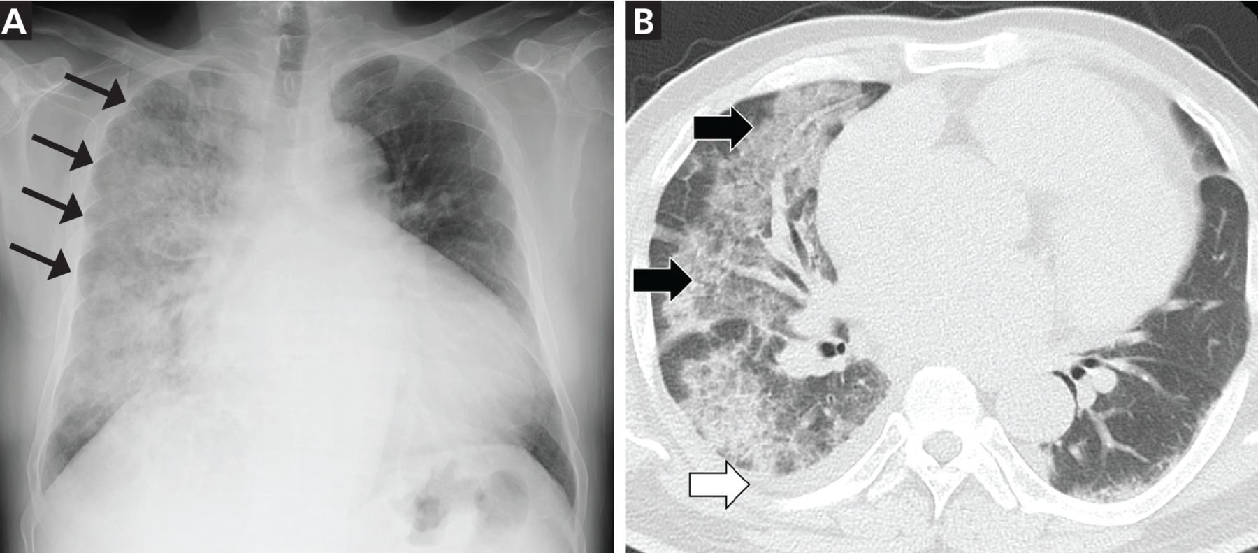
A 48-year-old man was transferred to our hospital for dyspnea. Although he had no significant medical history, he had been hospitalized for COVID-19 at a local healthcare facility 1 month earlier. Three weeks after discharge from that facility, he noted progressively worsening exertional dyspnea and fatigue. He returned to that facility, where he was afebrile at presentation and was diagnosed with bacterial pneumonia secondary to COVID-19 infection based on chest radiography and computed tomography that revealed a unilateral consolidation in the right lung and an inexplicable enlargement of the heart (Figure 1).

Figure 1
  • Download figure
  • Open in new tab
  • Download powerpoint
Figure 1

(Left) Chest radiography showed a right unilateral consolidation (arrows) and enlarged cardiac silhouette. (Right) Chest computed tomography revealed diffuse ground-glass opacities and consolidation (black arrows) together with a small amount of pleural effusion in the right lung (white arrow).

On admission to our facility, he presented with the following vital signs:

  • Clear level of consciousness

  • Blood pressure 134/96 mm Hg

  • Pulse rate 120 beats per minute

  • Body temperature 36.1°C (96.9°F)

  • Respiratory rate 24 breaths per minute

  • Oxygen saturation 91% on oxygen delivered by nasal cannula at 2 L per minute.

Physical examination revealed mild right jugular venous distention and mild pitting edema of the lower extremities. Coarse crackles were noted in the right lung, but no abnormal heart sounds were documented.

Although electrocardiography was unremarkable except for sinus tachycardia with no ST-segment or T-wave abnormalities, we considered heart failure in the differential diagnosis given his findings on physical examination and imaging. Transthoracic echocardiography revealed diffuse left ventricular hypokinesis with an ejection fraction of 20% and moderate mitral regurgitation toward the posterior left atrial wall. Additionally, his serum N-terminal probrain natriuretic peptide level was 6,469 pg/mL (reference range, 0–125 pg/mL). The patient was diagnosed with congestive heart failure and was placed on intravenous furosemide, nitrates, and noninvasive positive pressure ventilation. Initial therapy dramatically improved his dyspnea and chest radiograph within 72 hours after admission without the use of antibiotics (Figure 2). The patient made a full recovery and was discharged on hospital day 11.

Figure 2
  • Download figure
  • Open in new tab
  • Download powerpoint
Figure 2

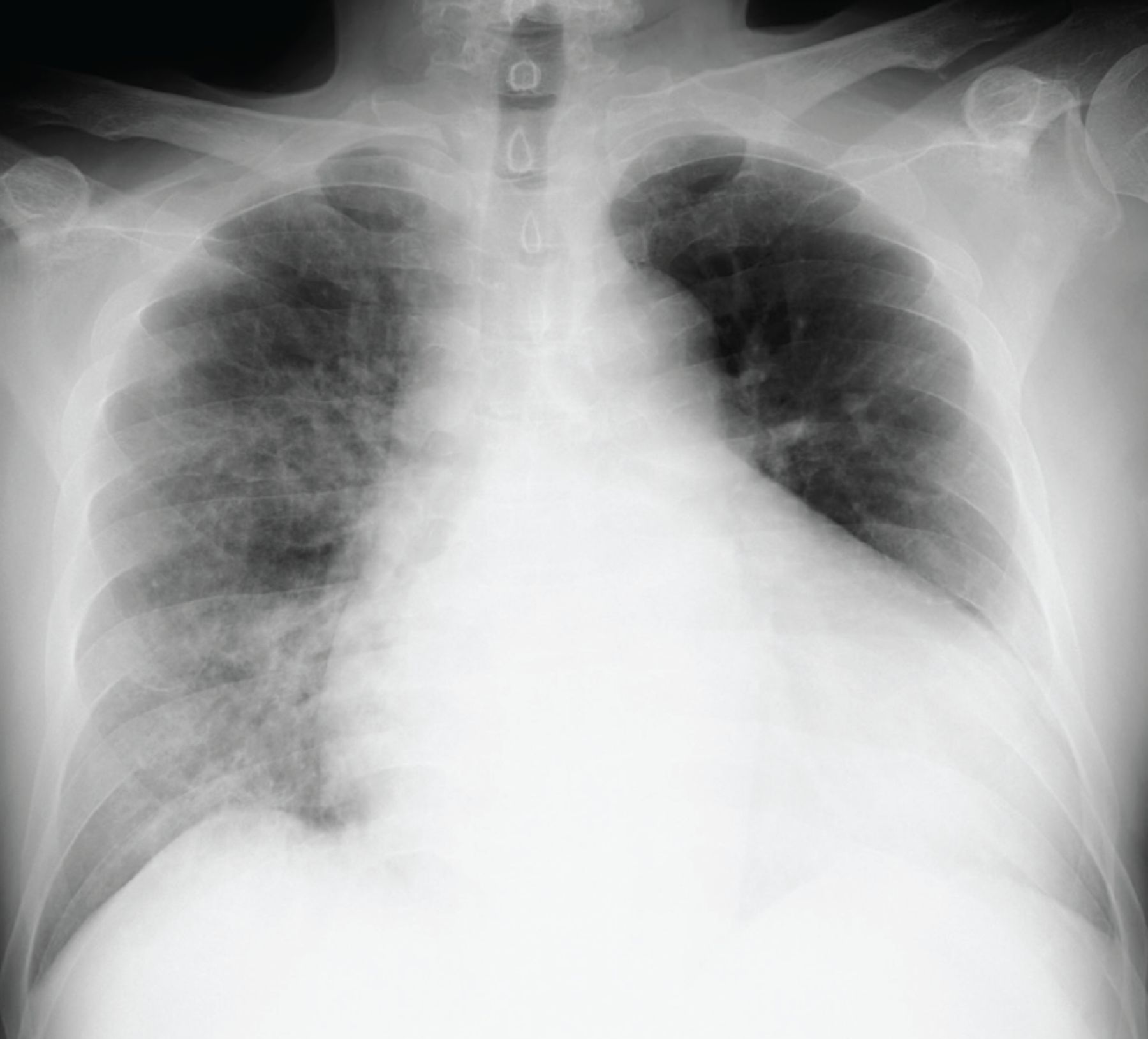
Chest radiograph 72 hours after admission and initiation of intravenous furosemide, nitrates, and noninvasive positive pressure ventilation. Unilateral consolidation in the right lung rapidly improved.

UNILATERAL PNEUMONIA WITH COVID-19

Approximately 1 out of every 10 patients with COVID-19 pneumonia presents with unilateral––as opposed to bilateral—pneumonia.1 Generally, one would consider viral or secondary bacterial pneumonia as the most likely diagnosis when a patient presents with unilateral consolidations on chest radiography. However, cardiogenic pulmonary edema accounts for 2.1% of unilateral pulmonary edema.2

On imaging, cardiogenic pulmonary edema generally appears as an opacity involving the right lung and is frequently attributed to severe mitral regurgitation.2 The direction of the mitral regurgitation toward the posterior left atrial wall could selectively impede the right pulmonary venous system,3,4 and the regurgitant flow may focally elevate the pressure in the right pulmonary vein.4 Our patient had retrograde blood flow toward the posterior left atrial wall. However, we could not confirm “severe” mitral regurgitation. Other factors that would affect the distribution of pulmonary edema include decubitus position of the patient (gravitational effect), position of the pulmonary veins, and preexisting lung disease.4

When encountering unilateral lung consolidations on chest imaging during the COVID-19 era, one should avoid the common pitfall of hastily attributing the finding to COVID-19 and instead should consider the full spectrum of differential diagnoses, including cardiogenic pulmonary edema.

DISCLOSURES

The authors report no relevant financial relationships which, in the context of their contributions, could be perceived as a potential conflict of interest.

  • Copyright © 2022 The Cleveland Clinic Foundation. All Rights Reserved.

REFERENCES

  1. ↵
    1. Wu J,
    2. Pan J,
    3. Teng D,
    4. Xu X,
    5. Feng J,
    6. Chen YC
    . Interpretation of CT signs of 2019 novel coronavirus (COVID-19) pneumonia. Eur Radiol 2020; 30(10):5455–5462. doi:10.1007/s00330-020-06915-5
    OpenUrlCrossRefPubMed
  2. ↵
    1. Attias D,
    2. Mansencal N,
    3. Auvert B, et al
    . Prevalence, characteristics, and outcomes of patients presenting with cardiogenic unilateral pulmonary edema. Circulation 2010; 122(11):1109–1115. doi:10.1161/CIRCULATIONAHA.109.934950
    OpenUrlAbstract/FREE Full Text
  3. ↵
    1. Miyatake K,
    2. Nimura Y,
    3. Sakakibara H, et al
    . Localisation and direction of mitral regurgitant flow in mitral orifice studied with combined use of ultrasonic pulsed Doppler technique and two dimensional echocardiography. Br Heart J 1982; 48(5):449–458. doi:10.1136/hrt.48.5.449
    OpenUrlAbstract/FREE Full Text
  4. ↵
    1. Gurney JW,
    2. Goodman LR
    . Pulmonary edema localized in the right upper lobe accompanying mitral regurgitation. Radiology 1989; 171(2):397–399. doi:10.1148/radiology.171.2.2704804
    OpenUrlCrossRefPubMed
PreviousNext
Back to top

In this issue

Cleveland Clinic Journal of Medicine: 89 (3)
Cleveland Clinic Journal of Medicine
Vol. 89, Issue 3
1 Mar 2022
  • Table of Contents
  • Table of Contents (PDF)
  • Index by author
  • Complete Issue (PDF)
Print
Download PDF
Article Alerts
Sign In to Email Alerts with your Email Address
Email Article

Thank you for your interest in spreading the word on Cleveland Clinic Journal of Medicine.

NOTE: We only request your email address so that the person you are recommending the page to knows that you wanted them to see it, and that it is not junk mail. We do not capture any email address.

Enter multiple addresses on separate lines or separate them with commas.
Unilateral pulmonary edema
(Your Name) has sent you a message from Cleveland Clinic Journal of Medicine
(Your Name) thought you would like to see the Cleveland Clinic Journal of Medicine web site.
CAPTCHA
This question is for testing whether or not you are a human visitor and to prevent automated spam submissions.
Citation Tools
Unilateral pulmonary edema
Yoshihiro Harano, Mikio Nakajima
Cleveland Clinic Journal of Medicine Mar 2022, 89 (3) 124-125; DOI: 10.3949/ccjm.89a.21046

Citation Manager Formats

  • BibTeX
  • Bookends
  • EasyBib
  • EndNote (tagged)
  • EndNote 8 (xml)
  • Medlars
  • Mendeley
  • Papers
  • RefWorks Tagged
  • Ref Manager
  • RIS
  • Zotero
Share
Unilateral pulmonary edema
Yoshihiro Harano, Mikio Nakajima
Cleveland Clinic Journal of Medicine Mar 2022, 89 (3) 124-125; DOI: 10.3949/ccjm.89a.21046
Reddit logo Twitter logo Facebook logo Mendeley logo
  • Tweet Widget
  • Facebook Like
  • Linkedin Share Button

Jump to section

  • Article
    • UNILATERAL PNEUMONIA WITH COVID-19
    • DISCLOSURES
    • REFERENCES
  • Figures & Data
  • Info & Metrics
  • PDF

Related Articles

  • No related articles found.
  • PubMed
  • Google Scholar

Cited By...

  • No citing articles found.
  • Google Scholar

More in this TOC Section

  • Mass under the lip
  • Oral leukoplakia and oral cancer
  • Median rhomboid glossitis caused by tongue-brushing
Show more The Clinical Picture

Similar Articles

Subjects

  • Infectious Diseases
  • Pulmonology
  • Imaging
  • Cardiology

Navigate

  • Current Issue
  • Past Issues
  • Supplements
  • Article Type
  • Specialty
  • CME/MOC Articles
  • CME/MOC Calendar
  • Media Kit

Authors & Reviewers

  • Manuscript Submission
  • Authors & Reviewers
  • Subscriptions
  • About CCJM
  • Contact Us
  • Cleveland Clinic Center for Continuing Education
  • Consult QD

Share your suggestions!

Copyright © 2023 The Cleveland Clinic Foundation. All rights reserved. The information provided is for educational purposes only. Use of this website is subject to the website terms of use and privacy policy. 

Powered by HighWire