


A 72-year-old woman was referred to the dermatology department with a 1-year history of itchy, erythematous plaques on the flexor aspects of her upper arms. The lesions had been previously diagnosed as contact allergic dermatitis and were treated with systemic and topical corticosteroids. The lesions would initially respond to treatment but would rapidly return after her prescriptions expired.
At presentation in the dermatology department, the patient had annular and polycyclic erythematous plaques with solitary papules on the periphery present on the flexor sides of the forearms (Figure 1). She declined biopsy but consented to skin scrapings. The scrapings showed the presence of hyphae, and mycological culture revealed Trichophyton rubrum. These findings and her history of immunosuppressant (ie, corticosteroid) therapy confirmed the diagnosis of tinea incognito, a localized, superficial dermatophyte infection that lacks the classic features of fungal infection as a result of immunosuppressant therapy. The patient received therapy with oral itraconazole 100 mg/day for 2 weeks and with topical miconazole, with full symptom resolution (Figure 2).
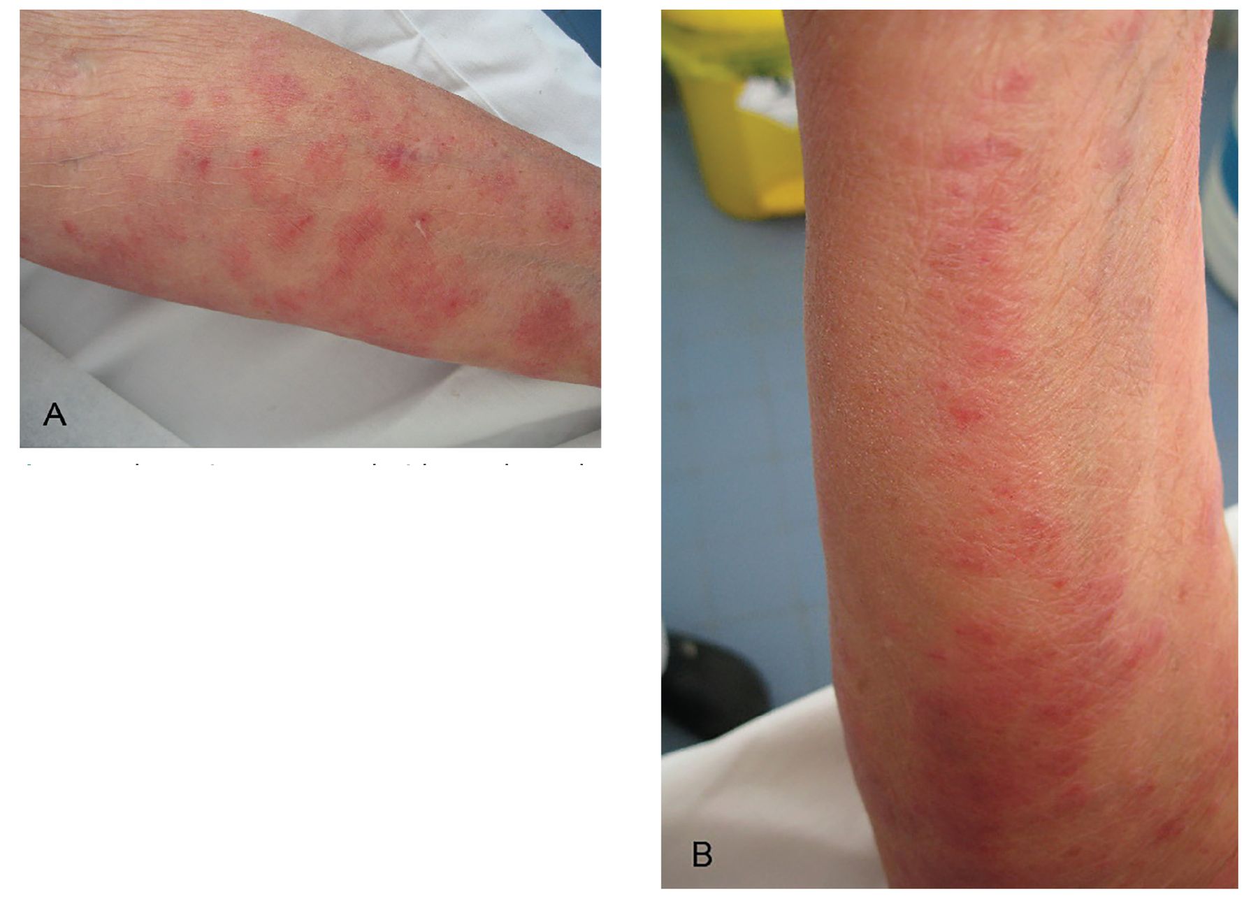
The patient presented with annular and polycyclic pruritic erythematous plaques localized on the flexor sides of (A) the left forearm and (B) the right forearm.

Complete improvement on both forearms after administration of antifungal agents.
SUPPRESSION OF THE LOCAL IMMUNE RESPONSE
Tinea incognito is often erroneously treated with corticosteroids, which suppress the local immune response, allowing the fungus to spread easily and creating an atypical clinical presentation.1,2
Because of the appearance of the lesions, we initially considered a diagnosis of systemic lupus erythematosus. However, the lesions were not present on the photo-exposed parts of the body that are characteristic for lupus erythematosus, and physical examination of the rest of her body was normal. Additionally, the patient stated that the skin lesions had not been directly exposed to the sun, which could have led to their appearance and worsening of the condition. Furthermore, antinuclear antibody assay for systemic lupus erythematosus was negative.
Other conditions in the differential diagnosis included eczema (which our patient had already been diagnosed with but was treated unsuccessfully), erythema annulare centrifugum (which was excluded with mycological examinations), psoriasis (her lesions were not characteristic or in typical distribution on the skin), and scabies (there was no evidence of skin burrows, and the itching was not dominant during the night and was not a dominant feature).1–4
Confirmatory tests
In the setting of nonresolving erythematous lesions in a patient treated with corticosteroids, skin scraping for fungal examination is recommended.3 A blunt scalpel is used to scrape the edges of a well-cleaned lesion. The scrapings are placed onto a slide covered with 10% potassium hydroxide and examined under a microscope.5 For tinea incognito, examination with low magnification (10 ×) and then higher magnification (40 ×) reveals the presence of fungal spores with or without hyphae.5
TAKE-HOME MESSAGE
When fungal infection is misdiagnosed and treated with corticosteroids, the normal cutaneous response is blunted, allowing the fungus to spread easily in the absence of significant erythema. The clinical presentation may suggest an infectious, paraneoplastic, allergic or autoimmune etiology.1 Therefore, if the appearance of a skin lesion changes or worsens during treatment with immunosuppressants, a diagnosis of tinea incognito should be considered.1,2
DISCLOSURES
The authors report no relevant financial relationships which, in the context of their contributions, could be perceived as a potential conflict of interest.
- Copyright © 2022 The Cleveland Clinic Foundation. All Rights Reserved.
In this issue


Jump to section
Related Articles
Cited By...
- No citing articles found.