
Main menu

  • Home
  • Content
    • Current Issue
    • Ahead of Print
    • Past Issues
    • Supplements
    • Article Type
  • Specialty
    • Articles by Specialty
  • CME/MOC
    • Articles
    • Calendar
  • Info For
    • Manuscript Submission
    • Authors & Reviewers
    • Subscriptions
    • About CCJM
    • Contact Us
    • Media Kit
  • Conversations with Leaders
  • Conference Coverage
    • Kidney Week 2024
    • CHEST 2024
    • ACR Convergence 2023
    • Kidney Week 2023
    • ObesityWeek 2023
    • IDWeek 2023
    • CHEST 2023
    • MDS 2023
    • IAS 2023
    • ACP 2023
    • AAN 2023
    • ACC / WCC 2023
    • AAAAI Meeting 2023
    • ACR Convergence 2022
    • Kidney Week 2022
    • AIDS 2022
  • Other Publications
    • www.clevelandclinic.org

User menu

  • Register
  • Log in

Search

  • Advanced search
Cleveland Clinic Journal of Medicine
  • Other Publications
    • www.clevelandclinic.org
  • Register
  • Log in
Cleveland Clinic Journal of Medicine

Advanced Search

  • Home
  • Content
    • Current Issue
    • Ahead of Print
    • Past Issues
    • Supplements
    • Article Type
  • Specialty
    • Articles by Specialty
  • CME/MOC
    • Articles
    • Calendar
  • Info For
    • Manuscript Submission
    • Authors & Reviewers
    • Subscriptions
    • About CCJM
    • Contact Us
    • Media Kit
  • Conversations with Leaders
  • Conference Coverage
    • Kidney Week 2024
    • CHEST 2024
    • ACR Convergence 2023
    • Kidney Week 2023
    • ObesityWeek 2023
    • IDWeek 2023
    • CHEST 2023
    • MDS 2023
    • IAS 2023
    • ACP 2023
    • AAN 2023
    • ACC / WCC 2023
    • AAAAI Meeting 2023
    • ACR Convergence 2022
    • Kidney Week 2022
    • AIDS 2022
The Clinical Picture

Skin-colored papules on the cheeks, acrochordons on the axillae

Irene López Riquelme, MD, María Ayala Blanca, MD, PhD and Elisabeth Gómez Moyano, MD, PhD
Cleveland Clinic Journal of Medicine June 2022, 89 (6) 297-299; DOI: https://doi.org/10.3949/ccjm.89a.21087
Irene López Riquelme
Department of Dermatology, Hospital Regional Universitario de Málaga, Málaga, Spain
  • Find this author on Google Scholar
  • Find this author on PubMed
  • Search for this author on this site
  • For correspondence: [email protected]
María Ayala Blanca
Department of Pathology, Hospital Regional Universitario de Málaga, Málaga, Spain
  • Find this author on Google Scholar
  • Find this author on PubMed
  • Search for this author on this site
Elisabeth Gómez Moyano
Department of Dermatology, Hospital Regional Universitario de Málaga, Málaga, Spain
  • Find this author on Google Scholar
  • Find this author on PubMed
  • Search for this author on this site
  • Article
  • Figures & Data
  • Info & Metrics
  • PDF
Loading

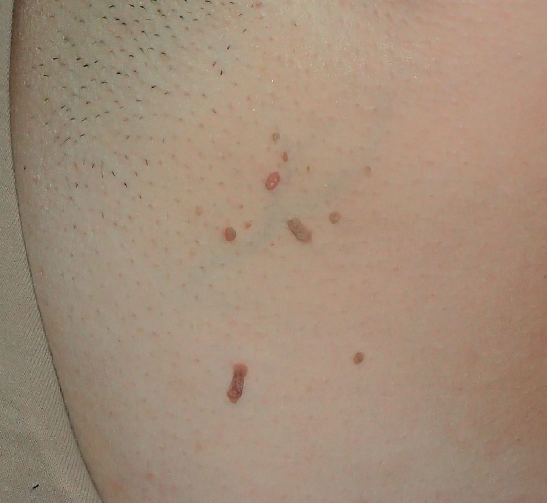
A 43-year-old woman presented to the dermatology department with multiple small skin-colored papules on her cheeks (Figure 1) and acrochordons (“skin tags”) on the axillae (Figure 2). She reported that they had appeared over the past few years. Her personal medical history was unremarkable, but she had a first-degree relative with renal carcinoma.

Figure 1
  • Download figure
  • Open in new tab
  • Download powerpoint
Figure 1

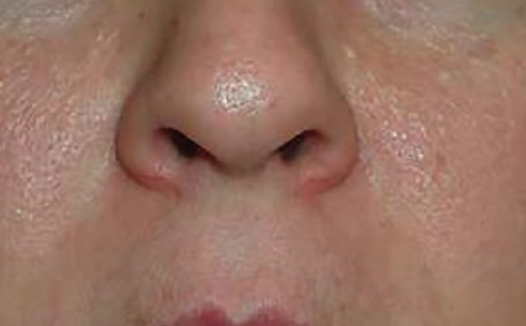
Multiple whitish to skin-colored papules on both cheeks.

Figure 2
  • Download figure
  • Open in new tab
  • Download powerpoint
Figure 2

Acrochordons on the axillae.

Biopsy of a papule revealed dermal follicular structures surrounded by a perifollicular fibrous sheath and a densely fibrous stroma, consistent with a fibrofolliculoma (Figure 3), ie, a benign hair follicle tumor pathognomonic for Birt-Hogg-Dubé (BHD) syndrome. Subsequent genetic testing found a mutation in the FLCN gene, confirming the diagnosis.

Figure 3
  • Download figure
  • Open in new tab
  • Download powerpoint
Figure 3

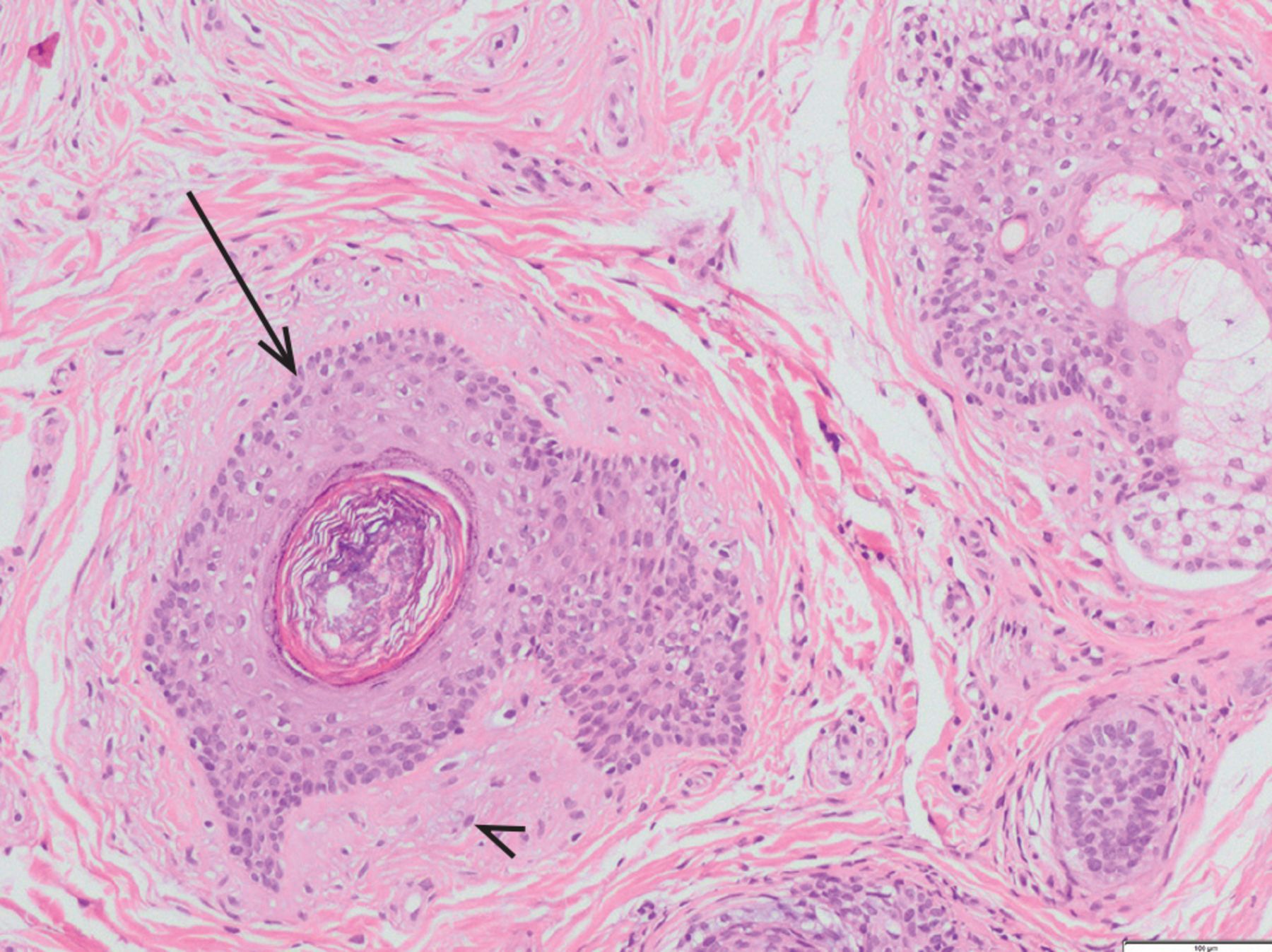
Skin biopsy showing dermal follicular structures (arrow) surrounded by a perifollicular fibrous sheath and a densely fibrous stroma (arrowhead), consistent with fibrofolliculoma (hematoxylin and eosin, × 400).

Computed tomography showed no evidence of lung cysts or kidney tumor. Regular cancer surveillance was instituted.

BHD SYNDROME: SKIN, LUNGS, KIDNEYS

BHD syndrome is an autosomal dominant genetic disorder that predisposes patients not only to characteristic skin lesions but also to the development of lung cysts, spontaneous pneumothorax, and kidney neoplasms. It is caused by inactivating mutations in the gene that codes for folliculin, a protein that likely acts as a tumor suppressor. Most studies suggest that folliculin plays a role in the mammalian target of rapamycin (mTOR) pathway.1,2

Prevalence of BHD syndrome is uncertain. It is likely underdiagnosed, especially as more than 150 pathogenic variants with variable penetrance have been described.3,4 Some studies suggest that BHD syndrome may be the cause of 5% to 10% of cases of apparent primary spontaneous pneumothorax.3

Skin manifestations often arise first

First clinical manifestations tend to appear in the second or third decade. Many patients with BHD syndrome (75% to 90% of White patients, but only 30% to 50% of Asian patients) present with multiple fibrofolliculomas.2,4 They are rarely seen in the general population.

BHD syndrome should be suspected in patients with more than 5 fibrofolliculomas. Histologically, they appear as epithelial cells arranged in rows growing from a central aberrant hair follicle, surrounded by a thick connective-tissue stroma.1

Other dermatologic manifestations may include trichodiscomas, perifollicular fibromas, and acrochordons. These benign tumors, including fibrofolliculomas, are considered hamartomas (ie, abnormal development of normal tissue) of the hair follicle. They may be indistinguishable from each other, and some argue that they are variants of the same lesion.4 Fibrofolliculomas and trichodiscomas present as multiple, small, whitish papules, most commonly on the face, neck, and upper trunk. Acrochordons are small, pedunculated outgrowths of epidermal and dermal tissue in the neck, eyelids, upper chest, and axillae, and they may be reported as fibrofolliculomas on histology.4

Pulmonary involvement commonly develops

Over 80% of patients develop multiple, bilateral pulmonary cysts in the fourth to fifth decades,1,2,4 with up to 38% of patients experiencing at least one pneumothorax.1 Larger number and size of cysts are associated with greater likelihood of spontaneous pneumothorax, but no clear association of BHD syndrome with pulmonary cancers has been found.4

Risk of renal carcinoma is markedly increased

Patients with BHD may have a 7-fold higher risk of renal cell cancer than the general population, with an estimated prevalence between 27% and 34%.1 Frequent histologic subtypes are chromophobe and oncocytic hybrid tumors, but they rarely show sarcomatoid transformation and generally have a favorable prognosis.5 Tumors in the same kidney may also be of different histologic types, but the significance of this for prognosis is not clear.2

MONITORING

An exact monitoring strategy has not been universally established. Risk of excessive radiation exposure from repeated computed tomography scans must be balanced with the relatively low risk of the slow-growing renal cancers associated with BHD syndrome: 12% to 34% of patients with BHD develop renal tumors.4

Most recommend periodic magnetic resonance imaging of the kidney, with the schedule (1 or 2 years) depending on past findings.6 Routine lung screening is not recommended.7 Periodic monitoring for cancers in other organs that may be associated with BHD syndrome—including the parotid, thyroid, and colon—is also recommended.7

TAKE-HOME POINTS

  • BHD syndrome is a genetic disorder caused by FLCN gene mutations that predispose patients to develop lung cysts, spontaneous pneumothorax, and renal neoplasms.

  • Multiple fibrofolliculomas and trichodiscomas are benign cutaneous lesions that provide a clue to BHD.

  • Upon diagnosis of BHD, screening with regular imaging is prognostically crucial for these patients, since identification of lung cysts and renal malignancies at early stages significantly improves life expectancy.

DISCLOSURES

The authors report no relevant financial relationships which, in the context of their contributions, could be perceived as a potential conflict of interest.

  • Copyright © 2022 The Cleveland Clinic Foundation. All Rights Reserved.

REFERENCES

  1. ↵
    1. Gupta N,
    2. Sunwoo BY,
    3. Kotloff RM
    . Birt-Hogg-Dubé syndrome. Clin Chest Med 2016; 37(3):475–486. doi:10.1016/j.ccm.2016.04.010
    OpenUrlCrossRef
  2. ↵
    1. Steinlein OK,
    2. Ertl-Wagner B,
    3. Ruzicka T,
    4. Sattler EC
    . Birt-Hogg-Dubé syndrome: an underdiagnosed genetic tumor syndrome. J Dtsch Dermatol Ges 2018; 16(3):278–283. doi:10.1111/ddg.13457
    OpenUrlCrossRef
  3. ↵
    1. Johannesma PC,
    2. Reinhard R,
    3. Kon Y, et al
    . Prevalence of Birt-Hogg-Dubé syndrome in patients with apparently primary spontaneous pneumothorax. Eur Respir J 2015; 45(4):1191–1194. doi:10.1183/09031936.00196914
    OpenUrlAbstract/FREE Full Text
  4. ↵
    1. Tong Y,
    2. Schneider JA,
    3. Coda AB,
    4. Hata TR,
    5. Cohen PR
    . Birt-Hogg-Dubé syndrome: a review of dermatological manifestations and other symptoms. Am J Clin Dermatol 2018; 19(1):87–101. doi:10.1007/s40257-017-0307-8
    OpenUrlCrossRef
  5. ↵
    1. Furuya M,
    2. Hasumi H,
    3. Yao M,
    4. Nagashima Y
    . Birt-Hogg-Dubé syndrome-associated renal cell carcinoma: histopathological features and diagnostic conundrum. Cancer Sci 2020; 111(1):15–22. doi:10.1111/cas.14255
    OpenUrlCrossRef
  6. ↵
    1. Stamatakis L,
    2. Metwalli AR,
    3. Middelton LA,
    4. Marston Linehan W
    . Diagnosis and management of BHD-associated kidney cancer. Fam Cancer 2013; 12(3):397–402. doi:10.1007/s10689-013-9657-4
    OpenUrlCrossRefPubMed
  7. ↵
    1. Adam MP,
    2. Ardinger HH,
    3. Pagon RA, et al.
    1. Sattler EC,
    2. Steinlein OK
    . Birt-Hogg-Dubé Syndrome. 2006 Feb 27 [Updated 2020 Jan 30]. In: Adam MP, Ardinger HH, Pagon RA, et al., eds. GeneReviews [Internet]. Seattle (WA): University of Washington, Seattle; 1993-2022. Table 5. [Recommended Surveillance for Individuals with Birt-Hogg-Dubé Syndrome]. Available from: https://www.ncbi.nlm.nih.gov/books/NBK1522/table/bhd.T.recommended_surveillance_for_indiv. Accessed May 17, 2022.
PreviousNext
Back to top

In this issue

Cleveland Clinic Journal of Medicine: 89 (6)
Cleveland Clinic Journal of Medicine
Vol. 89, Issue 6
1 Jun 2022
  • Table of Contents
  • Table of Contents (PDF)
  • Index by author
  • Complete Issue (PDF)
Print
Download PDF
Article Alerts
Sign In to Email Alerts with your Email Address
Email Article

Thank you for your interest in spreading the word on Cleveland Clinic Journal of Medicine.

NOTE: We only request your email address so that the person you are recommending the page to knows that you wanted them to see it, and that it is not junk mail. We do not capture any email address.

Enter multiple addresses on separate lines or separate them with commas.
Skin-colored papules on the cheeks, acrochordons on the axillae
(Your Name) has sent you a message from Cleveland Clinic Journal of Medicine
(Your Name) thought you would like to see the Cleveland Clinic Journal of Medicine web site.
CAPTCHA
This question is for testing whether or not you are a human visitor and to prevent automated spam submissions.
Citation Tools
Skin-colored papules on the cheeks, acrochordons on the axillae
Irene López Riquelme, María Ayala Blanca, Elisabeth Gómez Moyano
Cleveland Clinic Journal of Medicine Jun 2022, 89 (6) 297-299; DOI: 10.3949/ccjm.89a.21087

Citation Manager Formats

  • BibTeX
  • Bookends
  • EasyBib
  • EndNote (tagged)
  • EndNote 8 (xml)
  • Medlars
  • Mendeley
  • Papers
  • RefWorks Tagged
  • Ref Manager
  • RIS
  • Zotero
Share
Skin-colored papules on the cheeks, acrochordons on the axillae
Irene López Riquelme, María Ayala Blanca, Elisabeth Gómez Moyano
Cleveland Clinic Journal of Medicine Jun 2022, 89 (6) 297-299; DOI: 10.3949/ccjm.89a.21087
Twitter logo Facebook logo Mendeley logo
  • Tweet Widget
  • Facebook Like
  • Linkedin Share Button

Jump to section

  • Article
    • BHD SYNDROME: SKIN, LUNGS, KIDNEYS
    • MONITORING
    • TAKE-HOME POINTS
    • DISCLOSURES
    • REFERENCES
  • Figures & Data
  • Info & Metrics
  • PDF

Related Articles

  • No related articles found.
  • PubMed
  • Google Scholar

Cited By...

  • No citing articles found.
  • Google Scholar

More in this TOC Section

  • Tinea incognito
  • Prolonged venous filling time and dependent rubor in a patient with peripheral artery disease
  • Sarcoidosis with diffuse purplish erythematous plaques on the hands
Show more The Clinical Picture

Similar Articles

Subjects

  • Dermatology

Navigate

  • Current Issue
  • Past Issues
  • Supplements
  • Article Type
  • Specialty
  • CME/MOC Articles
  • CME/MOC Calendar
  • Media Kit

Authors & Reviewers

  • Manuscript Submission
  • Authors & Reviewers
  • Subscriptions
  • About CCJM
  • Contact Us
  • Cleveland Clinic Center for Continuing Education
  • Consult QD

Share your suggestions!

Copyright © 2025 The Cleveland Clinic Foundation. All rights reserved. The information provided is for educational purposes only. Use of this website is subject to the website terms of use and privacy policy. 

Powered by HighWire