


A 39-year-old man was referred to the dermatology clinic for nail bed discoloration on his right thumb. The discoloration had been present since the patient was 14 years old and had not changed in appearance.
Physical examination showed a brown-black linear band with regular borders extending from the proximal nail fold along the entire nail length (Figure 1). The patient reported no significant trauma to his fingers, and his medical history was unremarkable.
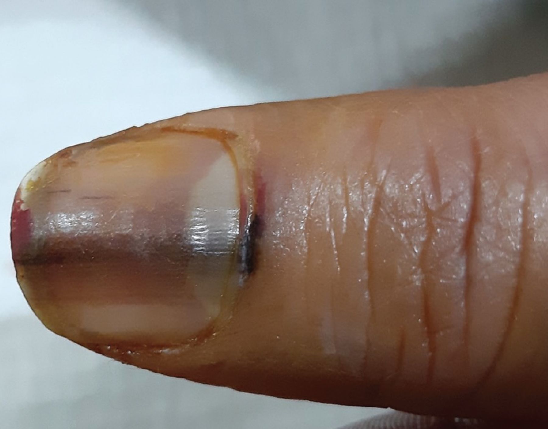
A brown-black linear band with regular borders extending from the proximal nail fold along the entire nail length of the patient’s right thumb.
Dermatoscopy revealed a linear, regular, brushy-patterned, brown-black band 3 mm wide, with homogeneity of color and thick pigmented parallel lines that were regular in width and spacing. Study of an incisional biopsy of the thumbnail matrix and the affected fold showed hyperpigmentation of the basal layer of the matrix and few melanocyte aggregations, without atypia. These features favored a diagnosis of acral lentiginous nevus.
However, because periungual pigmentation of the nail folds is also a feature of subungual melanoma (Hutchinson sign), immunohistochemical study was requested. Staining was positive for SOX10 in the epidermal melanocytes and for Ki67 in the parabasal epidermal cells. Additionally, the cyclin D1 marker was weakly positive in a few scattered cells in the upper dermis. Considering the results of pathology study, melanoma was excluded, and the diagnosis of acral lentiginous nevus presenting as pseudo-Hutchinson sign was made. The patient was referred to his primary care physician for routine follow-up.
DIFFERENTIAL DIAGNOSIS OF THE PSEUDO-HUTCHINSON SIGN
The Hutchinson nail sign, first described in 1886, is a periungual band of brown-black pigmentation extending from the nail matrix onto the surrounding tissue, usually due to progression of subungual melanoma.1 Pseudo-Hutchinson sign mimics Hutchinson sign but reflects a benign disease process. It is a diagnosis of exclusion, made only after malignancy is ruled out.2
Conditions associated with pseudo-Hutchinson sign include Bowen disease, subungual squamous cell carcinoma, fungal infection, ethnic (racial) melanonychia, Peutz-Jeghers syndrome, Laugier-Hunziker syndrome, systemic disease (systemic lupus erythematosus, scleroderma), radiation therapy, AIDS, congenital nevus, drug-induced dyschromia, malnutrition, chronic trauma, and subungual hematoma.2,3
ASSESSMENT OF NAIL BED DISCOLORATION
Assessment of a pigmented nail bed lesion should begin with a complete history of the lesion including duration, color changes, and trauma, as well as a medical history that includes medication use and a family history of melanoma. Factors that should prompt investigation for melanoma include linear band widths greater than 3 mm, linear bands wider proximally than distally, heterogeneous nail discoloration, irregular border of the discloration, nail plate dystrophy or ulceration and bleeding, or involvement of high-risk digits (ie, thumb, index finger, great toe). However, malignancy should not be ruled out based solely on the history and physical examination. Other methods such as dermatoscopy can be used.
Dermatoscopy is a principal tool for clinical assessment of skin lesions and should be used routinely for all skin lesions. Studies have shown that compared with examination with the naked eye, dermatoscopy increases the diagnostic accuracy of melanoma.4 Whereas a linear brushy pattern indicates a benign condition, a diffuse haphazard pattern suggests malignancy.5 For suspicious lesions, serial monitoring (every 2 to 3 months), biopsy (especially excision of the pigmented lesion), referral to a dermatologist, and a second look by an expert pathologist are highly recommended. Although biopsy even in skilled hands can lead to nail dystrophy, the risk does not outweigh the risk of failing to diagnose a malignant condition such as acral melanoma.
Additional evaluations
When these evaluations fail to confirm the diagnosis of melanoma but there is still suspicion, genetic studies can be used, especially since genetic mutations play a key role in development of cancer. For example, mutations of BRAF and NRAS are the 2 most common in melanoma tumors: 40% to 60% for BRAF, and 15% to 20% for NRAS. Other options for diagnosis include immunohistochemistry screening (to evaluate the rate of mitosis and proliferation), fluorescence in situ hybridization analysis, cytogenetic testing, and comparative genomic hybridization.6
DISCLOSURES
The authors report no relevant financial relationships which, in the context of their contributions, could be perceived as a potential conflict of interest.
- Copyright © 2022 The Cleveland Clinic Foundation. All Rights Reserved.
In this issue


Jump to section
Related Articles
Cited By...
- No citing articles found.