
Main menu

  • Home
  • Content
    • Current Issue
    • Ahead of Print
    • Past Issues
    • Supplements
    • Article Type
  • Specialty
    • Articles by Specialty
  • CME/MOC
    • Articles
    • Calendar
  • Info For
    • Manuscript Submission
    • Authors & Reviewers
    • Subscriptions
    • About CCJM
    • Contact Us
    • Media Kit
  • Conversations with Leaders
  • Conference Coverage
    • Kidney Week 2024
    • CHEST 2024
    • ACR Convergence 2023
    • Kidney Week 2023
    • ObesityWeek 2023
    • IDWeek 2023
    • CHEST 2023
    • MDS 2023
    • IAS 2023
    • ACP 2023
    • AAN 2023
    • ACC / WCC 2023
    • AAAAI Meeting 2023
    • ACR Convergence 2022
    • Kidney Week 2022
    • AIDS 2022
  • Other Publications
    • www.clevelandclinic.org

User menu

  • Register
  • Log in

Search

  • Advanced search
Cleveland Clinic Journal of Medicine
  • Other Publications
    • www.clevelandclinic.org
  • Register
  • Log in
Cleveland Clinic Journal of Medicine

Advanced Search

  • Home
  • Content
    • Current Issue
    • Ahead of Print
    • Past Issues
    • Supplements
    • Article Type
  • Specialty
    • Articles by Specialty
  • CME/MOC
    • Articles
    • Calendar
  • Info For
    • Manuscript Submission
    • Authors & Reviewers
    • Subscriptions
    • About CCJM
    • Contact Us
    • Media Kit
  • Conversations with Leaders
  • Conference Coverage
    • Kidney Week 2024
    • CHEST 2024
    • ACR Convergence 2023
    • Kidney Week 2023
    • ObesityWeek 2023
    • IDWeek 2023
    • CHEST 2023
    • MDS 2023
    • IAS 2023
    • ACP 2023
    • AAN 2023
    • ACC / WCC 2023
    • AAAAI Meeting 2023
    • ACR Convergence 2022
    • Kidney Week 2022
    • AIDS 2022
The Clinical Picture

Sarcoidosis with diffuse purplish erythematous plaques on the hands

Akihiro Orita, MD and Mitsuhito Ota, MD, PhD
Cleveland Clinic Journal of Medicine March 2025, 92 (3) 145-146; DOI: https://doi.org/10.3949/ccjm.92a.24022
Akihiro Orita
Department of Dermatology, Chitose City Hospital, Chitose, Japan
  • Find this author on Google Scholar
  • Find this author on PubMed
  • Search for this author on this site
Mitsuhito Ota
Department of Dermatology, Chitose City Hospital, Chitose, Japan
  • Find this author on Google Scholar
  • Find this author on PubMed
  • Search for this author on this site
  • For correspondence: [email protected]
  • Article
  • Figures & Data
  • Info & Metrics
  • PDF
Loading

A previously healthy 70-year-old woman presented in winter with a 1-month history of asymptomatic redness of the hands. She denied that the rash had been exacerbated by exposure to the cold temperatures. Physical examination revealed diffuse purplish erythematous plaques symmetrically distributed on the distal forearms and on the dorsal surface of the hands and fingers, with telangiectasia and swollen fingers (Figure 1). She had no other skin lesions.

Figure 1
  • Download figure
  • Open in new tab
  • Download powerpoint
Figure 1

Diffuse purplish erythema on the dorsal surface of the distal forearms, hands, and fingers, with telangiectasia and swollen fingers.

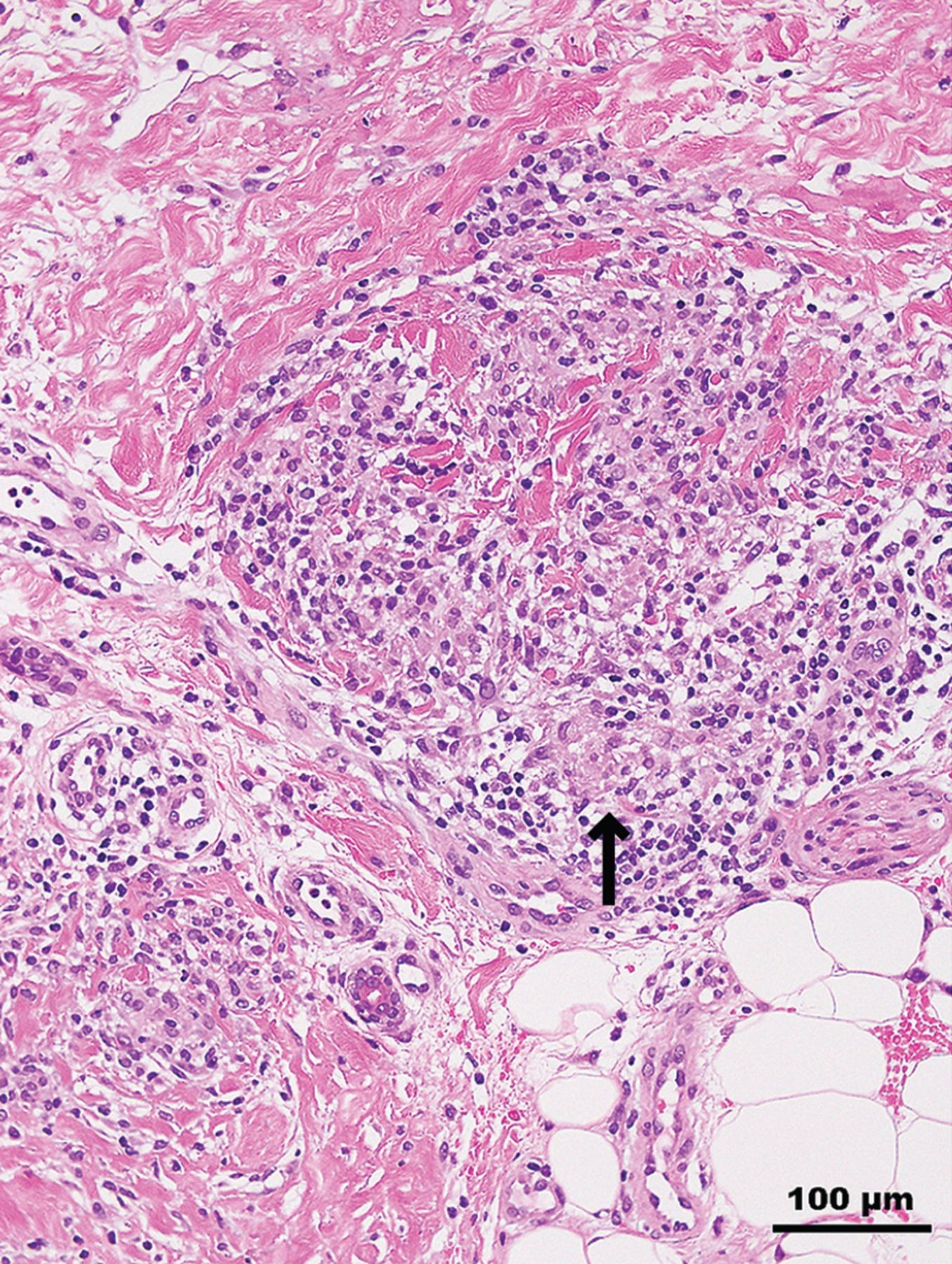
Histopathologic examination of a skin biopsy specimen showed noncaseating granulomatous infiltrates of epithelioid histiocytes with mild lymphocytic infiltrates in the dermis (Figure 2). Radiography of the hands found no bone lesions. Laboratory testing showed a serum angiotensin-converting enzyme level of 30.4 U/L (reference range 7.0–25.0 U/L). Systemic investigations identified supraclavicular and mediastinal lymphadenopathies and uveitis.

Figure 2
  • Download figure
  • Open in new tab
  • Download powerpoint
Figure 2

Histopathologic examination of a skin biopsy specimen revealed nodular histiocytic infiltration in the dermis, forming epithelioid granulomas (arrow) without evidence of necrosis (hematoxylin and eosin, original × 200).

A diagnosis of sarcoidosis was made, and treatment with oral prednisolone (0.5 mg/kg daily) was started. The skin lesions abated in 2 months, and the prednisolone was tapered off. The nodal lesions also have disappeared or regressed, and the uveitis has improved.

CUTANEOUS MANIFESTATIONS OF SARCOIDOSIS

Sarcoidosis is a systemic disorder of unknown etiology characterized by noncaseating epithelioid cell granulomas that mainly involve the lungs (about 90% of cases), mediastinal and peripheral lymph nodes, eyes, and skin.1 Involvement of the liver, spleen, central nervous system, heart, and bones occurs less often but is usually severe.

Cutaneous lesions specific for sarcoidosis, ie, those that display the histopathologic features of sarcoid granulomas, develop in up to 35% of patients with systemic sarcoidosis.2 Less than one-third of patients with cutaneous manifestations have isolated cutaneous sarcoidosis without any systemic features.1 The most common specific skin lesions include maculopapular sarcoidosis, nodular and plaque sarcoidosis, lupus pernio, scar or tattoo sarcoidosis, and subcutaneous sarcoidosis.2

Nonspecific skin lesions—those not caused by granulomas—also can occur in sarcoidosis and include erythema nodosum, prurigo, digital clubbing, erythema multiforme, pyoderma gangrenosum, and Sweet syndrome. Of these, erythema nodosum is the most common. However, there are many less common cutaneous manifestations of sarcoidosis, and, because of this varied morphology, sarcoidosis is considered one of the great imitators in dermatology.2

In this patient, the cutaneous lesion may have been lupus pernio, which causes indurated violaceous papulonodules and plaques on the nose, cheeks, and ears.3 The fingers and toes can be affected as well. To the best of our knowledge, lupus pernio occurring only on the hands has not been reported. Differential diagnoses in this patient included vascular disorders, such as chilblains, Raynaud phenomenon, and acrocyanosis.

Delays in the diagnosis of sarcoidosis are common given its varying presentations. Sarcoidosis is diagnosed more easily and earlier when skin lesions are present because the skin is easily accessible for histopathologic confirmation.4 However, this does not necessarily mean that it is easy to recognize a skin lesion as a manifestation of sarcoidosis. Because cutaneous sarcoidosis presents with varied morphology, it may present a diagnostic challenge.

A thorough skin examination and skin biopsy are necessary for diagnosing sarcoidosis. Laboratory tests can also be useful because at least 60% of patients diagnosed with sarcoidosis have an increased serum angiotensin-converting enzyme level.2 A systemic workup to evaluate the extent of involvement should be undertaken in all patients with cutaneous sarcoid lesions, even in those with minimal skin involvement.

DISCLOSURES

The authors report no relevant financial relationships which, in the context of their contributions, could be perceived as a potential conflict of interest.

  • Copyright © 2025 The Cleveland Clinic Foundation. All Rights Reserved.

REFERENCES

  1. ↵
    1. Islam S,
    2. Hossain MS,
    3. Murshed KM,
    4. Rahaman MFU,
    5. Azad MAK
    . Cutaneous sarcoidosis: a differential diagnosis to consider in undiagnosed skin lesions. Cureus 2023; 15(6):e39852. doi:10.7759/cureus.39852
    OpenUrlCrossRef
  2. ↵
    1. Griffiths C,
    2. Barker J,
    3. Bleiker T,
    4. Chalmers R,
    5. Creamer D
    1. Marcoval J,
    2. Mana J
    . Sarcoidosis. In: Griffiths C, Barker J, Bleiker T, Chalmers R, Creamer D, eds. Rook’s Textbook of Dermatology. 9th ed. United Kingdom: Wiley-Blackwell; 2016.
  3. ↵
    1. Popatia S,
    2. Wanat KA
    . Lupus pernio. JAMA Dermatol 2022; 158(4):446. doi:10.1001/jamadermatol.2021.6007
    OpenUrlCrossRefPubMed
  4. ↵
    1. Judson MA,
    2. Thompson BW,
    3. Rabin DL, et al
    . The diagnostic pathway to sarcoidosis. Chest 2003; 123(2):406–412. doi:10.1378/chest.123.2.406
    OpenUrlCrossRefPubMed
PreviousNext
Back to top

In this issue

Cleveland Clinic Journal of Medicine: 92 (3)
Cleveland Clinic Journal of Medicine
Vol. 92, Issue 3
1 Mar 2025
  • Table of Contents
  • Table of Contents (PDF)
  • Index by author
  • Complete Issue (PDF)
Print
Download PDF
Article Alerts
Sign In to Email Alerts with your Email Address
Email Article

Thank you for your interest in spreading the word on Cleveland Clinic Journal of Medicine.

NOTE: We only request your email address so that the person you are recommending the page to knows that you wanted them to see it, and that it is not junk mail. We do not capture any email address.

Enter multiple addresses on separate lines or separate them with commas.
Sarcoidosis with diffuse purplish erythematous plaques on the hands
(Your Name) has sent you a message from Cleveland Clinic Journal of Medicine
(Your Name) thought you would like to see the Cleveland Clinic Journal of Medicine web site.
CAPTCHA
This question is for testing whether or not you are a human visitor and to prevent automated spam submissions.
Citation Tools
Sarcoidosis with diffuse purplish erythematous plaques on the hands
Akihiro Orita, Mitsuhito Ota
Cleveland Clinic Journal of Medicine Mar 2025, 92 (3) 145-146; DOI: 10.3949/ccjm.92a.24022

Citation Manager Formats

  • BibTeX
  • Bookends
  • EasyBib
  • EndNote (tagged)
  • EndNote 8 (xml)
  • Medlars
  • Mendeley
  • Papers
  • RefWorks Tagged
  • Ref Manager
  • RIS
  • Zotero
Share
Sarcoidosis with diffuse purplish erythematous plaques on the hands
Akihiro Orita, Mitsuhito Ota
Cleveland Clinic Journal of Medicine Mar 2025, 92 (3) 145-146; DOI: 10.3949/ccjm.92a.24022
Twitter logo Facebook logo Mendeley logo
  • Tweet Widget
  • Facebook Like
  • Linkedin Share Button

Jump to section

  • Article
    • CUTANEOUS MANIFESTATIONS OF SARCOIDOSIS
    • DISCLOSURES
    • REFERENCES
  • Figures & Data
  • Info & Metrics
  • PDF

Related Articles

  • No related articles found.
  • PubMed
  • Google Scholar

Cited By...

  • No citing articles found.
  • Google Scholar

More in this TOC Section

  • Tinea incognito
  • Prolonged venous filling time and dependent rubor in a patient with peripheral artery disease
Show more The Clinical Picture

Similar Articles

Subjects

  • Dermatology
  • Pulmonology
  • Rheumatology

Navigate

  • Current Issue
  • Past Issues
  • Supplements
  • Article Type
  • Specialty
  • CME/MOC Articles
  • CME/MOC Calendar
  • Media Kit

Authors & Reviewers

  • Manuscript Submission
  • Authors & Reviewers
  • Subscriptions
  • About CCJM
  • Contact Us
  • Cleveland Clinic Center for Continuing Education
  • Consult QD

Share your suggestions!

Copyright © 2025 The Cleveland Clinic Foundation. All rights reserved. The information provided is for educational purposes only. Use of this website is subject to the website terms of use and privacy policy. 

Powered by HighWire