


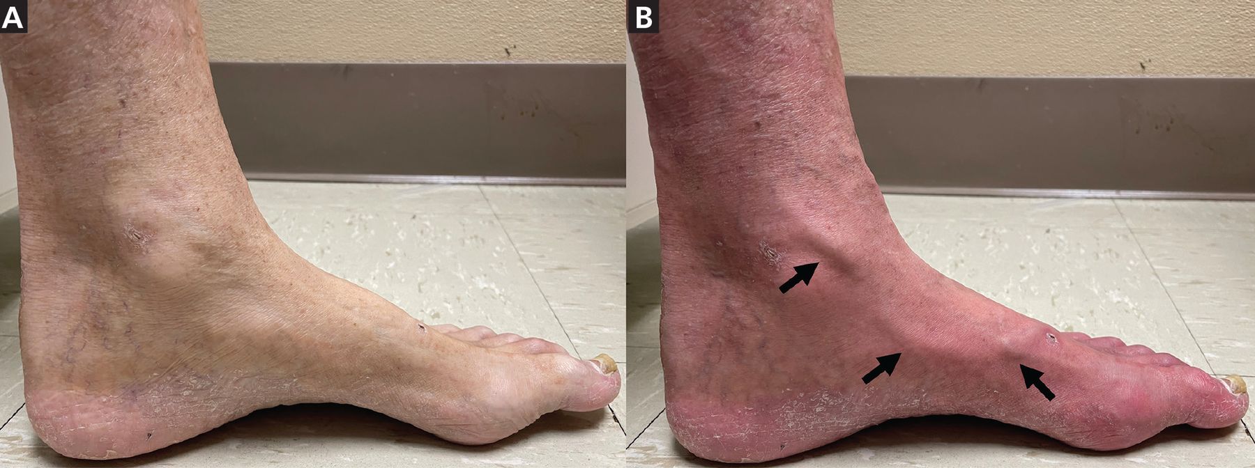
An 80-year-old man with a history of type 2 diabetes mellitus presented to the hospital with bilateral foot pain. Physical examination revealed absent bilateral posterior tibial and dorsalis pedis pulses and dusky erythema of both feet, which were cool to the touch. A prominent medial marginal vein on the left foot was identified (Figure 1), and the patient’s leg was elevated to 45 degrees above the hospital bed surface for 1 minute. The patient was then positioned sitting upright. The erythema returned, and the duration of time for the vein to rise above the level of the skin surface was recorded (45 seconds). Additional findings were elevation pallor (foot appeared pale when elevated) and dependent rubor (foot appeared red when patient was standing), with erythema spreading proximally from the toes.

The patient’s left leg was elevated 45 degrees above the examination table for 1 minute and then the patient was positioned sitting upright. (A) Elevation pallor was seen in the left foot immediately after the patient was repositioned sitting upright, and (B) 45 seconds later, the medial marginal vein of the left foot (black arrows) became visible and erythema of the entire foot was seen.
Because of concern for peripheral vascular disease, the patient was sent to the vascular laboratory for duplex ultrasonography to measure the ankle-brachial and toe-brachial indices. No definitive waveform in the posterior tibial artery was discernible, but the toe-brachial index of the left foot was 0.2 (normal value > 0.7). Computed tomography angiography subsequently revealed severe aortofemoral, femoro-popliteal, and peroneotibial arterial disease.
The patient was diagnosed with severe peripheral artery disease and referred for surgical revascularization.
THE ERYTHEMATOUS LOWER EXTREMITY
Erythematous lower extremities can present a diagnostic challenge.1 Disorders on the differential diagnosis of an erythematous lower limb include cellulitis, peripheral artery disease, venous stasis dermatitis, asteatotic eczema (also called xerotic eczema or eczema craquelé), irritant or allergic contact dermatitis, gout, and erythromelalgia. Because atrophic skin changes and distal limb hair loss have poor sensitivity and specificity for the diagnosis of peripheral artery disease, other clinical signs discovered on careful physical examination are essential to establishing the diagnosis.2,3
Clinical evaluation
First, feel for warmth of the erythematous skin, as a cool extremity in an area of redness argues against cellulitis, gout, and erythromelalgia. Second, absence of pulsation in the femoral, popliteal, or posterior tibialis and dorsalis pedis arteries increases the likelihood of peripheral artery disease. After attempting to identify visible veins, such as the medial and lateral marginal veins of the foot, raise the affected extremity to 45 degrees above the examination table for 1 minute and observe for elevation pallor, which is suggestive of peripheral artery disease. Upon returning the patient to an upright position with the foot dangling, record the time for the previously identified visible vein to rise above the skin surface level and observe for the return of dependent rubor in the affected limb.
Venous filling time can be prolonged in peripheral artery disease because diminished arterial flow leads to slower filling of the veins in the affected area. A study of patients with diabetes found that a venous filling time longer than 20 seconds was the most specific (94%) examination finding for peripheral artery disease.4 A prolonged venous filling time should not be observed in venous insufficiency because reflux into the lower leg veins causes rapid refilling of the veins. A normal or reduced venous filling time may be observed in patients with combined peripheral artery disease and venous insufficiency, which may explain the poor sensitivity (22%) of this measure if used alone to assess for peripheral artery disease.4
Dependent rubor is often present in severe peripheral artery disease due to reduced precapillary sphincter tone leading to passive dilation of the cutaneous capillary beds.
In summary, prolonged venous filling time is an indication of peripheral vascular disease with high specificity. In a patient with dependent rubor, its presence argues against the diagnosis of venous insufficiency.
DISCLOSURES
The authors report no relevant financial relationships which, in the context of their contributions, could be perceived as a potential conflict of interest.
- Copyright © 2025 The Cleveland Clinic Foundation. All Rights Reserved.
In this issue


Jump to section
Related Articles
Cited By...
- No citing articles found.