
Main menu

  • Home
  • Content
    • Current Issue
    • Ahead of Print
    • Past Issues
    • Supplements
    • Article Type
  • Specialty
    • Articles by Specialty
  • CME/MOC
    • Articles
    • Calendar
  • Info For
    • Manuscript Submission
    • Authors & Reviewers
    • Subscriptions
    • About CCJM
    • Contact Us
    • Media Kit
  • Conversations with Leaders
  • Conference Coverage
    • Kidney Week 2024
    • CHEST 2024
    • ACR Convergence 2023
    • Kidney Week 2023
    • ObesityWeek 2023
    • IDWeek 2023
    • CHEST 2023
    • MDS 2023
    • IAS 2023
    • ACP 2023
    • AAN 2023
    • ACC / WCC 2023
    • AAAAI Meeting 2023
    • ACR Convergence 2022
    • Kidney Week 2022
    • AIDS 2022
  • Other Publications
    • www.clevelandclinic.org

User menu

  • Register
  • Log in
  • Log out

Search

  • Advanced search
Cleveland Clinic Journal of Medicine
  • Other Publications
    • www.clevelandclinic.org
  • Register
  • Log in
  • Log out
Cleveland Clinic Journal of Medicine

Advanced Search

  • Home
  • Content
    • Current Issue
    • Ahead of Print
    • Past Issues
    • Supplements
    • Article Type
  • Specialty
    • Articles by Specialty
  • CME/MOC
    • Articles
    • Calendar
  • Info For
    • Manuscript Submission
    • Authors & Reviewers
    • Subscriptions
    • About CCJM
    • Contact Us
    • Media Kit
  • Conversations with Leaders
  • Conference Coverage
    • Kidney Week 2024
    • CHEST 2024
    • ACR Convergence 2023
    • Kidney Week 2023
    • ObesityWeek 2023
    • IDWeek 2023
    • CHEST 2023
    • MDS 2023
    • IAS 2023
    • ACP 2023
    • AAN 2023
    • ACC / WCC 2023
    • AAAAI Meeting 2023
    • ACR Convergence 2022
    • Kidney Week 2022
    • AIDS 2022
The Clinical Picture

Dactylitis from Mycobacterium intracellulare infection

Hayato Shimizu, MD, Takumi Hashimura, MD and Hiroaki Nishioka, MD, PhD
Cleveland Clinic Journal of Medicine July 2022, 89 (7) 360-362; DOI: https://doi.org/10.3949/ccjm.89a.21067
Hayato Shimizu
Department of General Internal Medicine, Kobe City Medical Center General Hospital, Kobe, Hyogo, Japan
  • Find this author on Google Scholar
  • Find this author on PubMed
  • Search for this author on this site
Takumi Hashimura
Department of Orthopaedic Surgery, Kobe City Medical Center General Hospital, Kobe, Hyogo, Japan
  • Find this author on Google Scholar
  • Find this author on PubMed
  • Search for this author on this site
Hiroaki Nishioka
Department of General Internal Medicine, Kobe City Medical Center General Hospital, Kobe, Hyogo, Japan
  • Find this author on Google Scholar
  • Find this author on PubMed
  • Search for this author on this site
  • For correspondence: [email protected]
  • Article
  • Figures & Data
  • Info & Metrics
  • PDF
Loading

A 69-year-old man was referred to our hospital with a 6-month history of progressive swelling in the right middle finger without systemic symptoms. There was no previous trauma or surgery to the finger. The patient had previously been diagnosed with rheumatoid arthritis treated with methotrexate and hypertension treated with cilnidipine (a calcium channel blocker).

Physical examination revealed non-tender, sausage-like swelling of the right middle finger (Figure 1) with no nail abnormality or rash suggestive of psoriasis. Laboratory testing revealed a white blood cell count of 5.4 × 10⁹/L (reference range 4.5–10.9) and a C-reactive protein level of 0.07 mg/dL (reference range 0.0–0.5). Hand radiography revealed soft-tissue swelling of the right middle finger without joint erosions or periostitis.

Figure 1
  • Download figure
  • Open in new tab
  • Download powerpoint
Figure 1

At presentation, the patient’s right middle finger was swollen, with a sausage-like appearance.

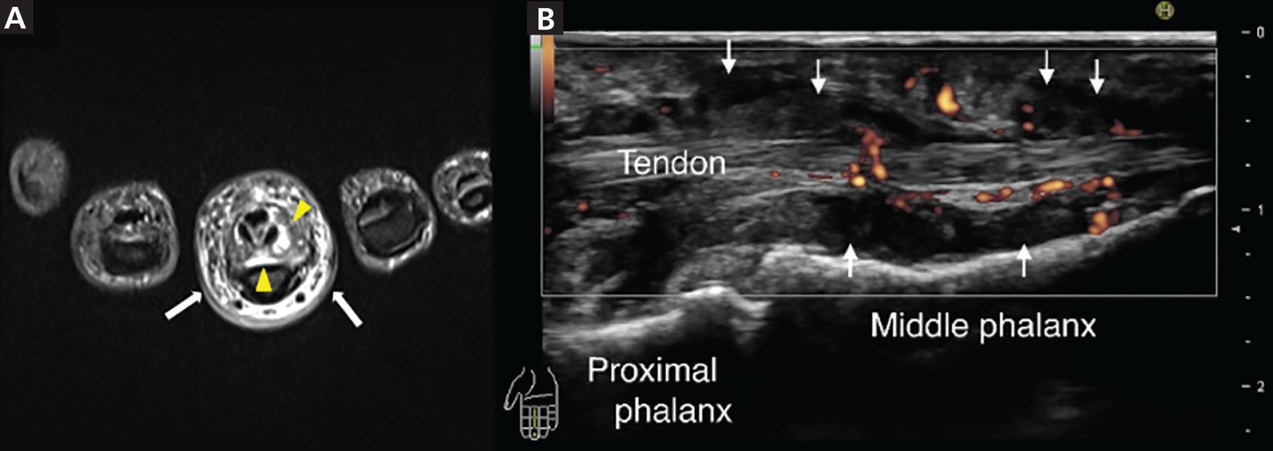
Magnetic resonance imaging (MRI) revealed soft-tissue swelling and thickening around the digital flexor tendon showing high intensity on short time inversion recovery (Figure 2A), and ultrasonography revealed low echoic area and power Doppler signals around the digital flexor tendon (Figure 2B), consistent with inflammation. Dactylitis is rare in patients with rheumatoid arthritis, and thus, the examination differed from that of typical rheumatoid arthritis. The patient did not show signs of other inflammatory diseases associated with dactylitis. We performed a biopsy around the digital flexor tendon of the affected finger to exclude infection.

Figure 2
  • Download figure
  • Open in new tab
  • Download powerpoint
Figure 2

(A) Axial magnetic resonance imaging set on short time inversion recovery showed a high signal around the digital flexor tendon of the right middle finger (yellow arrowheads) and in the subcutaneous tissue (white arrows). (B) Ultrasonography of the right middle finger (sagittal view) showed a low echoic area (white arrows) and power Doppler signals around the digital flexor tendon.

Histology revealed exudative synovitis with lymphocyte and plasma cell infiltration. Tissue culture initially yielded no bacteria; however, mycobacterial culture was positive for Mycobacterium intracellulare. The patient was diagnosed with dactylitis due to M intracellulare infection and treated with rifampin, ethambutol, and clarithromycin. The finger swelling markedly improved over 6 months.

DACTYLITIS

Dactylitis is the inflammation of a finger or toe with swelling of the entire digit rather than just a knuckle joint.1 Dactylitis can be classified based on the involved tissue of the digit, ie, bone only, bone and soft tissue, or soft tissue only. In addition, dactylitis may be classified according to the etiology as noninflammatory, inflammatory infectious, and inflammatory noninfectious.1

Spondyloarthritis dactylitis has been described as sausage-like in appearance and involves pain and swelling of the fingers or toes, mostly along the flexor tendons.1 Related disorders include psoriatic arthritis, ankylosing spondylitis, enteropathic arthritis, and reactive arthritis.1

MRI and ultrasonography2–4 show an increase in the volar bone-to-skin distance in dactylitic fingers that can be due to synovial thickening around the flexor tendons and may also show fluid collection in the tendon sheaths, indicating flexor tenosynovitis and adjacent soft-tissue swelling with small-joint synovitis.2–4

Although rare, tuberculous dactylitis can be the result of extrapulmonary tuberculosis with involvement of the digit and soft tissue.5 Radiographs typically show bone erosion (spina ventosa),6 and MRI shows that the lesion often extends to the soft tissue of the digit through cortical defects forming a sinus tract.5

The differential diagnosis of dactylitis includes syphilitic dactylitis, a manifestation of congenital syphilis; sarcoid dactylitis, with infiltration of the phalangeal soft tissue by noncaseating granulomas; sickle cell dactylitis, or “hand-foot syndrome,” associated with infarction of bone marrow, usually in children; and reactive distal dactylitis of the anterior fat pad, mostly due to group A beta-hemolytic streptococci.1 Dactylitis can also be associated with gout, resulting from deposition of monosodium urate crystals in the soft tissue of the involved digits1 and from infection.

MYCOBACTERIUM INTRACELLULARE INFECTION OF A FINGER

Nontuberculous mycobacteria (NTM) cause pulmonary disease in 90% of cases.7 NTM musculoskeletal infection is uncommon,7–9 but NTM tenosynovitis has been repeatedly reported.10–12 Wrists and hands are the most frequently reported sites of tenosynovitis, and some have described the presence of multiple granulomatous “rice bodies.”10–13 As seen in our patient, M intracellulare can infect a single digit and cause tenosynovitis-associated dactylitis.

Use of anti-tumor necrosis factor agents, high-dose corticosteroids, leflunomide, cyclophosphamide, azathioprine, cyclosporine, mycophenolate, and chlorambucil have been associated with NTM infection of soft tissue or joints.14

DISCLOSURES

The authors report no relevant financial relationships which, in the context of their contributions, could be perceived as a potential conflict of interest.

  • Copyright © 2022 The Cleveland Clinic Foundation. All Rights Reserved.

REFERENCES

  1. ↵
    1. Olivieri I,
    2. Scarano E,
    3. Padula A,
    4. Giasi V,
    5. Priolo F
    . Dactylitis, a term for different digit diseases. Scand J Rheumatol 2006; 35(5):333–340. doi:10.1080/03009740600906677
    OpenUrlCrossRefPubMed
  2. ↵
    1. Olivieri I,
    2. Barozzi L,
    3. Favaro L, et al
    . Dactylitis in patients with seronegative spondylarthropathy. Assessment by ultrasonography and magnetic resonance imaging. Arthritis Rheum 1996; 39(9):1524–1528. doi:10.1002/art.1780390912
    OpenUrlCrossRefPubMed
    1. Zenone T,
    2. Boibieux A,
    3. Tigaud S, et al
    . Non-tuberculous mycobacterial tenosynovitis: a review. Scand J Infect Dis 1999; 31(3):221–228. doi:10.1080/00365549950163482
    OpenUrlCrossRefPubMed
  3. ↵
    1. Bakewell CJ,
    2. Olivieri I,
    3. Aydin SZ, et al
    . Ultrasound and magnetic resonance imaging in the evaluation of psoriatic dactylitis: status and perspectives. J Rheumatol 2013; 40(12):1951–1957. doi:10.3899/jrheum.130643
    OpenUrlAbstract/FREE Full Text
  4. ↵
    1. Yoon CJ,
    2. Chung HW,
    3. Hong SH,
    4. Kim CJ,
    5. Kang HS
    . Case report: MR findings of tuberculous dactylitis. Eur J Radiol 2001; 39(3):163–167. doi:10.1016/s0720-048x(01)00327-8
    OpenUrlCrossRefPubMed
  5. ↵
    1. Bhaskar, Khonglah T,
    2. Bareh J
    . Tuberculous dactylitis (spina ventosa) with concomitant ipsilateral axillary scrofuloderma in an immunocompetent child: A rare presentation of skeletal tuberculosis. Adv Biomed Res 2013; 2(2):29. doi:10.4103/2277-9175.107993
    OpenUrlCrossRef
  6. ↵
    1. Park JW,
    2. Kim YS,
    3. Yoon JO, et al
    . Non-tuberculous mycobacterial infection of the musculoskeletal system: pattern of infection and efficacy of combined surgical/antimicrobial treatment. Bone Joint J 2014; 96-B(11):1561–1565. doi:10.1302/0301-620X.96B11.33427
    OpenUrlCrossRef
    1. Gundavda MK,
    2. Patil HG,
    3. Agashe VM,
    4. Soman R,
    5. Rodriques C,
    6. Deshpande RB
    . Nontuberculous mycobacterial infection of the musculoskeletal system in immunocompetent hosts. Indian J Orthop 2017; 51(2):205–212. doi:10.4103/0019-5413.201718
    OpenUrlCrossRefPubMed
  7. ↵
    1. Napaumpaiporn C,
    2. Katchamart W
    . Clinical manifestations and outcomes of musculoskeletal nontuberculous mycobacterial infections. Rheumatol Int 2019; 39(10):1783–1787. doi:10.1007/s00296-019-04392-8
    OpenUrlCrossRefPubMed
  8. ↵
    1. Piersimoni C,
    2. Scarparo C
    . Extrapulmonary infections associated with nontuberculous mycobacteria in immunocompetent persons. Emerg Infect Dis 2009; 15(9):1351–1358. doi:10.3201/eid1509.081259
    OpenUrlCrossRefPubMed
    1. Namkoong H,
    2. Fukumoto K,
    3. Hongo I,
    4. Hasegawa N
    . Refractory tenosynovitis with ‘rice bodies’ in the hand due to Mycobacterium intracellulare. Infection 2016; 44(3):393–394. doi:10.1007/s15010-015-0844-0
    OpenUrlCrossRef
  9. ↵
    1. Kwan M,
    2. Tupler R
    . Recurrent nontuberculous mycobacterial tenosynovitis. Ochsner J 2021; 21(1):86–89. doi:10.31486/toj.19.0010
    OpenUrlAbstract/FREE Full Text
  10. ↵
    1. Kreutz-Rodrigues L,
    2. Bakri K
    . Tenosynovitis due to mycobacterium avium complex. N Engl J Med 2019; 381(25):2461. doi:10.1056/NEJMicm1901520
    OpenUrlCrossRef
  11. ↵
    1. Brode SK,
    2. Jamieson FB,
    3. Ng R, et al
    . Increased risk of mycobacterial infections associated with anti-rheumatic medications. Thorax 2015; 70(7):677–682. doi:10.1136/thoraxjnl-2014-206470
    OpenUrlAbstract/FREE Full Text
PreviousNext
Back to top

In this issue

Cleveland Clinic Journal of Medicine: 89 (7)
Cleveland Clinic Journal of Medicine
Vol. 89, Issue 7
1 Jul 2022
  • Table of Contents
  • Table of Contents (PDF)
  • Index by author
  • Complete Issue (PDF)
Print
Download PDF
Article Alerts
Sign In to Email Alerts with your Email Address
Email Article

Thank you for your interest in spreading the word on Cleveland Clinic Journal of Medicine.

NOTE: We only request your email address so that the person you are recommending the page to knows that you wanted them to see it, and that it is not junk mail. We do not capture any email address.

Enter multiple addresses on separate lines or separate them with commas.
Dactylitis from Mycobacterium intracellulare infection
(Your Name) has sent you a message from Cleveland Clinic Journal of Medicine
(Your Name) thought you would like to see the Cleveland Clinic Journal of Medicine web site.
CAPTCHA
This question is for testing whether or not you are a human visitor and to prevent automated spam submissions.
Citation Tools
Dactylitis from Mycobacterium intracellulare infection
Hayato Shimizu, Takumi Hashimura, Hiroaki Nishioka
Cleveland Clinic Journal of Medicine Jul 2022, 89 (7) 360-362; DOI: 10.3949/ccjm.89a.21067

Citation Manager Formats

  • BibTeX
  • Bookends
  • EasyBib
  • EndNote (tagged)
  • EndNote 8 (xml)
  • Medlars
  • Mendeley
  • Papers
  • RefWorks Tagged
  • Ref Manager
  • RIS
  • Zotero
Share
Dactylitis from Mycobacterium intracellulare infection
Hayato Shimizu, Takumi Hashimura, Hiroaki Nishioka
Cleveland Clinic Journal of Medicine Jul 2022, 89 (7) 360-362; DOI: 10.3949/ccjm.89a.21067
Twitter logo Facebook logo Mendeley logo
  • Tweet Widget
  • Facebook Like
  • Linkedin Share Button

Jump to section

  • Article
    • DACTYLITIS
    • MYCOBACTERIUM INTRACELLULARE INFECTION OF A FINGER
    • DISCLOSURES
    • REFERENCES
  • Figures & Data
  • Info & Metrics
  • PDF

Related Articles

  • No related articles found.
  • PubMed
  • Google Scholar

Cited By...

  • Nontuberculous mycobacterial musculoskeletal infections: Recognizable, when suspected
  • Google Scholar

More in this TOC Section

  • Tinea incognito
  • Prolonged venous filling time and dependent rubor in a patient with peripheral artery disease
  • Sarcoidosis with diffuse purplish erythematous plaques on the hands
Show more The Clinical Picture

Similar Articles

Subjects

  • Infectious Diseases
  • Rheumatology

Navigate

  • Current Issue
  • Past Issues
  • Supplements
  • Article Type
  • Specialty
  • CME/MOC Articles
  • CME/MOC Calendar
  • Media Kit

Authors & Reviewers

  • Manuscript Submission
  • Authors & Reviewers
  • Subscriptions
  • About CCJM
  • Contact Us
  • Cleveland Clinic Center for Continuing Education
  • Consult QD

Share your suggestions!

Copyright © 2025 The Cleveland Clinic Foundation. All rights reserved. The information provided is for educational purposes only. Use of this website is subject to the website terms of use and privacy policy. 

Powered by HighWire