


A 54-year-old woman with alcohol use disorder presented to the emergency department with a 3-week history of progressively worsening fatigue, dyspnea on exertion, and easy bruising. She initially noticed a fingertip-size bruise on the right thigh that rapidly enlarged to involve both thighs, calves, and ankles.
She reported poor appetite, substantial weight loss, and inadequate nutrition with in tentional avoidance of vegetables, fruits, and red meat for over a year. She had no history of causes of bruising, including use of oral anticoagulants or antiplatelet medications.
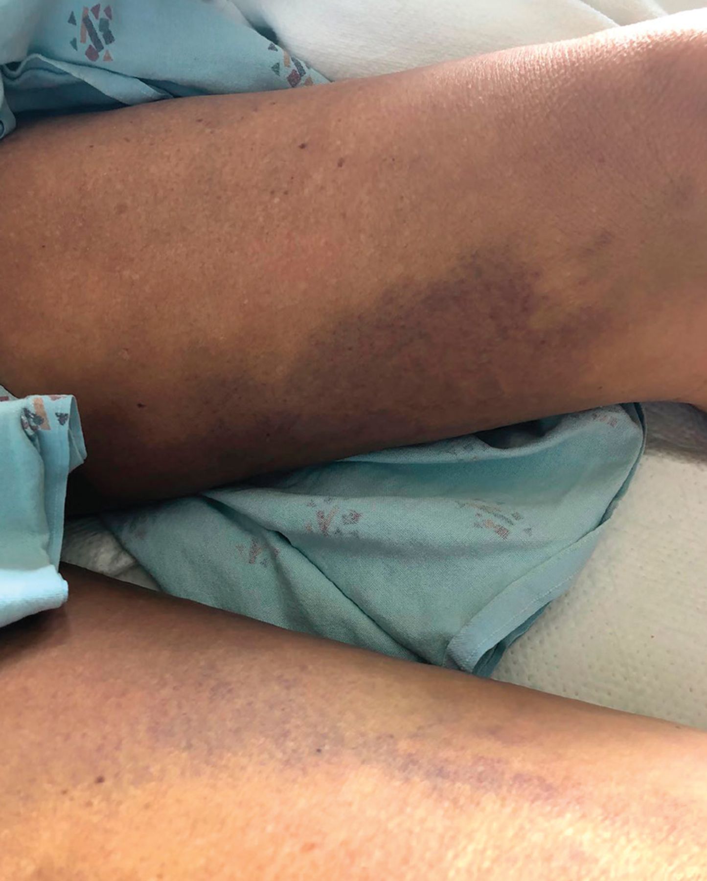
She appeared malnourished and frail. Her body weight was 94 lb. Physical examination revealed a cutaneous perifollicular hemorrhage, petechiae, and a large area of ecchymosis involving the thighs, calves, and ankles that was notably tender to palpation (Figures 1–3).

Perifollicular hemorrhage involving the anterior aspect of the lower left leg.

Ecchymosis involving the medial aspect of both thighs.
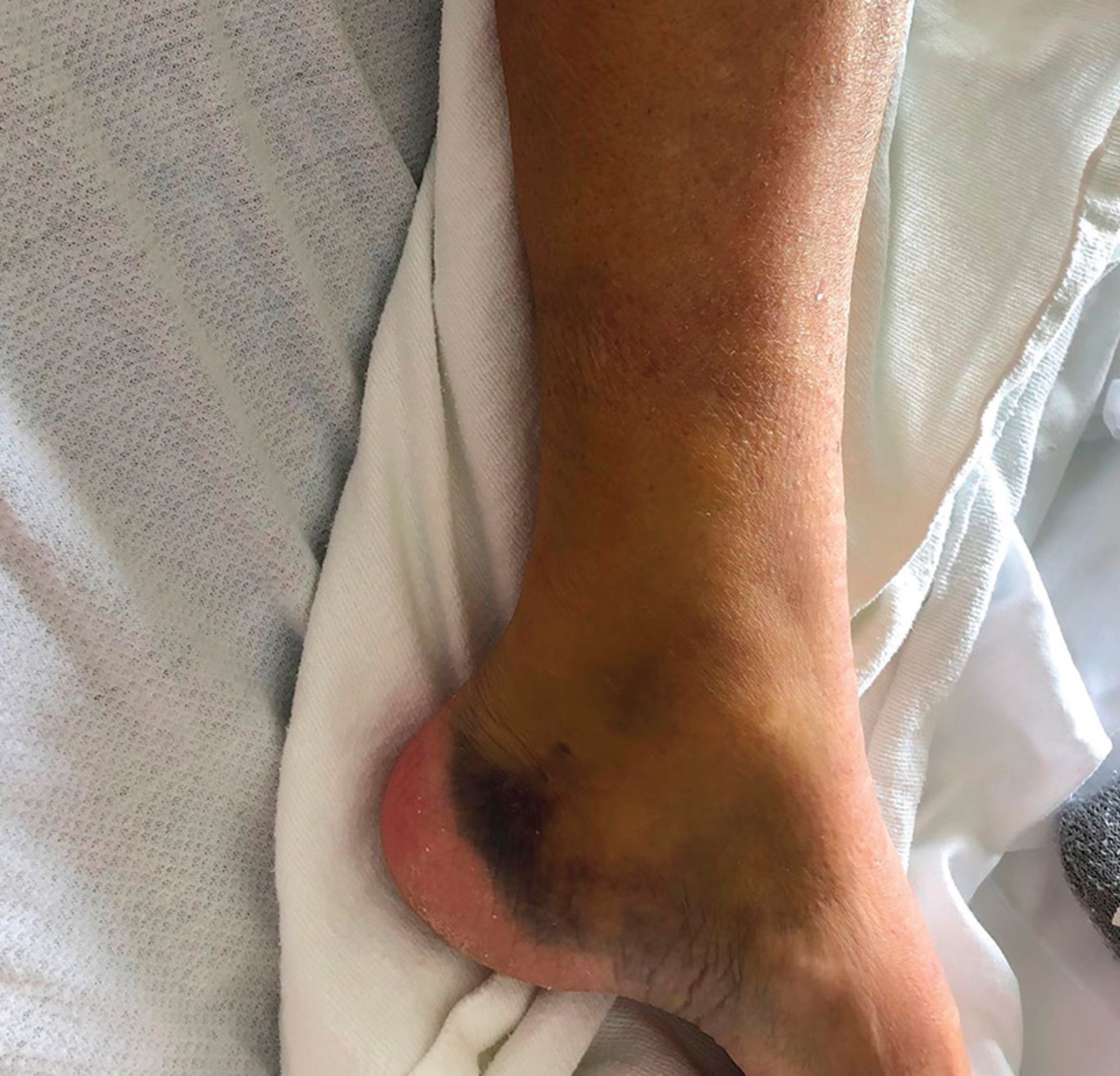
Atraumatic ecchymosis involving the left ankle area.
Laboratory testing results were significant for a hemoglobin concentration of 6.2 g/dL (reference range 12–16), mean corpuscular volume 84.5 (81–98), serum iron 14 μg/dL (37–145), iron saturation 5% (20%–50%), ferritin 90 ng/mL (13–150), soluble transferrin receptor–ferritin index 1.73 (> 1.4 has more than 90% sensitivity and specificity for the diagnosis of iron deficiency anemia), thiamine 47 nmol/L (70–180), 25-hydroxyvitamin D 15 ng/mL (> 30), and vitamin C less than 0.1 mg/dL (0.4–2). Platelet count, coagulation studies, and vitamin B12 and folate levels were normal. Fecal occult blood testing was negative. A clinical diagnosis of scurvy was established based on the history of a severely restricted diet, low serum vitamin C level, and resolution of physical findings after initiating oral vitamin C replacement.
High-dose oral vitamin C was administered and a vitamin C-rich diet was prescribed, leading to resolution of her symptoms and physical findings within 10 weeks. Iron, thiamine, and vitamin D were also supplemented.
THE CHANGING FACE OF THE SCURVY PATIENT
Scurvy, described as early as 1500 bce,1 results from severe dietary deficiency of l-ascorbic acid (vitamin C), a cofactor that humans must acquire from exogenous resources, primarily from fruits and vegetables. It is well documented in the literature that sailors who spent months at sea could avoid scurvy by consuming a diet rich in vegetables and fruits.2
Ascorbic acid is a cofactor for lysyl hydroxylase, an enzyme essential in hydroxylation of proline and lysine residues in the cross-link formation of collagen. It is also essential for iron absorption. In its absence, assembly of the collagen triple helix is incomplete, rendering collagen-dependent structures such as blood vessels unstable, leading to hemorrhagic manifestations and poor wound healing.3
Scurvy is now only rarely encountered in the United States. Risk factors include alcohol use disorder, malnutrition, malabsorption, cigarette use, and psychiatric disorders. Signs and symp toms tend to manifest when the body’s vitamin C stores drop below 300 mg. This can occur within as few as 1 to 3 months of absence of vitamin C from the diet.4
Clinical manifestations of scurvy can be divided into an early phase, characterized by nonspecific symptoms such as fatigue, malaise, and loss of appetite, and a late phase, with impaired wound healing, gingival bleeding, lower-extremity petechiae, and ecchymosis, along with symptoms secondary to tissue hemorrhage including bone pain and pseudoparalysis.5
The diagnosis of scurvy is primarily based on the history and physical examination,6 and is confirmed with undetectable vitamin C serum levels. Iron deficiency anemia and multivitamin deficiency often occur concurrently. Clinicians should have a high index of suspicion for scurvy in the patient with alcohol use disorder who presents with poor nutritional history, extensive bruising, and iron deficiency anemia.
- Copyright © 2020 The Cleveland Clinic Foundation. All Rights Reserved.
In this issue


Jump to section
Related Articles
Cited By...
- No citing articles found.