
Main menu

  • Home
  • Content
    • Current Issue
    • Ahead of Print
    • Past Issues
    • Supplements
    • Article Type
  • Specialty
    • Articles by Specialty
  • CME/MOC
    • Articles
    • Calendar
  • Info For
    • Manuscript Submission
    • Authors & Reviewers
    • Subscriptions
    • About CCJM
    • Contact Us
    • Media Kit
  • Conversations with Leaders
  • Conference Coverage
    • Kidney Week 2024
    • CHEST 2024
    • ACR Convergence 2023
    • Kidney Week 2023
    • ObesityWeek 2023
    • IDWeek 2023
    • CHEST 2023
    • MDS 2023
    • IAS 2023
    • ACP 2023
    • AAN 2023
    • ACC / WCC 2023
    • AAAAI Meeting 2023
    • ACR Convergence 2022
    • Kidney Week 2022
    • AIDS 2022
  • Other Publications
    • www.clevelandclinic.org

User menu

  • Register
  • Log in

Search

  • Advanced search
Cleveland Clinic Journal of Medicine
  • Other Publications
    • www.clevelandclinic.org
  • Register
  • Log in
Cleveland Clinic Journal of Medicine

Advanced Search

  • Home
  • Content
    • Current Issue
    • Ahead of Print
    • Past Issues
    • Supplements
    • Article Type
  • Specialty
    • Articles by Specialty
  • CME/MOC
    • Articles
    • Calendar
  • Info For
    • Manuscript Submission
    • Authors & Reviewers
    • Subscriptions
    • About CCJM
    • Contact Us
    • Media Kit
  • Conversations with Leaders
  • Conference Coverage
    • Kidney Week 2024
    • CHEST 2024
    • ACR Convergence 2023
    • Kidney Week 2023
    • ObesityWeek 2023
    • IDWeek 2023
    • CHEST 2023
    • MDS 2023
    • IAS 2023
    • ACP 2023
    • AAN 2023
    • ACC / WCC 2023
    • AAAAI Meeting 2023
    • ACR Convergence 2022
    • Kidney Week 2022
    • AIDS 2022
The Clinical Picture

Lacrimal gland involvement in a patient with sarcoidosis

Tanmayi Pai, MD, Mohamed S. Muneer, MD, Kafayat Oyemade, Diva R. Salomao, MD, Vivek Gupta, MD and Dana Harris, MD
Cleveland Clinic Journal of Medicine May 2022, 89 (5) 239-240; DOI: https://doi.org/10.3949/ccjm.89a.21037
Tanmayi Pai
Department of Internal Medicine, Mayo Clinic, Jacksonville, FL
  • Find this author on Google Scholar
  • Find this author on PubMed
  • Search for this author on this site
Mohamed S. Muneer
Department of Internal Medicine, OhioHealth Riverside Methodist Hospital, Columbus, OH
  • Find this author on Google Scholar
  • Find this author on PubMed
  • Search for this author on this site
  • For correspondence: [email protected] [email protected]
Kafayat Oyemade
Mayo Clinic Alix School of Medicine, Mayo Clinic, Rochester, MN
  • Find this author on Google Scholar
  • Find this author on PubMed
  • Search for this author on this site
Diva R. Salomao
Department of Laboratory Medicine and Pathology, Mayo Clinic, Rochester, MN
  • Find this author on Google Scholar
  • Find this author on PubMed
  • Search for this author on this site
Vivek Gupta
Department of Radiology, Mayo Clinic, Jacksonville, FL
  • Find this author on Google Scholar
  • Find this author on PubMed
  • Search for this author on this site
Dana Harris
Department of Internal Medicine, Mayo Clinic, Jacksonville, FL
  • Find this author on Google Scholar
  • Find this author on PubMed
  • Search for this author on this site
  • Article
  • Figures & Data
  • Info & Metrics
  • PDF
Loading

A 44-year-old woman presented to the primary care clinic with diplopia and swelling of the right eyelid that had increased over the past 2 weeks. She denied fevers, chills, headache, cough, shortness of breath, or rashes.

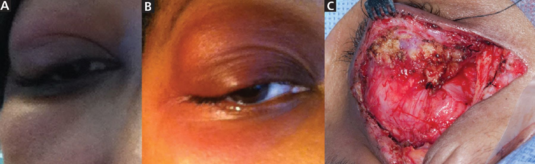
Physical examination confirmed right eyelid edema, with unilateral ptosis and proptosis (Figure 1). There was no pain with eye movements. She was prescribed doxycycline for suspected preseptal (periorbital) cellulitis. However, the eyelid swelling increased, and she was referred to an ophthalmologist for examination and imaging of the orbits.

Figure 1
  • Download figure
  • Open in new tab
  • Download powerpoint
Figure 1

(A) A cellphone photo taken by the patient shows right eyelid swelling approximately 1 week after symptom onset. (B) Eyelid swelling increased, with associated ptosis and proptosis, approximately 2 weeks after symptom onset. (C) Exposure of the lacrimal gland mass during orbitotomy.

Computed tomography (Figure 2) revealed abnormal soft tissue masses in the lacrimal glands of both eyes, with a larger mass in the lacrimal gland of the right eye, causing ptosis and downward displacement of the right globe. The patient underwent right anterior orbitotomy with biopsy of the right lacrimal gland. Vision and physical appearance of the left eye were not significantly affected.

Figure 2
  • Download figure
  • Open in new tab
  • Download powerpoint
Figure 2

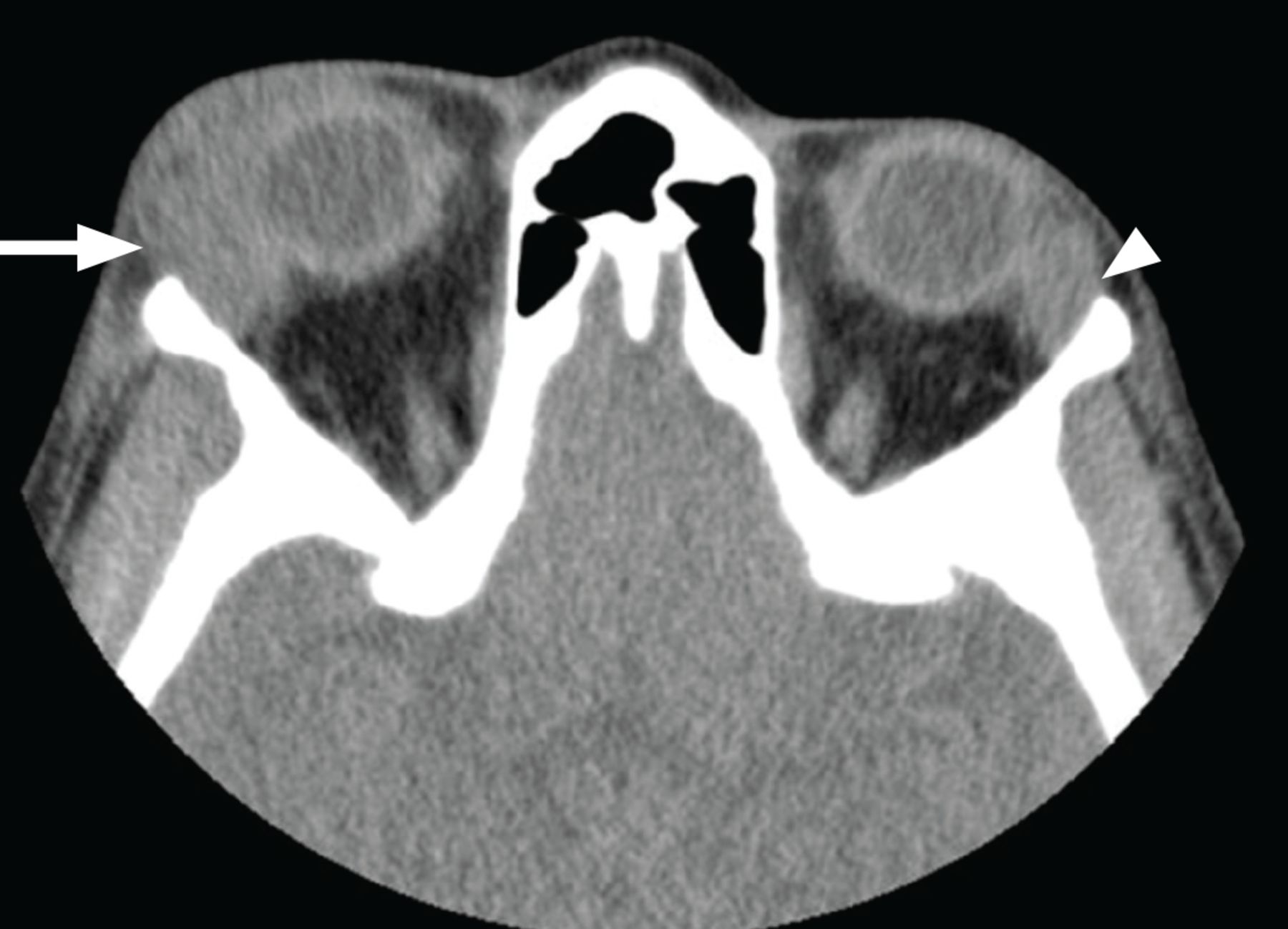
Axial computed tomography of the orbits showed homogeneously confluent, enlarged lacrimal glands, right (arrow) greater than left (arrowhead).

FURTHER EVALUATION PROVIDES DIAGNOSTIC CLUES

The differential diagnosis for the patient’s symptoms and presentation included infection, malignancy, and inflammatory disorders such as immunoglobulin G4-related disease and sarcoidosis. Biopsy of the right lacrimal gland demonstrated nonnecrotizing granulomatous inflammation, with well-formed granulomas. Histochemical staining for acid-fast bacilli and fungi was negative. There were no features concerning for malignancy. Testing for systemic inflammatory disease—computed tomography of the chest, C-reactive protein, and sedimentation rate—was nondiagnostic.

The diagnosis of sarcoidosis is based on 3 major criteria designated by the American Thoracic Society: a clinical presentation compatible with sarcoidosis (eg, lacrimal gland swelling, as in this patient), nonnecrotizing granulomatous inflammation in a tissue sample, and exclusion of other etiologies of granulomatous disease.1 As other causes of granulomatous inflammation were felt to be less likely, sarcoidosis was favored as the cause of the patient’s lacrimal enlargement. Given the absence of systemic symptoms and normal results on chest computed tomography, disease involvement was initially considered to be isolated to extra ocular tissue, and the patient was diagnosed with extraocular sarcoidosis using International Workshop on Ocular Sarcoidosis criteria.2

ISOLATED LACRIMAL GLAND INVOLVEMENT IN SARCOIDOSIS

Sarcoidosis may affect any organ, but the lungs are usually involved. Patients frequently present with ocular involvement, which is more common in female and African American patients.3,4

The American Thoracic Society recommends a baseline eye examination for all patients diagnosed with systemic sarcoidosis.1 Sarcoidosis may affect any part of the eye and its adnexa, presenting most commonly with uveitis, dry eyes, and conjunctival nodules.3 The lacrimal gland is also often affected.5 Significant enlargement of the lacrimal gland leads to the effects observed in our patient, ie, eyelid swelling, ptosis, and globe displacement.

Isolated lacrimal involvement is unusual. Collison et al,6 in a small case series, concluded that although most patients with extraocular orbital sarcoidosis eventually develop systemic sarcoidosis, there are rare cases in which there is no evidence of systemic disease at the time of biopsy.6

Our patient eventually developed systemic sarcoidosis with biopsy-proven cutaneous lesions 10 months after the onset of extraocular symptoms.

MANAGEMENT OF OCULAR SARCOIDOSIS

Management of ocular sarcoidosis centers on initial systemic corticosteroid and immunosuppressive therapy, with or without excision.2 Our patient’s symptoms progressed on prednisone at a high dose of 80 mg and hydroxychloroquine. She has been maintained on oral methotrexate monotherapy at a weekly dose of 10 mg with folic acid supplementation.

DISCLOSURES

The authors report no relevant financial relationships which, in the context of their contributions, could be perceived as a potential conflict of interest.

  • Copyright © 2022 The Cleveland Clinic Foundation. All Rights Reserved.

REFERENCES

  1. ↵
    1. Crouser ED,
    2. Maier LA,
    3. Wilson KC, et al
    . Diagnosis and detection of sarcoidosis. An official American Thoracic Society clinical practice guideline. Am J Respir Crit Care Med 2020; 201(8):e26–e51. doi:10.1164/rccm.202002-0251ST
    OpenUrlCrossRefPubMed
  2. ↵
    1. Pasadhika S,
    2. Rosenbaum JT
    . Ocular sarcoidosis. Clin Chest Med 2015; 36(4):669–683. doi:10.1016/j.ccm.2015.08.009
    OpenUrlCrossRef
  3. ↵
    1. Obenauf CD,
    2. Shaw HE,
    3. Sydnor CF,
    4. Klintworth GK
    . Sarcoidosis and its ophthalmic manifestations. Am J Ophthalmol 1978; 86(5):648–655. doi:10.1016/0002-9394(78)90184-8
    OpenUrlCrossRefPubMed
  4. ↵
    1. Llanos O,
    2. Hamzeh N
    . Sarcoidosis. Med Clin North Am 2019; 103(3):527–534. doi:10.1016/j.mcna.2018.12.011
    OpenUrlCrossRefPubMed
  5. ↵
    1. Mavrikakis I,
    2. Rootman J
    . Diverse clinical presentations of orbital sarcoid. Am J Ophthalmol 2007; 144(5):769–775. doi:10.1016/j.ajo.2007.07.019
    OpenUrlCrossRefPubMed
  6. ↵
    1. Collison JM,
    2. Miller NR,
    3. Green WR
    . Involvement of orbital tissues by sarcoid. Am J Ophthalmol 1986; 102(3):302–307. doi:10.1016/0002-9394(86)90002-4
    OpenUrlCrossRefPubMed
PreviousNext
Back to top

In this issue

Cleveland Clinic Journal of Medicine: 89 (5)
Cleveland Clinic Journal of Medicine
Vol. 89, Issue 5
1 May 2022
  • Table of Contents
  • Table of Contents (PDF)
  • Index by author
  • Complete Issue (PDF)
Print
Download PDF
Article Alerts
Sign In to Email Alerts with your Email Address
Email Article

Thank you for your interest in spreading the word on Cleveland Clinic Journal of Medicine.

NOTE: We only request your email address so that the person you are recommending the page to knows that you wanted them to see it, and that it is not junk mail. We do not capture any email address.

Enter multiple addresses on separate lines or separate them with commas.
Lacrimal gland involvement in a patient with sarcoidosis
(Your Name) has sent you a message from Cleveland Clinic Journal of Medicine
(Your Name) thought you would like to see the Cleveland Clinic Journal of Medicine web site.
CAPTCHA
This question is for testing whether or not you are a human visitor and to prevent automated spam submissions.
Citation Tools
Lacrimal gland involvement in a patient with sarcoidosis
Tanmayi Pai, Mohamed S. Muneer, Kafayat Oyemade, Diva R. Salomao, Vivek Gupta, Dana Harris
Cleveland Clinic Journal of Medicine May 2022, 89 (5) 239-240; DOI: 10.3949/ccjm.89a.21037

Citation Manager Formats

  • BibTeX
  • Bookends
  • EasyBib
  • EndNote (tagged)
  • EndNote 8 (xml)
  • Medlars
  • Mendeley
  • Papers
  • RefWorks Tagged
  • Ref Manager
  • RIS
  • Zotero
Share
Lacrimal gland involvement in a patient with sarcoidosis
Tanmayi Pai, Mohamed S. Muneer, Kafayat Oyemade, Diva R. Salomao, Vivek Gupta, Dana Harris
Cleveland Clinic Journal of Medicine May 2022, 89 (5) 239-240; DOI: 10.3949/ccjm.89a.21037
Twitter logo Facebook logo Mendeley logo
  • Tweet Widget
  • Facebook Like
  • Linkedin Share Button

Jump to section

  • Article
    • FURTHER EVALUATION PROVIDES DIAGNOSTIC CLUES
    • ISOLATED LACRIMAL GLAND INVOLVEMENT IN SARCOIDOSIS
    • MANAGEMENT OF OCULAR SARCOIDOSIS
    • DISCLOSURES
    • REFERENCES
  • Figures & Data
  • Info & Metrics
  • PDF

Related Articles

  • No related articles found.
  • PubMed
  • Google Scholar

Cited By...

  • No citing articles found.
  • Google Scholar

More in this TOC Section

  • Tinea incognito
  • Prolonged venous filling time and dependent rubor in a patient with peripheral artery disease
  • Sarcoidosis with diffuse purplish erythematous plaques on the hands
Show more The Clinical Picture

Similar Articles

Subjects

  • Emergency Medicine
  • Imaging
  • Oncology
  • Vascular Medicine

Navigate

  • Current Issue
  • Past Issues
  • Supplements
  • Article Type
  • Specialty
  • CME/MOC Articles
  • CME/MOC Calendar
  • Media Kit

Authors & Reviewers

  • Manuscript Submission
  • Authors & Reviewers
  • Subscriptions
  • About CCJM
  • Contact Us
  • Cleveland Clinic Center for Continuing Education
  • Consult QD

Share your suggestions!

Copyright © 2025 The Cleveland Clinic Foundation. All rights reserved. The information provided is for educational purposes only. Use of this website is subject to the website terms of use and privacy policy. 

Powered by HighWire