
Main menu

  • Home
  • Content
    • Current Issue
    • Ahead of Print
    • Past Issues
    • Supplements
    • Article Type
  • Specialty
    • Articles by Specialty
  • CME/MOC
    • Articles
    • Calendar
  • Info For
    • Manuscript Submission
    • Authors & Reviewers
    • Subscriptions
    • About CCJM
    • Contact Us
    • Media Kit
  • Conversations with Leaders
  • Conference Coverage
    • Kidney Week 2024
    • CHEST 2024
    • ACR Convergence 2023
    • Kidney Week 2023
    • ObesityWeek 2023
    • IDWeek 2023
    • CHEST 2023
    • MDS 2023
    • IAS 2023
    • ACP 2023
    • AAN 2023
    • ACC / WCC 2023
    • AAAAI Meeting 2023
    • ACR Convergence 2022
    • Kidney Week 2022
    • AIDS 2022
  • Other Publications
    • www.clevelandclinic.org

User menu

  • Register
  • Log in

Search

  • Advanced search
Cleveland Clinic Journal of Medicine
  • Other Publications
    • www.clevelandclinic.org
  • Register
  • Log in
Cleveland Clinic Journal of Medicine

Advanced Search

  • Home
  • Content
    • Current Issue
    • Ahead of Print
    • Past Issues
    • Supplements
    • Article Type
  • Specialty
    • Articles by Specialty
  • CME/MOC
    • Articles
    • Calendar
  • Info For
    • Manuscript Submission
    • Authors & Reviewers
    • Subscriptions
    • About CCJM
    • Contact Us
    • Media Kit
  • Conversations with Leaders
  • Conference Coverage
    • Kidney Week 2024
    • CHEST 2024
    • ACR Convergence 2023
    • Kidney Week 2023
    • ObesityWeek 2023
    • IDWeek 2023
    • CHEST 2023
    • MDS 2023
    • IAS 2023
    • ACP 2023
    • AAN 2023
    • ACC / WCC 2023
    • AAAAI Meeting 2023
    • ACR Convergence 2022
    • Kidney Week 2022
    • AIDS 2022
The Clinical Picture

Asymptomatic oral plaques and erosion

Li-wen Zhang, MD, Wen-ju Wang, MD, Tao Chen, MD, PhD and Rong-hua Xu, MD
Cleveland Clinic Journal of Medicine July 2023, 90 (7) 399-400; DOI: https://doi.org/10.3949/ccjm.90a.22077
Li-wen Zhang
Department of Dermatovenereology, Chengdu Second People’s Hospital, Chengdu, Sichuan, China
  • Find this author on Google Scholar
  • Find this author on PubMed
  • Search for this author on this site
Wen-ju Wang
Department of Dermatovenereology, Chengdu Second People’s Hospital, Chengdu, Sichuan, China
  • Find this author on Google Scholar
  • Find this author on PubMed
  • Search for this author on this site
Tao Chen
Department of Dermatovenereology, Chengdu Second People’s Hospital, Chengdu, Sichuan, China
  • Find this author on Google Scholar
  • Find this author on PubMed
  • Search for this author on this site
  • For correspondence: [email protected] [email protected]
Rong-hua Xu
Institute of Dermatology, Chengdu Second People’s Hospital, Chengdu, Sichuan, China
  • Find this author on Google Scholar
  • Find this author on PubMed
  • Search for this author on this site
  • Article
  • Figures & Data
  • Info & Metrics
  • PDF
Loading

A 47-year-old male presented with a 4-week history of asymptomatic plaques and erosion on the left buccal mucosa and lower lip (Figure 1). He reported seeing a stomatologist, but the prescribed treatment was ineffective. He was then referred to the dermatology department because similar lesions later appeared in genital and perineal regions (Figure 2). He had several high-risk sexual behaviors in the past year.

Figure 1
  • Download figure
  • Open in new tab
  • Download powerpoint
Figure 1

(A) An indurated and painless nodule and plaque on the lower lip. (B) Multiple off-white plaques and erosion on the left buccal mucosa.

Figure 2
  • Download figure
  • Open in new tab
  • Download powerpoint
Figure 2

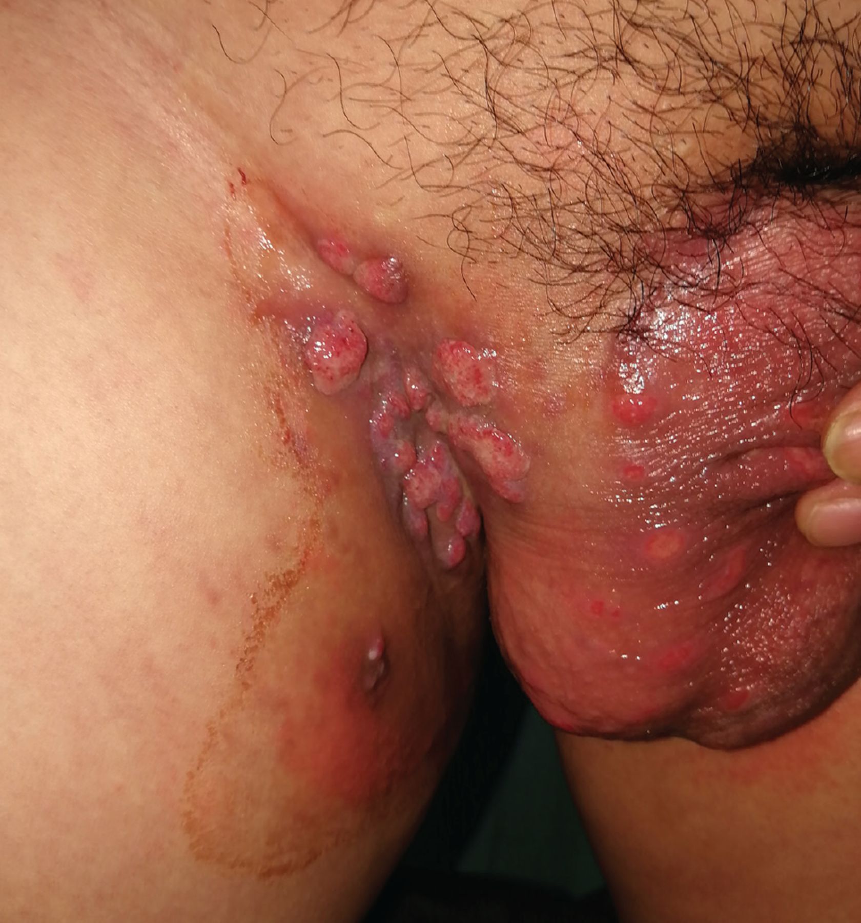
The lesions in the inguinal and genital regions at presentation.

Treponema pallidum particle agglutination testing was positive, and the toluidine red unheated serum test titer was high at 1:32. These results along with the presence of oral and genital-perineal condyloma lata confirmed the diagnosis of secondary syphilis. The patient was treated with benzathine penicillin G 2.4 million units intramuscularly once a week for 3 weeks. His lesions entirely resolved at 6 weeks.

ACQUIRED SECONDARY SYPHILIS

Syphilis has resurfaced in recent years, with oral manifestations occurring at an increased rate.1 The clinical diagnosis of acquired oral syphilis is challenging due to its diverse manifestations. It is more common in young and middle-aged men.1,2

Primary oral syphilis is characterized by a chancre, a single painless ulcerated oral lesion on the lip, labial commissure, or tongue.2,3 In most patients, primary oral syphilis is accompanied by nontender regional lymph-adenopathy.2,3 The majority of cases of oral syphilis represent the secondary stage of syphilis.1,2 Secondary oral syphilis usually presents as multiple subacute erosive or ulcerative lesions, mucous patches on the tongue, nodular lesions, and leukokeratotic lesions.4 Tertiary syphilis is a painless localized granuloma that presents as hardened, nodular, or ulcerated lesions on the hard palate or the dorsal surface of the tongue.3

BROAD DIFFERENTIAL DIAGNOSIS

The range of differential diagnoses is wide and includes infectious diseases, potentially malignant oral disorders, malignant neoplasms, and immune-mediated diseases.1,2 Painful lesions that need to be considered include aphthous ulcer, traumatic ulcer, Behçet disease, inflammatory bowel disease, pemphigus vulgaris, erythema multiforme, candidiasis, and leishmaniasis.1 The differential for painless lesions includes frictional keratosis, necrotizing sialometaplasia, lichen planus, sarcoidosis, tuberculosis, blastomycosis, leukoplakia, erythroplakia, squamous cell carcinoma, and soft-tissue and mesenchymal tumor.1

VALUABLE CLUES TO DIAGNOSIS

Symptoms of oral syphilis are nonspecific, so serological studies remain the gold standard.2,3 However, we need to be aware that syphilis serology may be nonreactive in the very early stage and falsely negative due to the prozone reaction or acquired immunodeficiency syndrome. In the absence of serological evidence, the valuable clues are based on a complete medical history (unprotected sexual and orogenital contact, multiple partners, men who have sex with men, drug use, and history of other sexually transmitted diseases), careful physical examination (extraoral syphilitic manifestations), and timely pathological examination (diffuse or perivascular infiltration of lymphocytes and plasma cells). It needs to be noted that dark-field microscopic examination, immunohistochemical techniques, and silver staining are not specific to oral syphilis diagnosis due to the presence of commensal treponemes.5

Promoting awareness of oral syphilis is vital for early diagnosis, treatment, and prevention of onward transmission.

DISCLOSURES

The authors report no relevant financial relationships which, in the context of their contributions, could be perceived as a potential conflict of interest.

  • Copyright © 2023 The Cleveland Clinic Foundation. All Rights Reserved.

REFERENCES

  1. ↵
    1. Schuch LF,
    2. da Silva KD,
    3. de Arruda JAA, et al
    . Forty cases of acquired oral syphilis and a review of the literature. Int J Oral Maxillofac Surg 2019; 48(5):635–643. doi:10.1016/j.ijom.2018.10.023
    OpenUrlCrossRef
  2. ↵
    1. de Andrade BAB,
    2. de Arruda JAA,
    3. Gilligan G, et al
    . Acquired oral syphilis: a multicenter study of 339 patients from South America. Oral Dis 2022; 28(6):1561–1572. doi:10.1111/odi.13963
    OpenUrlCrossRef
  3. ↵
    1. de Arruda JAA,
    2. do Valle IB,
    3. Mesquita RA,
    4. Silva TA
    . Oral syphilis. J Am Acad Dermatol 2021; 84(2):e101–e102. doi:10.1016/j.jaad.2020.09.067
    OpenUrlCrossRef
  4. ↵
    1. Lampros A,
    2. Seta V,
    3. Gerhardt P,
    4. Isnard C,
    5. Husson C,
    6. Dupin N
    . Oral forms of secondary syphilis: an illustration of the pitfalls set by the great imitator. J Am Acad Dermatol 2021; 84(2):348–353. doi:10.1016/j.jaad.2020.04.089
    OpenUrlCrossRef
  5. ↵
    1. Rajlawat BP,
    2. Evans-Jones J,
    3. Triantafyllou A,
    4. Varga E,
    5. Field EA
    . A solitary oral ulcer. Clin Exp Dermatol 2011; 36(2):217–219. doi:10.1111/j.1365-2230.2010.03879.x
    OpenUrlCrossRefPubMed
PreviousNext
Back to top

In this issue

Cleveland Clinic Journal of Medicine: 90 (7)
Cleveland Clinic Journal of Medicine
Vol. 90, Issue 7
1 Jul 2023
  • Table of Contents
  • Table of Contents (PDF)
  • Index by author
  • Complete Issue (PDF)
Print
Download PDF
Article Alerts
Sign In to Email Alerts with your Email Address
Email Article

Thank you for your interest in spreading the word on Cleveland Clinic Journal of Medicine.

NOTE: We only request your email address so that the person you are recommending the page to knows that you wanted them to see it, and that it is not junk mail. We do not capture any email address.

Enter multiple addresses on separate lines or separate them with commas.
Asymptomatic oral plaques and erosion
(Your Name) has sent you a message from Cleveland Clinic Journal of Medicine
(Your Name) thought you would like to see the Cleveland Clinic Journal of Medicine web site.
CAPTCHA
This question is for testing whether or not you are a human visitor and to prevent automated spam submissions.
Citation Tools
Asymptomatic oral plaques and erosion
Li-wen Zhang, Wen-ju Wang, Tao Chen, Rong-hua Xu
Cleveland Clinic Journal of Medicine Jul 2023, 90 (7) 399-400; DOI: 10.3949/ccjm.90a.22077

Citation Manager Formats

  • BibTeX
  • Bookends
  • EasyBib
  • EndNote (tagged)
  • EndNote 8 (xml)
  • Medlars
  • Mendeley
  • Papers
  • RefWorks Tagged
  • Ref Manager
  • RIS
  • Zotero
Share
Asymptomatic oral plaques and erosion
Li-wen Zhang, Wen-ju Wang, Tao Chen, Rong-hua Xu
Cleveland Clinic Journal of Medicine Jul 2023, 90 (7) 399-400; DOI: 10.3949/ccjm.90a.22077
Twitter logo Facebook logo Mendeley logo
  • Tweet Widget
  • Facebook Like
  • Linkedin Share Button

Jump to section

  • Article
    • ACQUIRED SECONDARY SYPHILIS
    • BROAD DIFFERENTIAL DIAGNOSIS
    • VALUABLE CLUES TO DIAGNOSIS
    • DISCLOSURES
    • REFERENCES
  • Figures & Data
  • Info & Metrics
  • PDF

Related Articles

  • No related articles found.
  • PubMed
  • Google Scholar

Cited By...

  • No citing articles found.
  • Google Scholar

More in this TOC Section

  • Tinea incognito
  • Prolonged venous filling time and dependent rubor in a patient with peripheral artery disease
  • Sarcoidosis with diffuse purplish erythematous plaques on the hands
Show more The Clinical Picture

Similar Articles

Subjects

  • Dermatology
  • Infectious Diseases

Navigate

  • Current Issue
  • Past Issues
  • Supplements
  • Article Type
  • Specialty
  • CME/MOC Articles
  • CME/MOC Calendar
  • Media Kit

Authors & Reviewers

  • Manuscript Submission
  • Authors & Reviewers
  • Subscriptions
  • About CCJM
  • Contact Us
  • Cleveland Clinic Center for Continuing Education
  • Consult QD

Share your suggestions!

Copyright © 2025 The Cleveland Clinic Foundation. All rights reserved. The information provided is for educational purposes only. Use of this website is subject to the website terms of use and privacy policy. 

Powered by HighWire