
Main menu

  • Home
  • Content
    • Current Issue
    • Ahead of Print
    • Past Issues
    • Supplements
    • Article Type
  • Specialty
    • Articles by Specialty
  • CME/MOC
    • Articles
    • Calendar
  • Info For
    • Manuscript Submission
    • Authors & Reviewers
    • Subscriptions
    • About CCJM
    • Contact Us
    • Media Kit
  • Conversations with Leaders
  • Conference Coverage
    • Kidney Week 2024
    • CHEST 2024
    • ACR Convergence 2023
    • Kidney Week 2023
    • ObesityWeek 2023
    • IDWeek 2023
    • CHEST 2023
    • MDS 2023
    • IAS 2023
    • ACP 2023
    • AAN 2023
    • ACC / WCC 2023
    • AAAAI Meeting 2023
    • ACR Convergence 2022
    • Kidney Week 2022
    • AIDS 2022
  • Other Publications
    • www.clevelandclinic.org

User menu

  • Register
  • Log in

Search

  • Advanced search
Cleveland Clinic Journal of Medicine
  • Other Publications
    • www.clevelandclinic.org
  • Register
  • Log in
Cleveland Clinic Journal of Medicine

Advanced Search

  • Home
  • Content
    • Current Issue
    • Ahead of Print
    • Past Issues
    • Supplements
    • Article Type
  • Specialty
    • Articles by Specialty
  • CME/MOC
    • Articles
    • Calendar
  • Info For
    • Manuscript Submission
    • Authors & Reviewers
    • Subscriptions
    • About CCJM
    • Contact Us
    • Media Kit
  • Conversations with Leaders
  • Conference Coverage
    • Kidney Week 2024
    • CHEST 2024
    • ACR Convergence 2023
    • Kidney Week 2023
    • ObesityWeek 2023
    • IDWeek 2023
    • CHEST 2023
    • MDS 2023
    • IAS 2023
    • ACP 2023
    • AAN 2023
    • ACC / WCC 2023
    • AAAAI Meeting 2023
    • ACR Convergence 2022
    • Kidney Week 2022
    • AIDS 2022
The Clinical Picture

Persistent erosions of the glans penis and foreskin

Zheng Gu, MD and Dong-Lai Ma, MD, PhD
Cleveland Clinic Journal of Medicine August 2024, 91 (8) 463-464; DOI: https://doi.org/10.3949/ccjm.91a.23085
Zheng Gu
Beijing University of Chinese Medicine, Beijing, China; Department of Dermatology, China-Japan Friendship Hospital, Beijing, China
  • Find this author on Google Scholar
  • Find this author on PubMed
  • Search for this author on this site
Dong-Lai Ma
Department of Dermatology, Peking Union Medical College Hospital, Beijing, China; State Key Laboratory of Complex, Severe, and Rare Diseases, Beijing, China; Clinical Professor, Chinese Academy of Medical Sciences and Peking Union Medical College, Beijing, China; National Clinical Research Center for Dermatologic and Immunologic Diseases, Beijing, China
  • Find this author on Google Scholar
  • Find this author on PubMed
  • Search for this author on this site
  • For correspondence: [email protected]
  • Article
  • Figures & Data
  • Info & Metrics
  • PDF
Loading

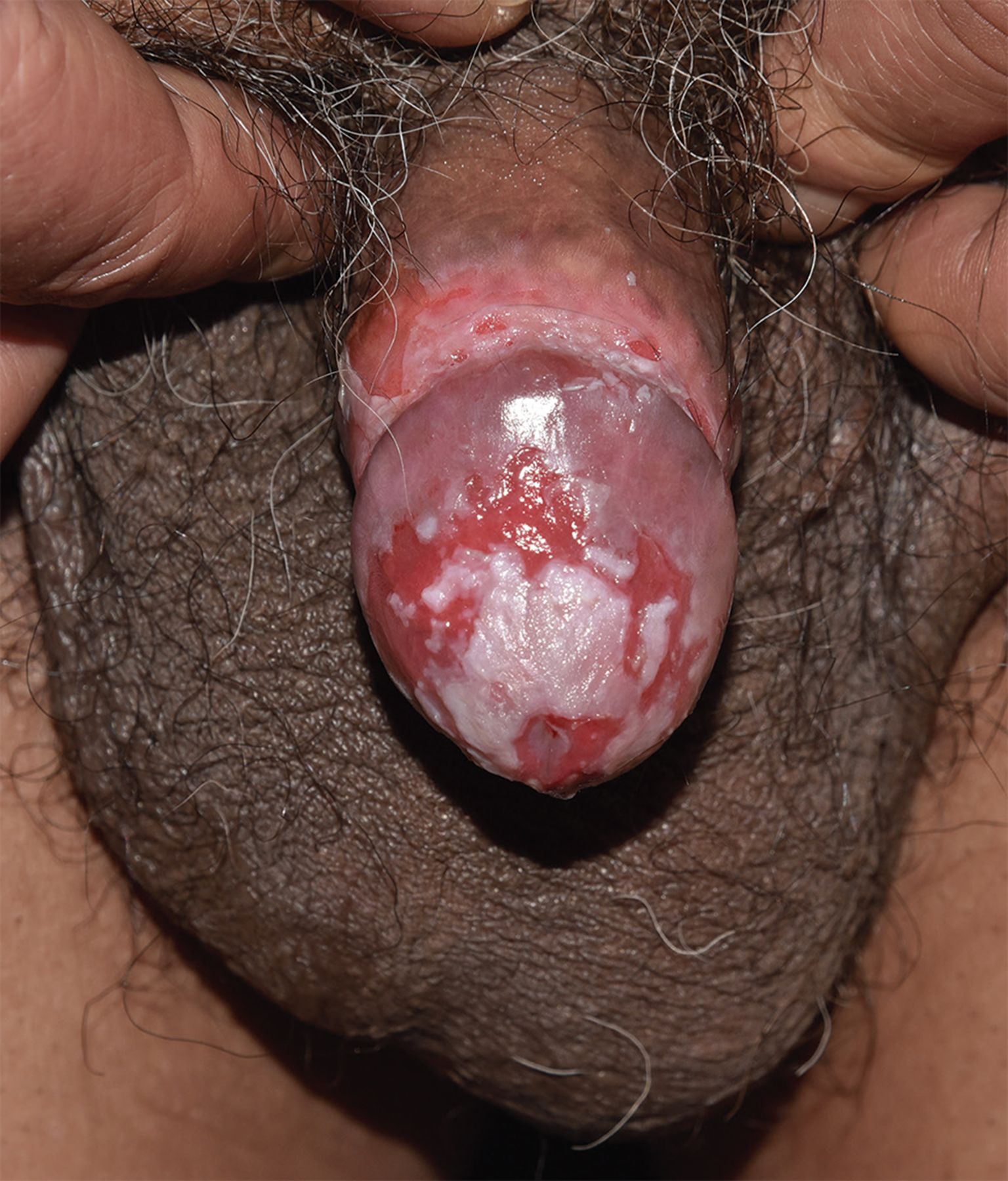
A 66-year-old man presented with an 8-month history of persistent erosions of the glans penis and foreskin with slight itching and pain. Physical examination revealed erythema and superficial erosions on the glans penis and foreskin (Figure 1). Similar lesions were not found on the skin or oral mucosa elsewhere. Tests for syphilis were negative.

Figure 1
  • Download figure
  • Open in new tab
  • Download powerpoint
Figure 1

Erythema and superficial erosions on the glans penis and foreskin before treatment.

Biopsy taken from the foreskin showed suprabasal bullae with acantholysis. Direct immunofluorescence was negative for deposition of immunoglobulin (Ig) G, IgA, and IgM and complement C3 in the epidermal cells and basement membrane bands. However, indirect immunofluorescence tests showed that antispinous intercellular desmoglein antibodies were deposited in the interspinous cell reticulum (using monkey esophagus as a deposition substrate) at a titer of 1:320. No antibasement membrane zone antibodies (important autoantibodies in the diagnosis of bullous pemphigoid) were found.

The patient was diagnosed with localized pemphigus vulgaris. The erosions improved significantly after 2 months of treatment with oral prednisolone at an initial dose of 30 mg daily (Figure 2).

Figure 2
  • Download figure
  • Open in new tab
  • Download powerpoint
Figure 2

The erosions improved significantly after treatment.

PEMPHIGUS

Pemphigus encompasses a group of rare autoimmune disorders characterized by the development of flaccid blisters and erosions on the skin and mucous membranes.1 These blisters are fragile and can easily rupture, leading to open sores and erosions. The majority of patients present with pemphigus vulgaris.2 Pemphigus vulgaris can affect the skin or mucous membranes throughout the body, including the chest, back, head, and, in severe cases, the whole body, but oral involvement often occurs first. Lesions may localize to a single body site such as the nose, cheeks, or penis, which can easily lead to misdiagnosis.

Other subtypes of pemphigus include pemphigus foliaceus and rare pemphigus variants like paraneoplastic pemphigus and IgA pemphigus. Pemphigus foliaceus manifests with skin lesions, usually without mucosal involvement.1 Patients with paraneoplastic pemphigus have known or potential tumors, usually of lymphoid tissue. Pain and severe oral and conjunctival erosions are the main features. The staining patterns on direct and indirect immunofluorescence differ in paraneoplastic pemphigus and classical pemphigus and can be used to distinguish between them.2

The differential diagnosis

Pemphigus should be distinguished from bullous pemphigoid, severe erythema multiforme, and drug-induced bullosa epidermolysis. Persistent erosions on the glans and foreskin of the penis are often encountered and have a wide differential, including syphilis, herpes simplex virus infection, candida balanitis, lichen planus, psoriasis, other autoimmune diseases, trauma, and skin cancer.3 Pemphigus vulgaris can be differentiated from these diseases through histopathology, immunofluorescence, and autoimmune serum titers.2,4

Diagnosis and treatment

Diagnosis is based on clinical presentation, histopathology showing intraepidermal acantholysis, and either positive findings on direct immunofluorescence (ie, IgG or complement C3 deposits at the surface of keratinocytes) or detection of serum autoantibodies against epithelial cell surface.4,5 Samples for biopsy should be taken from normal-appearing skin immediately adjacent to a lesion; sampling inflamed or blistered skin may lead to false-negative results on direct immunofluorescence5 because the inflammatory process associated with pemphigus can damage immune deposits.1

First-line treatments are corticosteroids and anti-CD20 monoclonal antibodies.4 In patients with moderate to severe disease, combination therapy may be used to improve efficacy and reduce the dose of glucocorticoids at the start of treatment or when the effect of glucocorticoids alone is not significant. First-line immunosuppressants are azathioprine and mycophenolate mofetil.

The initial dose of glucocorticoids depends on the type and severity of disease. The absence of new blisters indicates that the dose is adequate. Conversely, the dosage should be increased or other immunosuppressive agents added if new blisters appear. Once disease control is observed, the dosage should be reduced slowly and gradually to prevent recurrence. Withdrawal of systemic corticosteroids may be proposed in patients in complete remission on minimal therapy.2

DISCLOSURES

The authors report no relevant financial relationships which, in the context of their contributions, could be perceived as a potential conflict of interest.

  • Copyright © 2024 The Cleveland Clinic Foundation. All Rights Reserved.

REFERENCES

  1. ↵
    1. Malik AM,
    2. Tupchong S,
    3. Huang S,
    4. Are A,
    5. Hsu S,
    6. Motaparthi K
    . An updated review of pemphigus diseases. Medicina (Kaunas) 2021; 57(10):1080. doi:10.3390/medicina57101080
    OpenUrlCrossRef
  2. ↵
    1. Schmidt E,
    2. Kasperkiewicz M,
    3. Joly P
    . Pemphigus. Lancet 2019; 394(10201):882–894. doi:10.1016/S0140-6736(19)31778-7
    OpenUrlCrossRef
  3. ↵
    1. Su O,
    2. Dizman D,
    3. Ozkaya DB,
    4. Yildiz P,
    5. Demirkesen C,
    6. Onsun N
    . Pemphigus vulgaris localised exclusively to the penis. Indian J Dermatol Venereol Leprol 2015; 81(3):298–299. doi:10.4103/0378-6323.154794
    OpenUrlCrossRef
  4. ↵
    1. Murrell DF,
    2. Peña S,
    3. Joly P, et al
    . Diagnosis and management of pemphigus: recommendations of an international panel of experts. J Am Acad Dermatol 2020; 82(3):575–585.e1. doi:10.1016/j.jaad.2018.02.021
    OpenUrlCrossRef
  5. ↵
    1. Giurdanella F,
    2. Diercks GF,
    3. Jonkman MF,
    4. Pas HH
    . Laboratory diagnosis of pemphigus: direct immunofluorescence remains the gold standard. Br J Dermatol 2016; 175(1):185–186. doi:10.1111/bjd.14408
    OpenUrlCrossRefPubMed
PreviousNext
Back to top

In this issue

Cleveland Clinic Journal of Medicine: 91 (8)
Cleveland Clinic Journal of Medicine
Vol. 91, Issue 8
1 Aug 2024
  • Table of Contents
  • Table of Contents (PDF)
  • Index by author
  • Complete Issue (PDF)
Print
Download PDF
Article Alerts
Sign In to Email Alerts with your Email Address
Email Article

Thank you for your interest in spreading the word on Cleveland Clinic Journal of Medicine.

NOTE: We only request your email address so that the person you are recommending the page to knows that you wanted them to see it, and that it is not junk mail. We do not capture any email address.

Enter multiple addresses on separate lines or separate them with commas.
Persistent erosions of the glans penis and foreskin
(Your Name) has sent you a message from Cleveland Clinic Journal of Medicine
(Your Name) thought you would like to see the Cleveland Clinic Journal of Medicine web site.
CAPTCHA
This question is for testing whether or not you are a human visitor and to prevent automated spam submissions.
Citation Tools
Persistent erosions of the glans penis and foreskin
Zheng Gu, Dong-Lai Ma
Cleveland Clinic Journal of Medicine Aug 2024, 91 (8) 463-464; DOI: 10.3949/ccjm.91a.23085

Citation Manager Formats

  • BibTeX
  • Bookends
  • EasyBib
  • EndNote (tagged)
  • EndNote 8 (xml)
  • Medlars
  • Mendeley
  • Papers
  • RefWorks Tagged
  • Ref Manager
  • RIS
  • Zotero
Share
Persistent erosions of the glans penis and foreskin
Zheng Gu, Dong-Lai Ma
Cleveland Clinic Journal of Medicine Aug 2024, 91 (8) 463-464; DOI: 10.3949/ccjm.91a.23085
Twitter logo Facebook logo Mendeley logo
  • Tweet Widget
  • Facebook Like
  • Linkedin Share Button

Jump to section

  • Article
    • PEMPHIGUS
    • DISCLOSURES
    • REFERENCES
  • Figures & Data
  • Info & Metrics
  • PDF

Related Articles

  • No related articles found.
  • PubMed
  • Google Scholar

Cited By...

  • No citing articles found.
  • Google Scholar

More in this TOC Section

  • Tinea incognito
  • Prolonged venous filling time and dependent rubor in a patient with peripheral artery disease
  • Sarcoidosis with diffuse purplish erythematous plaques on the hands
Show more The Clinical Picture

Similar Articles

Subjects

  • Dermatology
  • Infectious Diseases

Navigate

  • Current Issue
  • Past Issues
  • Supplements
  • Article Type
  • Specialty
  • CME/MOC Articles
  • CME/MOC Calendar
  • Media Kit

Authors & Reviewers

  • Manuscript Submission
  • Authors & Reviewers
  • Subscriptions
  • About CCJM
  • Contact Us
  • Cleveland Clinic Center for Continuing Education
  • Consult QD

Share your suggestions!

Copyright © 2025 The Cleveland Clinic Foundation. All rights reserved. The information provided is for educational purposes only. Use of this website is subject to the website terms of use and privacy policy. 

Powered by HighWire