
Main menu

  • Home
  • Content
    • Current Issue
    • Ahead of Print
    • Past Issues
    • Supplements
    • Article Type
  • Specialty
    • Articles by Specialty
  • CME/MOC
    • Articles
    • Calendar
  • Info For
    • Manuscript Submission
    • Authors & Reviewers
    • Subscriptions
    • About CCJM
    • Contact Us
    • Media Kit
  • Conversations with Leaders
  • Conference Coverage
    • Kidney Week 2024
    • CHEST 2024
    • ACR Convergence 2023
    • Kidney Week 2023
    • ObesityWeek 2023
    • IDWeek 2023
    • CHEST 2023
    • MDS 2023
    • IAS 2023
    • ACP 2023
    • AAN 2023
    • ACC / WCC 2023
    • AAAAI Meeting 2023
    • ACR Convergence 2022
    • Kidney Week 2022
    • AIDS 2022
  • Other Publications
    • www.clevelandclinic.org

User menu

  • Register
  • Log in

Search

  • Advanced search
Cleveland Clinic Journal of Medicine
  • Other Publications
    • www.clevelandclinic.org
  • Register
  • Log in
Cleveland Clinic Journal of Medicine

Advanced Search

  • Home
  • Content
    • Current Issue
    • Ahead of Print
    • Past Issues
    • Supplements
    • Article Type
  • Specialty
    • Articles by Specialty
  • CME/MOC
    • Articles
    • Calendar
  • Info For
    • Manuscript Submission
    • Authors & Reviewers
    • Subscriptions
    • About CCJM
    • Contact Us
    • Media Kit
  • Conversations with Leaders
  • Conference Coverage
    • Kidney Week 2024
    • CHEST 2024
    • ACR Convergence 2023
    • Kidney Week 2023
    • ObesityWeek 2023
    • IDWeek 2023
    • CHEST 2023
    • MDS 2023
    • IAS 2023
    • ACP 2023
    • AAN 2023
    • ACC / WCC 2023
    • AAAAI Meeting 2023
    • ACR Convergence 2022
    • Kidney Week 2022
    • AIDS 2022
The Clinical Picture

Skin manifestations in a patient with acute bacterial infective endocarditis

Basar Aykent, MD and Ozlem Yilmaz, MD
Cleveland Clinic Journal of Medicine November 2024, 91 (11) 657-659; DOI: https://doi.org/10.3949/ccjm.91a.24066
Basar Aykent
Department of Internal Medicine, Division of Nephrology, Istanbul Sisli Hamidiye Etfal Training and Research Hospital, Istanbul, Turkey
  • Find this author on Google Scholar
  • Find this author on PubMed
  • Search for this author on this site
Ozlem Yilmaz
Department of Internal Medicine, Division of Geriatrics, Istanbul Training and Research Hospital, Istanbul, Turkey
  • Find this author on Google Scholar
  • Find this author on PubMed
  • Search for this author on this site
  • For correspondence: [email protected]
  • Article
  • Figures & Data
  • Info & Metrics
  • PDF
Loading

A 50-year-old woman with end-stage renal disease caused by secondary amyloidosis resulting from familial Mediterranean fever had been receiving routine hemodialysis for 9 years when her arteriovenous fistula became ineffective. Efforts to create another fistula were unsuccessful, and a tunneled dialysis catheter was placed.

See related article, page 661

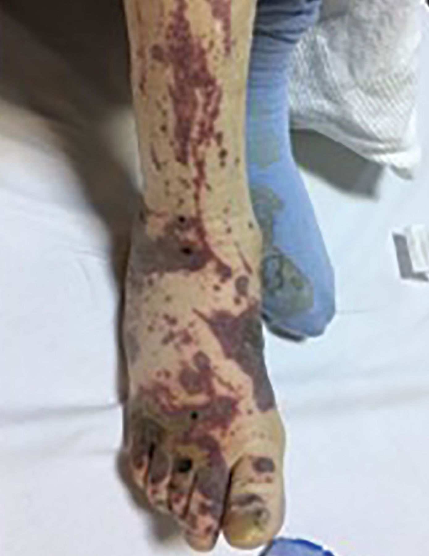
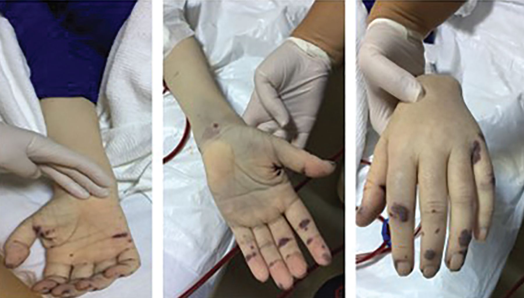
During a standard hemodialysis session about 5 months later, she experienced the onset of rigors, accompanied by the appearance of painless, inflamed purpuric lesions, which developed into petechiae. The petechiae were located on the nose, with a cluster at the tip of the nose, and on the legs (Figure 1). Painless, erythematous hemorrhagic lesions (Janeway lesions) and ecchymotic areas were observed on the dorsum of the hands and feet, palmar surfaces, ankles, and anterior tibial surfaces, and there were palpable purpura on the legs and painful Osler nodes on the fingers (Figure 2 and Figure 3).

Figure 1
  • Download figure
  • Open in new tab
  • Download powerpoint
Figure 1

Petechiae clustered on the tip of the nose.

Figure 2
  • Download figure
  • Open in new tab
  • Download powerpoint
Figure 2

Painless erythematous and hemorrhagic purpura and ecchymotic areas on the anterior surface of the ankle and shin.

Figure 3
  • Download figure
  • Open in new tab
  • Download powerpoint
Figure 3

Janeway lesions and ecchymotic areas on the palms and dorsum of the patient’s hands. Finger lesions, or Osler nodes, are typically painful.

The patient was admitted to the hospital. On examination, her temperature was 38.5°C (101.3°F). A new grade 3 systolic murmur not previously present was detected over the aorta. Laboratory testing showed a neutrophilic leukocytosis (white blood cell count 23 × 109/L [reference range 4–11], neutrophils 11 × 109/L [1.5–8.0]).

Blood cultures were obtained from the peripheral veins and catheter at 2-hour intervals, and vancomycin and ceftazidime were started empirically to cover likely causes of catheter-associated bacteremia. After 3 days of antibiotic therapy, the fever persisted, the patient developed severe headache, confusion, and lethargy, and her clinical condition deteriorated further. Diffusion-weighted magnetic resonance imaging of the brain showed diffusion restriction, areas of edema, and sharply defined bright areas in the white matter. A preliminary diagnosis of septic embolism was made.

Infective endocarditis due to metastatic catheter infection was suspected given the skin findings, newly developed systolic murmur, and catheter infection resistant to antibiotic treatment. Blood cultures grew methicillin-resistant Staphylococcus aureus, and treatment was changed according to the antibiogram results. Transesophageal echocardiogram showed a 2 × 2-cm mobile vegetation in the aortic valve.

The patient was diagnosed with acute bacterial infective endocarditis. She developed heart failure and cerebral septic embolism during follow-up and passed away in the second week after diagnosis.

SKIN MANIFESTATIONS OF INFECTIVE ENDOCARDITIS

This patient was diagnosed with acute bacterial infective endocarditis after exhibiting signs such as persistent fever, aortic murmur, and skin lesions including Janeway lesions and Osler nodes, classic skin manifestations of endocarditis.1 Our patient had 2 major criteria for the diagnosis of infective endocarditis (presence of a lesion compatible with endocarditis on echocardiography, growth of microorganisms compatible with infective endocarditis, such as S aureus, in 2 blood cultures) and 3 minor criteria (condition that predisposes to infective endocarditis [hemodialysis catheter], fever > 38°C [100.4°F], vascular phenomena [septic cerebral infarction, Janeway lesions]).2

Janeway lesions are painless erythematous or hemorrhagic lesions found on the palms and soles, while Osler nodes are painful petechial lesions found on the fingers and toes.3 Janeway lesions are more common in acute bacterial endocarditis, and Osler nodes are more often seen in subacute endocarditis.1,3

The palpable purpura on the patient’s legs were more consistent with a vasculitis process, likely secondary to the increased immune complex activation and deposition occurring in infectious endocarditis.4 The cause of Osler nodes and Janeway lesions in endocarditis remains controversial, as studies have reported different histologic findings and both positive and negative results on lesion culture.1,3 These differences may reflect the timing of biopsy, with earlier biopsy showing septic emboli with microabscess formation and later biopsy showing an inflammatory response to the microabscess and negative culture.1

DISCLOSURES

The authors report no relevant financial relationships which, in the context of their contributions, could be perceived as a potential conflict of interest.

  • Copyright © 2024 The Cleveland Clinic Foundation. All Rights Reserved.

REFERENCES

  1. ↵
    1. Gomes RT,
    2. Tiberto LR,
    3. Bello VN,
    4. Lima MA,
    5. Nai GA,
    6. Abreu MA
    . Dermatologic manifestations of infective endocarditis. An Bras Dermatol 2016; 91(5 suppl 1):92–94. doi:10.1590/abd1806-4841.20164718
    OpenUrlCrossRefPubMed
  2. ↵
    1. Li JS,
    2. Sexton DJ,
    3. Mick N, et al
    . Proposed modifications to the Duke criteria for the diagnosis of infective endocarditis. Clin Infect Dis 2000; 30(4):633–638. doi:10.1086/313753
    OpenUrlCrossRefPubMed
  3. ↵
    1. Parashar K,
    2. Daveluy S
    . Osler node and Janeway lesions. In: StatPearls. Treasure Island, FL: StatPearls Publishing; 2024.
  4. ↵
    1. Holland TL,
    2. Baddour LM,
    3. Bayer AS,
    4. Hoen B,
    5. Miro JM,
    6. Fowler VG Jr.
    . Infective endocarditis. Nat Rev Dis Primers 2016; 2:16059. doi:10.1038/nrdp.2016.59
    OpenUrlCrossRefPubMed
PreviousNext
Back to top

In this issue

Cleveland Clinic Journal of Medicine: 91 (11)
Cleveland Clinic Journal of Medicine
Vol. 91, Issue 11
1 Nov 2024
  • Table of Contents
  • Table of Contents (PDF)
  • Index by author
  • Complete Issue (PDF)
Print
Download PDF
Article Alerts
Sign In to Email Alerts with your Email Address
Email Article

Thank you for your interest in spreading the word on Cleveland Clinic Journal of Medicine.

NOTE: We only request your email address so that the person you are recommending the page to knows that you wanted them to see it, and that it is not junk mail. We do not capture any email address.

Enter multiple addresses on separate lines or separate them with commas.
Skin manifestations in a patient with acute bacterial infective endocarditis
(Your Name) has sent you a message from Cleveland Clinic Journal of Medicine
(Your Name) thought you would like to see the Cleveland Clinic Journal of Medicine web site.
CAPTCHA
This question is for testing whether or not you are a human visitor and to prevent automated spam submissions.
Citation Tools
Skin manifestations in a patient with acute bacterial infective endocarditis
Basar Aykent, Ozlem Yilmaz
Cleveland Clinic Journal of Medicine Nov 2024, 91 (11) 657-659; DOI: 10.3949/ccjm.91a.24066

Citation Manager Formats

  • BibTeX
  • Bookends
  • EasyBib
  • EndNote (tagged)
  • EndNote 8 (xml)
  • Medlars
  • Mendeley
  • Papers
  • RefWorks Tagged
  • Ref Manager
  • RIS
  • Zotero
Share
Skin manifestations in a patient with acute bacterial infective endocarditis
Basar Aykent, Ozlem Yilmaz
Cleveland Clinic Journal of Medicine Nov 2024, 91 (11) 657-659; DOI: 10.3949/ccjm.91a.24066
Twitter logo Facebook logo Mendeley logo
  • Tweet Widget
  • Facebook Like
  • Linkedin Share Button

Jump to section

  • Article
    • SKIN MANIFESTATIONS OF INFECTIVE ENDOCARDITIS
    • DISCLOSURES
    • REFERENCES
  • Figures & Data
  • Info & Metrics
  • PDF

Related Articles

  • The history of blood cultures: From the research laboratory to the bedside
  • PubMed
  • Google Scholar

Cited By...

  • The history of blood cultures: From the research laboratory to the bedside
  • Its time for a little history of medicine--introducing a new feature in CCJM
  • Google Scholar

More in this TOC Section

  • Tinea incognito
  • Prolonged venous filling time and dependent rubor in a patient with peripheral artery disease
  • Sarcoidosis with diffuse purplish erythematous plaques on the hands
Show more The Clinical Picture

Similar Articles

Subjects

  • Cardiology
  • Infectious Diseases
  • Physical Exam

Navigate

  • Current Issue
  • Past Issues
  • Supplements
  • Article Type
  • Specialty
  • CME/MOC Articles
  • CME/MOC Calendar
  • Media Kit

Authors & Reviewers

  • Manuscript Submission
  • Authors & Reviewers
  • Subscriptions
  • About CCJM
  • Contact Us
  • Cleveland Clinic Center for Continuing Education
  • Consult QD

Share your suggestions!

Copyright © 2025 The Cleveland Clinic Foundation. All rights reserved. The information provided is for educational purposes only. Use of this website is subject to the website terms of use and privacy policy. 

Powered by HighWire