
Main menu

  • Home
  • Content
    • Current Issue
    • Ahead of Print
    • Past Issues
    • Supplements
    • Article Type
  • Specialty
    • Articles by Specialty
  • CME/MOC
    • Articles
    • Calendar
  • Info For
    • Manuscript Submission
    • Authors & Reviewers
    • Subscriptions
    • About CCJM
    • Contact Us
    • Media Kit
  • Conversations with Leaders
  • Conference Coverage
    • Kidney Week 2024
    • CHEST 2024
    • ACR Convergence 2023
    • Kidney Week 2023
    • ObesityWeek 2023
    • IDWeek 2023
    • CHEST 2023
    • MDS 2023
    • IAS 2023
    • ACP 2023
    • AAN 2023
    • ACC / WCC 2023
    • AAAAI Meeting 2023
    • ACR Convergence 2022
    • Kidney Week 2022
    • AIDS 2022
  • Other Publications
    • www.clevelandclinic.org

User menu

  • Register
  • Log in

Search

  • Advanced search
Cleveland Clinic Journal of Medicine
  • Other Publications
    • www.clevelandclinic.org
  • Register
  • Log in
Cleveland Clinic Journal of Medicine

Advanced Search

  • Home
  • Content
    • Current Issue
    • Ahead of Print
    • Past Issues
    • Supplements
    • Article Type
  • Specialty
    • Articles by Specialty
  • CME/MOC
    • Articles
    • Calendar
  • Info For
    • Manuscript Submission
    • Authors & Reviewers
    • Subscriptions
    • About CCJM
    • Contact Us
    • Media Kit
  • Conversations with Leaders
  • Conference Coverage
    • Kidney Week 2024
    • CHEST 2024
    • ACR Convergence 2023
    • Kidney Week 2023
    • ObesityWeek 2023
    • IDWeek 2023
    • CHEST 2023
    • MDS 2023
    • IAS 2023
    • ACP 2023
    • AAN 2023
    • ACC / WCC 2023
    • AAAAI Meeting 2023
    • ACR Convergence 2022
    • Kidney Week 2022
    • AIDS 2022
The Clinical Picture

Heartburn or heart attack? A mimic of MI

Willis S. Bowman, MD, Amir Farid, MD and Paul Aronowitz, MD
Cleveland Clinic Journal of Medicine September 2017, 84 (9) 664-665; DOI: https://doi.org/10.3949/ccjm.84a.16093
Willis S. Bowman
Department of Internal Medicine, University of California Davis Medical Center, Sacramento, CA
  • Find this author on Google Scholar
  • Find this author on PubMed
  • Search for this author on this site
  • For correspondence: [email protected]
Amir Farid
Department of Internal Medicine, University of California Davis Medical Center, Sacramento, CA
  • Find this author on Google Scholar
  • Find this author on PubMed
  • Search for this author on this site
Paul Aronowitz
Health Sciences Clinical Professor of Medicine, Department of Internal Medicine, University of California Davis Medical Center, Sacramento, CA
  • Find this author on Google Scholar
  • Find this author on PubMed
  • Search for this author on this site
  • Article
  • Figures & Data
  • Info & Metrics
  • PDF
Loading

A 71-year-old man with a history of hypertension, 4 prior myocardial infarctions (MIs), and well- compensated ischemic cardiomyopathy presented to the emergency department after 2 episodes of sharp pain in the left upper abdomen and chest. The episodes lasted 1 to 2 minutes and were not relieved by rest. Their location was similar to that of the pain he experienced with his MIs. He could not identify any exacerbating or ameliorating factors. The pain had resolved without specific therapy before he arrived.

He reported polydipsia and constipation over the past 2 weeks and generalized muscle weakness and acute exacerbations of chronic back pain in the past 2 days. Neither he nor a friend who accompanied him noticed any confusion. He had been taking as many as 15 calcium carbonate tablets a day for 6 weeks to self-treat dyspepsia refractory to once-daily ranitidine, and hydrochlorothiazide for his hypertension for 3 weeks.

FURTHER EVALUATION, CARDIOLOGY CONSULT

On physical examination, he had diffuse weakness, dry mucous membranes, and an irregular heart rhythm.

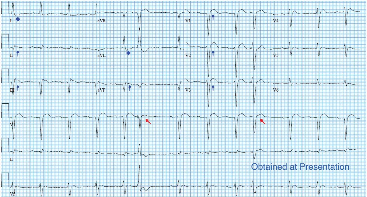
Electrocardiography (ECG) (Figure 1) showed ST- segment elevation in leads V1, V2, V3, II, III, and aVF and ST-segment depression in leads I and aVL. The corrected QT interval was 360 ms, compared with 426 ms 4 months earlier (Figure 2).

FIGURE 1
  • Download figure
  • Open in new tab
  • Download powerpoint
FIGURE 1

Electrocardiography at presentation showed a corrected QT interval of 360 msec. In addition to ST-segment elevations (blue arrows) and ST-segment depressions (blue diamonds), varying ectopic beats (red arrows) were present that were absent on previous studies.

FIGURE 2
  • Download figure
  • Open in new tab
  • Download powerpoint
FIGURE 2

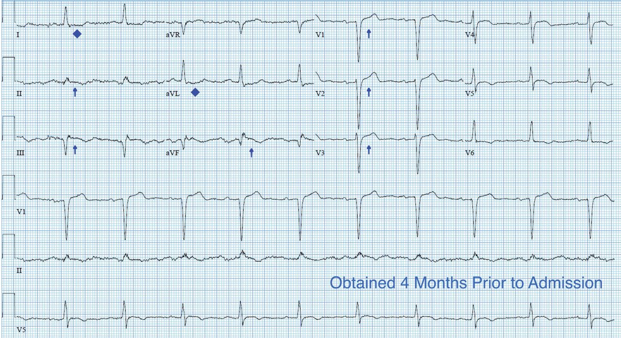
An electrocardiogram 4 months earlier showed no ST-segment elevations (blue arrows) or ST-segment depressions (blue diamonds).

Laboratory testing showed the following:

  • Troponin I 0.11 ng/mL (reference range < 0.04); repeated, it was 0.12 ng/mL

  • Serum creatinine 3.4 mg/dL (0.44–1.27) (9 months earlier it had been 0.99 mg/dL)

  • Serum calcium 17.3 mg/dL (8.6–10.5)

  • Parathyroid hormone 9 pg/mL (12–88)

  • Serum bicarbonate 33 mmol/L (24–32); 2 weeks earlier, it had been 27 mmol/L.

A cardiology consult was ordered out of concern for ST-elevation MI (STEMI). The cardiology consult team did not recommend coronary angiography because the patient’s chest pain had resolved spontaneously, its presentation was atypical, and the results of laboratory studies indicated acute kidney injury, a relative contraindication to angiography.

DIAGNOSIS: MILK-ALKALI SYNDROME

The diagnosis, based on the presentation and the results of the workup, was milk-alkali syndrome complicated by recent hydrochlorothiazide use. This syndrome consists of the triad of hypercalcemia, metabolic alkalosis, and acute kidney injury, all due to excessive ingestion of calcium and alkali, usually calcium carbonate.

His hydrochlorothiazide and calcium carbonate were discontinued. He was given intravenous normal saline and subcutaneous calcitonin, and his serum calcium level came down to 11.5 mg/dL within the next 24 hours. His dyspepsia was treated with pantoprazole.

The patient had no further episodes of chest pain, and the cardiology consult team again recommended against coronary angiography. Repeat ECG after the hypercalcemia resolved showed results identical to those 4 months before his admission. Two months later, his serum calcium level was 9.4 mg/dL and his creatinine level was 1.24 mg/dL.

A MIMIC OF STEMI

In numerous reported cases, these electrocardiographic findings coupled with chest pain led to misdiagnosis of STEMI.1–3 While STEMI and occasionally hypercalcemia can cause ST elevation, hypercalcemia causes a significant shortening of the corrected QT interval that is not associated with STEMI.4,5

Ultimately, the diagnosis of MI involves clinical, laboratory, and ECG findings, and if a strong clinical suspicion for myocardial ischemia exists, STEMI cannot reliably be distinguished from hypercalcemia by ECG alone. It is nonetheless important to be aware of this complication of hypercalcemia to avoid unnecessary cardiac interventions.

  • Copyright © 2017 The Cleveland Clinic Foundation. All Rights Reserved.

REFERENCES

  1. ↵
    1. Ashizawa N,
    2. Arakawa S,
    3. Koide Y,
    4. Toda G,
    5. Seto S,
    6. Yano K
    . Hypercalcemia due to vitamin D intoxication with clinical features mimicking acute myocardial infarction. Intern Med 2003; 42:340–344.
    OpenUrlCrossRefPubMed
    1. Nishi SP,
    2. Barbagelata NA,
    3. Atar S,
    4. Birnbaum Y,
    5. Tuero E
    . Hypercalcemia-induced ST-segment elevation mimicking acute myocardial infarction. J Electrocardiol 2006; 39:298–300.
    OpenUrlCrossRefPubMed
  2. ↵
    1. Turnham S,
    2. Kilickap M,
    3. Kilinc S
    . ST segment elevation mimicking acute myocardial infarction in hypercalcemia. Heart 2005; 91:999.
    OpenUrlFREE Full Text
  3. ↵
    1. Nierenberg DW,
    2. Ransil BJ
    . Q-aTc interval as a clinical indicator of hypercalcemia. Am J Cardiol 1979; 44:243–248.
    OpenUrlCrossRefPubMed
  4. ↵
    1. Ahmed R,
    2. Hashiba K
    . Reliability of QT intervals as indicators of clinical hypercalcemia. Clin Cardiol 1988; 11:395–400.
    OpenUrlCrossRefPubMed
PreviousNext
Back to top

In this issue

Cleveland Clinic Journal of Medicine: 84 (9)
Cleveland Clinic Journal of Medicine
Vol. 84, Issue 9
1 Sep 2017
  • Table of Contents
  • Table of Contents (PDF)
  • Index by author
Print
Download PDF
Article Alerts
Sign In to Email Alerts with your Email Address
Email Article

Thank you for your interest in spreading the word on Cleveland Clinic Journal of Medicine.

NOTE: We only request your email address so that the person you are recommending the page to knows that you wanted them to see it, and that it is not junk mail. We do not capture any email address.

Enter multiple addresses on separate lines or separate them with commas.
Heartburn or heart attack? A mimic of MI
(Your Name) has sent you a message from Cleveland Clinic Journal of Medicine
(Your Name) thought you would like to see the Cleveland Clinic Journal of Medicine web site.
CAPTCHA
This question is for testing whether or not you are a human visitor and to prevent automated spam submissions.
Citation Tools
Heartburn or heart attack? A mimic of MI
Willis S. Bowman, Amir Farid, Paul Aronowitz
Cleveland Clinic Journal of Medicine Sep 2017, 84 (9) 664-665; DOI: 10.3949/ccjm.84a.16093

Citation Manager Formats

  • BibTeX
  • Bookends
  • EasyBib
  • EndNote (tagged)
  • EndNote 8 (xml)
  • Medlars
  • Mendeley
  • Papers
  • RefWorks Tagged
  • Ref Manager
  • RIS
  • Zotero
Share
Heartburn or heart attack? A mimic of MI
Willis S. Bowman, Amir Farid, Paul Aronowitz
Cleveland Clinic Journal of Medicine Sep 2017, 84 (9) 664-665; DOI: 10.3949/ccjm.84a.16093
Twitter logo Facebook logo Mendeley logo
  • Tweet Widget
  • Facebook Like
  • Linkedin Share Button

Jump to section

  • Article
    • FURTHER EVALUATION, CARDIOLOGY CONSULT
    • DIAGNOSIS: MILK-ALKALI SYNDROME
    • A MIMIC OF STEMI
    • REFERENCES
  • Figures & Data
  • Info & Metrics
  • PDF

Related Articles

  • No related articles found.
  • PubMed
  • Google Scholar

Cited By...

  • No citing articles found.
  • Google Scholar

More in this TOC Section

  • Tinea incognito
  • Prolonged venous filling time and dependent rubor in a patient with peripheral artery disease
  • Sarcoidosis with diffuse purplish erythematous plaques on the hands
Show more The Clinical Picture

Similar Articles

Subjects

  • Cardiology
  • Emergency Medicine
  • Endocrinology
  • Gastroenterology

Navigate

  • Current Issue
  • Past Issues
  • Supplements
  • Article Type
  • Specialty
  • CME/MOC Articles
  • CME/MOC Calendar
  • Media Kit

Authors & Reviewers

  • Manuscript Submission
  • Authors & Reviewers
  • Subscriptions
  • About CCJM
  • Contact Us
  • Cleveland Clinic Center for Continuing Education
  • Consult QD

Share your suggestions!

Copyright © 2025 The Cleveland Clinic Foundation. All rights reserved. The information provided is for educational purposes only. Use of this website is subject to the website terms of use and privacy policy. 

Powered by HighWire