
Main menu

  • Home
  • Content
    • Current Issue
    • Ahead of Print
    • Past Issues
    • Supplements
    • Article Type
  • Specialty
    • Articles by Specialty
  • CME/MOC
    • Articles
    • Calendar
  • Info For
    • Manuscript Submission
    • Authors & Reviewers
    • Subscriptions
    • About CCJM
    • Contact Us
    • Media Kit
  • Conversations with Leaders
  • Conference Coverage
    • Kidney Week 2024
    • CHEST 2024
    • ACR Convergence 2023
    • Kidney Week 2023
    • ObesityWeek 2023
    • IDWeek 2023
    • CHEST 2023
    • MDS 2023
    • IAS 2023
    • ACP 2023
    • AAN 2023
    • ACC / WCC 2023
    • AAAAI Meeting 2023
    • ACR Convergence 2022
    • Kidney Week 2022
    • AIDS 2022
  • Other Publications
    • www.clevelandclinic.org

User menu

  • Register
  • Log in

Search

  • Advanced search
Cleveland Clinic Journal of Medicine
  • Other Publications
    • www.clevelandclinic.org
  • Register
  • Log in
Cleveland Clinic Journal of Medicine

Advanced Search

  • Home
  • Content
    • Current Issue
    • Ahead of Print
    • Past Issues
    • Supplements
    • Article Type
  • Specialty
    • Articles by Specialty
  • CME/MOC
    • Articles
    • Calendar
  • Info For
    • Manuscript Submission
    • Authors & Reviewers
    • Subscriptions
    • About CCJM
    • Contact Us
    • Media Kit
  • Conversations with Leaders
  • Conference Coverage
    • Kidney Week 2024
    • CHEST 2024
    • ACR Convergence 2023
    • Kidney Week 2023
    • ObesityWeek 2023
    • IDWeek 2023
    • CHEST 2023
    • MDS 2023
    • IAS 2023
    • ACP 2023
    • AAN 2023
    • ACC / WCC 2023
    • AAAAI Meeting 2023
    • ACR Convergence 2022
    • Kidney Week 2022
    • AIDS 2022
The Clinical Picture

Whiplash-shaped acute rash

Lidia Maroñas-Jiménez, MD, Alejandro Lobato-Berezo, MD, Ramon Pigem, MD, Diana Menis, MD and Sara Palencia-Pérez, MD, PhD
Cleveland Clinic Journal of Medicine June 2016, 83 (6) 417-418; DOI: https://doi.org/10.3949/ccjm.83a.15062
Lidia Maroñas-Jiménez
Department of Dermatology, Hospital 12 de Octubre, and i+12 Research Institute, Universidad Complutense, Madrid, Spain
  • Find this author on Google Scholar
  • Find this author on PubMed
  • Search for this author on this site
  • For correspondence: [email protected]
Alejandro Lobato-Berezo
Department of Dermatology, Hospital Severo Ochoa, Leganés, Madrid, Spain
  • Find this author on Google Scholar
  • Find this author on PubMed
  • Search for this author on this site
Ramon Pigem
Department of Dermatology, Hospital Clínic, Barcelona, Spain
  • Find this author on Google Scholar
  • Find this author on PubMed
  • Search for this author on this site
Diana Menis
Department of Dermatology, Hospital 12 de Octubre, Madrid, Spain
  • Find this author on Google Scholar
  • Find this author on PubMed
  • Search for this author on this site
Sara Palencia-Pérez
Department of Dermatology, Hospital 12 de Octubre, Madrid, Spain
  • Find this author on Google Scholar
  • Find this author on PubMed
  • Search for this author on this site
  • Article
  • Figures & Data
  • Info & Metrics
  • PDF
Loading

Apreviously healthy 32-year-old man presented to the emergency room with a persistent, nonpruritic rash on his trunk, which had suddenly appeared 2 days after he ate Chinese food.

Physical examination revealed multiple cross-linked linear plaques that appeared like scratches over his chest, back, and shoulders (Figures 1 and 2). He had no dermatographism, and his scalp, nails, palms, and soles were not affected. He had no signs of lymphadenopathy or systemic involvement.

FIGURE 1
  • Download figure
  • Open in new tab
  • Download powerpoint
FIGURE 1

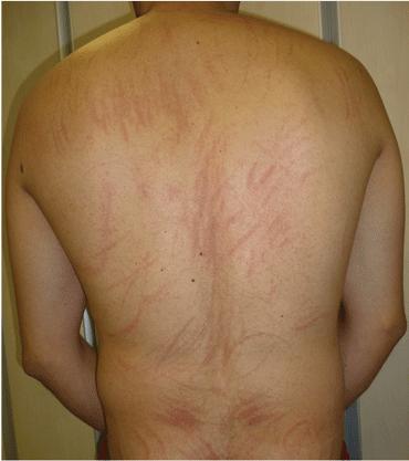
A widespread streaked rash with a scratch-like appearance over the patient’s back.

FIGURE 2
  • Download figure
  • Open in new tab
  • Download powerpoint
FIGURE 2

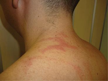
Closer inspection of the lesions showed intensely erythematous linear plaques with a pseudo vesicular surface.

Basic blood and urinary laboratory testing, blood cultures, and serologic studies showed normal or negative results.

Given the presentation and results of initial testing, his rash was diagnosed as flagellate erythema, likely due to shiitake mushroom intake. The diagnosis does not require histopathologic confirmation.

The rash resolved spontaneously over the next 2 weeks with use of a topical emollient and without scarring or residual hyperpigmentation.

FLAGELLATE ERYTHEMA

Flagellate erythema is a peculiar cutaneous eruption characterized by the progressive or sudden onset of parallel linear or curvilinear plaques, most commonly on the trunk. The plaques are typically arranged in a scratch pattern resembling marks left by the lashes of a whip.1 In contrast to other itchy dermatoses and neurotic excoriations that may present with self-induced linear marks, flagellate erythema appears spontaneously.

Drug-related causes, disease associations

Originally described in association with bleomycin treatment, flagellate erythema is currently considered a distinct feature of several dermatologic and systemic disorders, and therefore the ability to recognize it is valuable in daily practice.2 In addition to bleomycin analogues and anticancer agents such as peplomycin,1 bendamustine,3 and docetax-el,4 physicians should consider shiitake dermatitis5 and other less commonly reported associations such as dermatomyositis,6 lupus,7 Still disease,8 and parvovirus infection.9

Diagnostic features

The diagnosis of flagellate erythema is mainly based on the morphologic features of the clinical lesions.1 Shiitake dermatitis and flagellate erythema related to rheumatologic disease usually present with more inflammatory and erythematous plaques. Chemotherapy-induced flagellate rash typically has a violaceous or purpuric coloration, which tends to leave notice able hyperpigmentation for several months.2

Skin biopsy may be necessary to distinguish it from similar-looking dermatoses with different histologic findings, such as dermatographism, phytophotodermatitis, erythema gyratum repens, and factitious dermatoses, which may require specific treatments or be related to important underlying pathology.1,2

Treatment

Treatment includes both specific treatment of the underlying cause and symptomatic care of the skin with topical emollients and, in cases of associated pruritus, oral antihistamines. The patient should also be reassured about the self-healing nature of shiitake dermatitis rash.5

  • Copyright © 2016 The Cleveland Clinic Foundation. All Rights Reserved.

REFERENCES

  1. ↵
    1. Yamamoto T,
    2. Nishioka K
    . Flagellate erythema. Int J Dermatol 2006; 45:627–631.
    OpenUrlPubMed
  2. ↵
    1. Bhushan P,
    2. Manjul P,
    3. Baliyan V
    . Flagellate dermatoses. Indian J Der-matol Venereol Leprol 2014; 80:149–152.
    OpenUrl
  3. ↵
    1. Mahmoud BH,
    2. Eide MJ
    . Bendamustine-induced “flagellate dermatitis”. Dermatol Online J 2012; 18:12.
    OpenUrlPubMed
  4. ↵
    1. Tallon B,
    2. Lamb S
    . Flagellate erythema induced by docetaxel. Clin Exp Dermatol 2008; 33:276–277.
    OpenUrlCrossRefPubMed
  5. ↵
    1. Adler MJ,
    2. Larsen WG
    . Clinical variability of shiitake dermatitis. J Am Acad Dermatol 2012; 67:140–141.
    OpenUrl
  6. ↵
    1. Jara M,
    2. Amérigo J,
    3. Duce S,
    4. Borbujo J
    . Dermatomyositis and flagellate erythema. Clin Exp Dermatol 1996; 21:440–441.
    OpenUrlCrossRefPubMed
  7. ↵
    1. Niiyama S,
    2. Katsuoka K
    . Systemic lupus erythematosus with flagellate erythema. Eur J Dermatol 2012; 22:808–809.
    OpenUrl
  8. ↵
    1. Ciliberto H,
    2. Kumar MG,
    3. Musiek A
    . Flagellate erythema in a patient with fever. JAMA Dermatol 2013; 149:1425–1426.
    OpenUrl
  9. ↵
    1. Miguélez A,
    2. Dueñas J,
    3. Hervás D,
    4. Hervás JA,
    5. Salva F,
    6. Martín-Santiago A
    . Flagellate erythema in parvovirus B19 infection. Int J Dermatol 2014; 53:e583–e585.
    OpenUrl
PreviousNext
Back to top

In this issue

Cleveland Clinic Journal of Medicine: 83 (6)
Cleveland Clinic Journal of Medicine
Vol. 83, Issue 6
1 Jun 2016
  • Table of Contents
  • Table of Contents (PDF)
  • Index by author
Print
Download PDF
Article Alerts
Sign In to Email Alerts with your Email Address
Email Article

Thank you for your interest in spreading the word on Cleveland Clinic Journal of Medicine.

NOTE: We only request your email address so that the person you are recommending the page to knows that you wanted them to see it, and that it is not junk mail. We do not capture any email address.

Enter multiple addresses on separate lines or separate them with commas.
Whiplash-shaped acute rash
(Your Name) has sent you a message from Cleveland Clinic Journal of Medicine
(Your Name) thought you would like to see the Cleveland Clinic Journal of Medicine web site.
CAPTCHA
This question is for testing whether or not you are a human visitor and to prevent automated spam submissions.
Citation Tools
Whiplash-shaped acute rash
Lidia Maroñas-Jiménez, Alejandro Lobato-Berezo, Ramon Pigem, Diana Menis, Sara Palencia-Pérez
Cleveland Clinic Journal of Medicine Jun 2016, 83 (6) 417-418; DOI: 10.3949/ccjm.83a.15062

Citation Manager Formats

  • BibTeX
  • Bookends
  • EasyBib
  • EndNote (tagged)
  • EndNote 8 (xml)
  • Medlars
  • Mendeley
  • Papers
  • RefWorks Tagged
  • Ref Manager
  • RIS
  • Zotero
Share
Whiplash-shaped acute rash
Lidia Maroñas-Jiménez, Alejandro Lobato-Berezo, Ramon Pigem, Diana Menis, Sara Palencia-Pérez
Cleveland Clinic Journal of Medicine Jun 2016, 83 (6) 417-418; DOI: 10.3949/ccjm.83a.15062
Twitter logo Facebook logo Mendeley logo
  • Tweet Widget
  • Facebook Like
  • Linkedin Share Button

Jump to section

  • Article
    • FLAGELLATE ERYTHEMA
    • REFERENCES
  • Figures & Data
  • Info & Metrics
  • PDF

Related Articles

  • No related articles found.
  • PubMed
  • Google Scholar

Cited By...

  • No citing articles found.
  • Google Scholar

More in this TOC Section

  • Tinea incognito
  • Prolonged venous filling time and dependent rubor in a patient with peripheral artery disease
  • Sarcoidosis with diffuse purplish erythematous plaques on the hands
Show more The Clinical Picture

Similar Articles

Subjects

  • Allergy/Immunology
  • Dermatology

Navigate

  • Current Issue
  • Past Issues
  • Supplements
  • Article Type
  • Specialty
  • CME/MOC Articles
  • CME/MOC Calendar
  • Media Kit

Authors & Reviewers

  • Manuscript Submission
  • Authors & Reviewers
  • Subscriptions
  • About CCJM
  • Contact Us
  • Cleveland Clinic Center for Continuing Education
  • Consult QD

Share your suggestions!

Copyright © 2025 The Cleveland Clinic Foundation. All rights reserved. The information provided is for educational purposes only. Use of this website is subject to the website terms of use and privacy policy. 

Powered by HighWire