
Main menu

  • Home
  • Content
    • Current Issue
    • Ahead of Print
    • Past Issues
    • Supplements
    • Article Type
  • Specialty
    • Articles by Specialty
  • CME/MOC
    • Articles
    • Calendar
  • Info For
    • Manuscript Submission
    • Authors & Reviewers
    • Subscriptions
    • About CCJM
    • Contact Us
    • Media Kit
  • Conversations with Leaders
  • Conference Coverage
    • Kidney Week 2024
    • CHEST 2024
    • ACR Convergence 2023
    • Kidney Week 2023
    • ObesityWeek 2023
    • IDWeek 2023
    • CHEST 2023
    • MDS 2023
    • IAS 2023
    • ACP 2023
    • AAN 2023
    • ACC / WCC 2023
    • AAAAI Meeting 2023
    • ACR Convergence 2022
    • Kidney Week 2022
    • AIDS 2022
  • Other Publications
    • www.clevelandclinic.org

User menu

  • Register
  • Log in

Search

  • Advanced search
Cleveland Clinic Journal of Medicine
  • Other Publications
    • www.clevelandclinic.org
  • Register
  • Log in
Cleveland Clinic Journal of Medicine

Advanced Search

  • Home
  • Content
    • Current Issue
    • Ahead of Print
    • Past Issues
    • Supplements
    • Article Type
  • Specialty
    • Articles by Specialty
  • CME/MOC
    • Articles
    • Calendar
  • Info For
    • Manuscript Submission
    • Authors & Reviewers
    • Subscriptions
    • About CCJM
    • Contact Us
    • Media Kit
  • Conversations with Leaders
  • Conference Coverage
    • Kidney Week 2024
    • CHEST 2024
    • ACR Convergence 2023
    • Kidney Week 2023
    • ObesityWeek 2023
    • IDWeek 2023
    • CHEST 2023
    • MDS 2023
    • IAS 2023
    • ACP 2023
    • AAN 2023
    • ACC / WCC 2023
    • AAAAI Meeting 2023
    • ACR Convergence 2022
    • Kidney Week 2022
    • AIDS 2022
The Clinical Picture

Multiple linear subcutaneous nodules

Gitesh U. Sawatkar, MD, Dipankar De, MD, Uma Nahar Saikia, MD and Sanjeev Handa, MD, FRCP (EDIN)
Cleveland Clinic Journal of Medicine June 2016, 83 (6) 414-416; DOI: https://doi.org/10.3949/ccjm.83a.15058
Gitesh U. Sawatkar
Department of Dermatology, Venereology, and Leprology, Postgraduate Institute of Medical Education and Research, Chandigarh, India
  • Find this author on Google Scholar
  • Find this author on PubMed
  • Search for this author on this site
Dipankar De
Department of Dermatology, Venereology, and Leprology, Postgraduate Institute of Medical Education and Research, Chandigarh, India
  • Find this author on Google Scholar
  • Find this author on PubMed
  • Search for this author on this site
  • For correspondence: [email protected]
Uma Nahar Saikia
Department of Histopathology, Postgraduate Institute of Medical Education and Research, Chandigarh, India
  • Find this author on Google Scholar
  • Find this author on PubMed
  • Search for this author on this site
Sanjeev Handa
Department of Dermatology, Venereology, and Leprology, Postgraduate Institute of Medical Education and Research, Chandigarh, India
  • Find this author on Google Scholar
  • Find this author on PubMed
  • Search for this author on this site
  • Article
  • Figures & Data
  • Info & Metrics
  • PDF
Loading

A 34-year-old woman sought consultation at our clinic for an asymptomatic swelling on her right foot that had been growing very slowly over the last 15 years. She said she had presented to other health-care facilities, but no diagnosis had been made and no treatment had been offered.

Examination revealed a linear swelling extending from the lower third to the mid-dorsal surface of the right foot (Figure 1). Palpation revealed multiple, closely set nodules arranged in a linear fashion. This finding along with the history raised the suspicion of neurofibroma and other conditions in the differential diagnosis, eg, pure neuritic Hansen disease, phaeohyphomycosis, and palisaded neutrophilic granulomatous dermatitis. The rest of the mucocutaneous examination results were normal. No café-au-lait spots, axillary freckling, or other swelling suggestive of neurofibroma was seen. She had no family history of mucocutaneous disease or other systemic disorder.

FIGURE 1
  • Download figure
  • Open in new tab
  • Download powerpoint
FIGURE 1

A linear swelling extended from the lower third to the mid-dorsal aspect of the right foot.

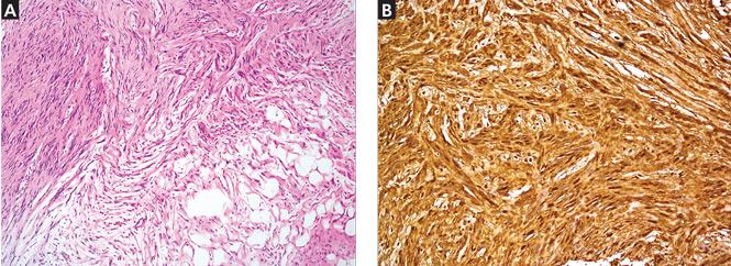
Because of the suspicion of neurofibromatosis, slit-lamp examination of the eyes was done to rule out Lisch nodules, a common feature of neurofibromatosis; the results were normal. Plain radiography of the right foot showed only soft-tissue swelling. Magnetic resonance imaging with contrast, done to determine the extent of the lesions, revealed multiple dumbbell-shaped lesions with homogeneous enhancement (Figure 2). Histopathologic study of a biopsy specimen of the lesions showed tumor cells in the dermis. The cells were long, with elongated nuclei with pointed ends, arranged in long and short fascicles—an appearance characteristic of neurofibroma. Areas of hypocellularity and hypercellularity were seen, and on S100 protein immunostaining, the tumor cells showed strong nuclear and cytoplasmic positivity (Figure 3).

FIGURE 2
  • Download figure
  • Open in new tab
  • Download powerpoint
FIGURE 2

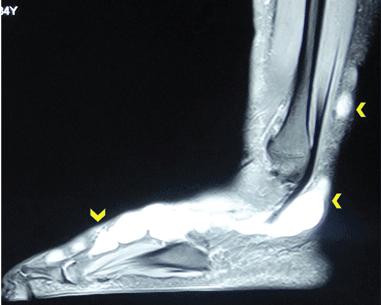
T2-weighted magnetic resonance imaging with contrast showed a hyperintense lesion along the posterolateral aspect of the right foot in the subcutaneous space up to the proximal end of the proximal phalanx of the fourth toe in the location of the sural nerve.

FIGURE 3
  • Download figure
  • Open in new tab
  • Download powerpoint
FIGURE 3

The photomicrograph (A) shows tumor composed of spindle cells with pointed ends that infiltrate adjacent fat (hematoxylin and eosin, × 100). Tumor cells (B) show strong nuclear and cytoplasmic positivity (S100 protein immunostaining, × 200).

The histologic evaluation confirmed neurofibroma. The specific diagnosis of sporadic solitary neurofibroma was made based on the onset of the lesions, the number of lesions (one in this patient), and the absence of features suggestive of neurofibromatosis.

SPORADIC SOLITARY NEUROFIBROMA

Neurofibroma is a common tumor of the peripheral nerve sheath and, when present with features such as café-au-lait spots, axillary freckling, and characteristic bone changes, it is pathognomic of neurofibromatosis type 1.1 But solitary neurofibromas can occur sporadically in the absence of other features of neurofibromatosis.

Sporadic solitary neurofibroma arises from small nerves, is benign in nature, and carries a lower rate of malignant transformation than its counterpart that occurs in the setting of neurofibromatosis.2 Though sporadic solitary neurofibroma can occur in any part of the body, it is commonly seen on the head and neck, and occasionally on the presacral and parasacral space, thigh, intrascrotal area,3 the ankle and foot,4,5 and the subungual region.6 A series of 397 peripheral neural sheath tumors examined over 30 years showed 55 sporadic solitary neurofibromas occurring in the brachial plexus region, 45 in the upper extremities, 10 in the pelvic plexus, and 31 in the lower extremities.7

Management of sporadic solitary neurofibroma depends on the patient’s discomfort. For asymptomatic lesions, serial observation is all that is required. Complete surgical excision including the parent nerve is the treatment for large lesions. More research is needed to define the potential role of drugs such as pirfenidone and tipifarnib.

THE DIFFERENTIAL DIAGNOSIS

Sporadic solitary neurofibroma can masquerade as pure neuritic Hansen disease (leprosy), phaeohyphomycosis, and palisaded neutrophilic granulomatous dermatitis. The absence of neural symptoms and no evidence of trophic changes exclude pure neuritic Hansen disease. Phaeohyphomycosis clinically presents as a single cyst that may evolve into pigmented plaques,8 and the diagnosis relies on the presence of fungus in tissue. The absence of cystic changes clinically and fungi histopathologically in this patient did not favor phaeohyphomycosis. Palisaded neutrophilic granulomatous dermatitis is characterized clinically by cordlike skin lesions (the “rope sign”) and is accompanied by extracutaneous, mostly articular features. Histopathologically, it shows intense neutrophilic infiltrate and interstitial histiocytic infiltrate along with collagen degeneration. The absence of extracutaneous and classical histologic features negated this possibility in this patient.

Though sporotrichosis and cutaneous atypical mycobacterial infections may present in linear fashion following the course of the lymphatic vessels, the absence of epidermal changes after a disease course of 15 years and the absence of granulomatous infiltrate in histopathology excluded these possibilities in this patient.

The patient was referred to a plastic surgeon, and the lesions were successfully resected. She did not return for additional review after that.

  • Copyright © 2016 The Cleveland Clinic Foundation. All Rights Reserved.

REFERENCES

  1. ↵
    1. Hirbe AC,
    2. Gutmann DH
    . Neurofibromatosis type 1: a multidisciplinary approach to care. Lancet Neurol 2014; 13:834–843.
    OpenUrlCrossRefPubMed
  2. ↵
    1. Pulathan Z,
    2. Imamoglu M,
    3. Cay A,
    4. Guven YK
    . Intermittent claudication due to right common femoral artery compression by a solitary neurofibroma. Eur J Pediatr 2005; 164:463–465.
    OpenUrlPubMed
  3. ↵
    1. Hosseini MM,
    2. Geramizadeh B,
    3. Shakeri S,
    4. Karimi MH
    . Intrascrotal solitary neurofibroma: a case report and review of the literature. Urol Ann 2012; 4:119–121.
    OpenUrlCrossRefPubMed
  4. ↵
    1. Carvajal JA,
    2. Cuartas E,
    3. Qadir R,
    4. Levi AD,
    5. Temple HT
    . Peripheral nerve sheath tumors of the foot and ankle. Foot Ankle Int 2011; 32:163–167.
    OpenUrlPubMed
  5. ↵
    1. Tahririan MA,
    2. Hekmatnia A,
    3. Ahrar H,
    4. Heidarpour M,
    5. Hekmatnia F
    . Solitary giant neurofibroma of thigh. Adv Biomed Res 2014; 3:158.
    OpenUrl
  6. ↵
    1. Huajun J,
    2. Wei Q,
    3. Ming L,
    4. Chongyang F,
    5. Weiguo Z,
    6. Decheng L
    . Solitary subungual neurofibroma in the right first finger. Int J Dermatol 2012; 51:335–338.
    OpenUrlPubMed
  7. ↵
    1. Kim DH,
    2. Murovic JA,
    3. Tiel RL,
    4. Moes G,
    5. Kline DG
    . A series of 397 peripheral neural sheath tumors: 30-year experience at Louisiana State University Health Sciences Center. J Neurosurg 2005; 102:246–255.
    OpenUrlCrossRefPubMed
  8. ↵
    1. Garnica M,
    2. Nucci M,
    3. Queiroz-Telles F
    . Difficult mycoses of the skin: advances in the epidemiology and management of eumycetoma, phaeohyphomycosis and chromoblastomycosis. Curr Opin Infect Dis 2009; 22:559–563.
    OpenUrlCrossRefPubMed
PreviousNext
Back to top

In this issue

Cleveland Clinic Journal of Medicine: 83 (6)
Cleveland Clinic Journal of Medicine
Vol. 83, Issue 6
1 Jun 2016
  • Table of Contents
  • Table of Contents (PDF)
  • Index by author
Print
Download PDF
Article Alerts
Sign In to Email Alerts with your Email Address
Email Article

Thank you for your interest in spreading the word on Cleveland Clinic Journal of Medicine.

NOTE: We only request your email address so that the person you are recommending the page to knows that you wanted them to see it, and that it is not junk mail. We do not capture any email address.

Enter multiple addresses on separate lines or separate them with commas.
Multiple linear subcutaneous nodules
(Your Name) has sent you a message from Cleveland Clinic Journal of Medicine
(Your Name) thought you would like to see the Cleveland Clinic Journal of Medicine web site.
CAPTCHA
This question is for testing whether or not you are a human visitor and to prevent automated spam submissions.
Citation Tools
Multiple linear subcutaneous nodules
Gitesh U. Sawatkar, Dipankar De, Uma Nahar Saikia, Sanjeev Handa
Cleveland Clinic Journal of Medicine Jun 2016, 83 (6) 414-416; DOI: 10.3949/ccjm.83a.15058

Citation Manager Formats

  • BibTeX
  • Bookends
  • EasyBib
  • EndNote (tagged)
  • EndNote 8 (xml)
  • Medlars
  • Mendeley
  • Papers
  • RefWorks Tagged
  • Ref Manager
  • RIS
  • Zotero
Share
Multiple linear subcutaneous nodules
Gitesh U. Sawatkar, Dipankar De, Uma Nahar Saikia, Sanjeev Handa
Cleveland Clinic Journal of Medicine Jun 2016, 83 (6) 414-416; DOI: 10.3949/ccjm.83a.15058
Twitter logo Facebook logo Mendeley logo
  • Tweet Widget
  • Facebook Like
  • Linkedin Share Button

Jump to section

  • Article
    • SPORADIC SOLITARY NEUROFIBROMA
    • THE DIFFERENTIAL DIAGNOSIS
    • REFERENCES
  • Figures & Data
  • Info & Metrics
  • PDF

Related Articles

  • No related articles found.
  • PubMed
  • Google Scholar

Cited By...

  • No citing articles found.
  • Google Scholar

More in this TOC Section

  • Tinea incognito
  • Prolonged venous filling time and dependent rubor in a patient with peripheral artery disease
  • Sarcoidosis with diffuse purplish erythematous plaques on the hands
Show more The Clinical Picture

Similar Articles

Subjects

  • Allergy/Immunology
  • Dermatology
  • Imaging
  • Infectious Diseases
  • Neurology
  • Oncology
  • Rheumatology

Navigate

  • Current Issue
  • Past Issues
  • Supplements
  • Article Type
  • Specialty
  • CME/MOC Articles
  • CME/MOC Calendar
  • Media Kit

Authors & Reviewers

  • Manuscript Submission
  • Authors & Reviewers
  • Subscriptions
  • About CCJM
  • Contact Us
  • Cleveland Clinic Center for Continuing Education
  • Consult QD

Share your suggestions!

Copyright © 2025 The Cleveland Clinic Foundation. All rights reserved. The information provided is for educational purposes only. Use of this website is subject to the website terms of use and privacy policy. 

Powered by HighWire