
Main menu

  • Home
  • Content
    • Current Issue
    • Ahead of Print
    • Past Issues
    • Supplements
    • Article Type
  • Specialty
    • Articles by Specialty
  • CME/MOC
    • Articles
    • Calendar
  • Info For
    • Manuscript Submission
    • Authors & Reviewers
    • Subscriptions
    • About CCJM
    • Contact Us
    • Media Kit
  • Conversations with Leaders
  • Conference Coverage
    • Kidney Week 2024
    • CHEST 2024
    • ACR Convergence 2023
    • Kidney Week 2023
    • ObesityWeek 2023
    • IDWeek 2023
    • CHEST 2023
    • MDS 2023
    • IAS 2023
    • ACP 2023
    • AAN 2023
    • ACC / WCC 2023
    • AAAAI Meeting 2023
    • ACR Convergence 2022
    • Kidney Week 2022
    • AIDS 2022
  • Other Publications
    • www.clevelandclinic.org

User menu

  • Register
  • Log in

Search

  • Advanced search
Cleveland Clinic Journal of Medicine
  • Other Publications
    • www.clevelandclinic.org
  • Register
  • Log in
Cleveland Clinic Journal of Medicine

Advanced Search

  • Home
  • Content
    • Current Issue
    • Ahead of Print
    • Past Issues
    • Supplements
    • Article Type
  • Specialty
    • Articles by Specialty
  • CME/MOC
    • Articles
    • Calendar
  • Info For
    • Manuscript Submission
    • Authors & Reviewers
    • Subscriptions
    • About CCJM
    • Contact Us
    • Media Kit
  • Conversations with Leaders
  • Conference Coverage
    • Kidney Week 2024
    • CHEST 2024
    • ACR Convergence 2023
    • Kidney Week 2023
    • ObesityWeek 2023
    • IDWeek 2023
    • CHEST 2023
    • MDS 2023
    • IAS 2023
    • ACP 2023
    • AAN 2023
    • ACC / WCC 2023
    • AAAAI Meeting 2023
    • ACR Convergence 2022
    • Kidney Week 2022
    • AIDS 2022
The Clinical Picture

‘Air-raising’: An air-fluid level in the right subphrenic region

Anupam Gupta, MD and Oleg Epelbaum, MD
Cleveland Clinic Journal of Medicine September 2016, 83 (9) 638-640; DOI: https://doi.org/10.3949/ccjm.83a.15162
Anupam Gupta
Division of Pulmonary and Critical Care Medicine, Elmhurst Hospital Center, Icahn School of Medicine at Mount Sinai, Elmhurst, NY
  • Find this author on Google Scholar
  • Find this author on PubMed
  • Search for this author on this site
  • For correspondence: [email protected]
Oleg Epelbaum
Division of Pulmonary and Critical Care Medicine, Elmhurst Hospital Center, Icahn School of Medicine at Mount Sinai, Elmhurst, NY
  • Find this author on Google Scholar
  • Find this author on PubMed
  • Search for this author on this site
  • Article
  • Figures & Data
  • Info & Metrics
  • PDF
Loading

A 39-year-old filipino man presented with nausea, vomiting, and abdominal pain of 2 weeks’ duration. He did not report trauma, and he had no history of medical illness or surgery.

On arrival, his blood pressure was 123/83 mm Hg, pulse 122 beats per minute, respiratory rate 18 breaths per minute, and temperature 100.7°F (38.1°C). On physical examination, he exhibited marked tenderness of the right upper quadrant on palpation. The abdomen was otherwise soft with no guarding or rebound tenderness.

Results of initial laboratory testing were as follows:

  • Leukocyte count 17.0 × 10⁹/L (reference range 4.5–11.0)

  • Serum glucose 558 mg/dL without ketoacidosis

  • Aspartate aminotransferase 109 U/L (2–40)

  • Alanine aminotranferase 28 U/L (2–50)

  • Total serum bilirubin 4.0 mg/dL (0.0–1.5).

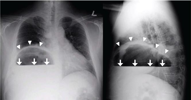
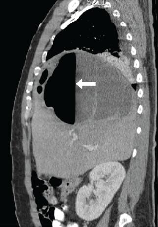
Plain chest radiography showed dramatic elevation of the right hemidiaphragm with a large subphrenic air-fluid level (Figure 1). Abdominal computed tomography (CT) demonstrated a multiloculated hepatic abscess 18 × 13.5 cm subjacent to the diaphragm (Figure 2). Cultures of blood and the abscess yielded Klebsiella pneumoniae. The patient recovered after percutaneous drainage and a course of ceftriaxone.

FIGURE 1
  • Download figure
  • Open in new tab
  • Download powerpoint
FIGURE 1

Plain chest radiography showed a dramatic elevation of the right hemidiaphragm (arrowheads), with a large subphrenic air-fluid level (arrows). The air-fluid level was characterized by a sharp horizontal demarcation between the lighter gas component floating at the top and the heavier fluid component settling on the bottom.

FIGURE 2
  • Download figure
  • Open in new tab
  • Download powerpoint
FIGURE 2

Abdominal computed tomography showed a multiloculated hepatic abscess 18 × 13.5 cm subjacent to the diaphragm (arrow).

PRIMARY KLEBSIELLA LIVER ABSCESS

K pneumoniae, a gram-negative aerobic encapsulated bacillus of the normal human intestinal flora, is closely related to Escherichia coli, historically the most frequent bacterial cause of pyogenic liver abscess.1 Over the last 30 years, K pneumoniae has eclipsed E coli as the most common causative agent, with the epicenter of this trend being located in Taiwan and South Korea, perhaps because rates of fecal Klebsiella carriage in that region are particularly high.1,2

Concurrently, there has been increasing recognition—initially across Asia, but lately in Europe and the Western Hemisphere—of the so-called invasive Klebsiella liver abscess (KLA) syndrome, virtually unique to the hypervirulent K1 and K2 capsular serotypes of K pneumoniae prevalent in Asia.3–6 This community-acquired syndrome is characterized by hematogenous deposition of the organism at distant sites, such as the lung, soft tissues, central nervous system, and eyes. Impairment of phagocytic function, as occurs in diabetes mellitus, and the resistance to phagocytosis conferred by the K1 and K2 serotypes have been identified as predisposing factors for dissemination.7,8 The mucoid phenotype of K pneumoniae, very common in Asian isolates of the K1 and K2 serotypes, is also associated with hypervirulence and extrahepatic spread, presumably through evasion of phagocytosis and complement-mediated opsonization.2,9

Our patient’s risk factors for KLA were his Asian origin and uncontrolled diabetes. No evidence of remote infection was detected during his hospitalization.

HEMIDIAPHRAGM ELEVATION

Acquired hemidiaphragm elevation is most commonly unilateral and typically represents an incidental radiologic finding attributable to paralysis of the corresponding diaphragm after phrenic nerve injury caused by trauma, surgery, or infection. Unilateral diaphragmatic paralysis is classically confirmed by performing a fluoroscopic sniff test, which is positive if the affected hemidiaphragm is observed in real time to paradoxically move upward during forced inhalation.10 This condition is usually asymptomatic at rest but could cause exertional dyspnea and contribute to ventilatory failure when pulmonary disease coexists.11

Occasionally, as in our patient, hemidiaphragm elevation is part of the presentation of active abdominal pathology that displaces the corresponding hemidiaphragm cephalad by mass effect. Examples of such space-occupying abdominal lesions include infections, malignancy, hepatosplenomegaly, and pneumoperitoneum from a ruptured viscus. Pneumoperitoneum is suggested by the presence of an air crescent immediately subjacent to the affected hemidiaphragm on an upright radiograph accompanied by peritoneal signs.

Although there was subphrenic air on this patient’s initial chest radiograph, it was actually part of an air-fluid level without associated peritoneal signs. An air-fluid level is characterized by a sharp horizontal demarcation between the lighter gas component floating at the top and the heavier fluid component settling on the bottom (Figure 1). The subsequent CT excluded free intra-abdominal air while identifying a large hepatic abscess as the cause of hemidiaphragm elevation. In trauma victims, CT is also helpful in ruling out diaphragmatic rupture, which can have a similar radiographic appearance.12

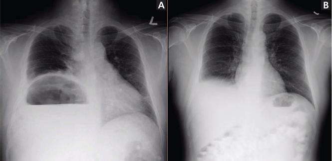
Our patient’s presentation was a reminder that an elevated hemidiaphragm may refl ect abdominal pathology and that subphrenic air in this context need not be either “free” or a surgical emergency. Drainage of the abscess restored the normal position of our patient’s right hemidiaphragm (Figure 3).

FIGURE 3
  • Download figure
  • Open in new tab
  • Download powerpoint
FIGURE 3

Compared with the initial radiograph (A), the subsequent frontal radiograph (B) shows the reduction in the height of the right hemidiaphragm after drainage of the abscess.

  • Copyright © 2016 The Cleveland Clinic Foundation. All Rights Reserved.

REFERENCES

  1. ↵
    1. Huang CJ,
    2. Pitt HA,
    3. Lipsett PA,
    4. et al
    . Pyogenic hepatic abscess: changing trends over 42 years. Ann Surg 1996; 223:600–607.
    OpenUrlCrossRefPubMed
  2. ↵
    1. Lin YT,
    2. Siu LK,
    3. Lin JC,
    4. et al
    . Seroepidemiology of Klebsiella pneumoniae colonizing the intestinal tract of healthy Chinese and overseas Chinese adults in Asian countries. BMC Microbiol 2012; 12:13.
    OpenUrlCrossRefPubMed
  3. ↵
    1. Wang JH,
    2. Liu YC,
    3. Lee SS,
    4. et al
    . Primary liver abscess due to Klebsiella pneumoniae in Taiwan. Clin Infect Dis 1998; 26:1434–1438.
    OpenUrlCrossRefPubMed
    1. Pastagia M,
    2. Arumugam V
    . Klebsiella pneumoniae liver abscesses in a public hospital in Queens, New York. Travel Med Infect Dis 2008; 6:228–233.
    OpenUrlCrossRefPubMed
    1. Rahimian J,
    2. Wilson T,
    3. Oram V,
    4. Holzman RS
    . Pyogenic liver abscess: recent trends in etiology and mortality. Clin Infect Dis 2004; 39:1654–1659.
    OpenUrlCrossRefPubMed
  4. ↵
    1. Moore R,
    2. O’Shea D,
    3. Geoghegan T,
    4. Mallon PW,
    5. Sheehan G
    . Community-acquired Klebsiella pneumoniae liver abscess: an emerging infection in Ireland and Europe. Infection 2013; 41:681–686.
    OpenUrlCrossRefPubMed
  5. ↵
    1. Lecube A,
    2. Pachón G,
    3. Petriz J,
    4. Hernández C,
    5. Simó R
    . Phagocytic activity is impaired in type 2 diabetes mellitus and increases after metabolic improvement. PLoS One 2011; 6:e23366.
    OpenUrlCrossRefPubMed
  6. ↵
    1. Lin JC,
    2. Siu LK,
    3. Fung CP,
    4. et al
    . Impaired phagocytosis of capsular serotypes K1 or K2 Klebsiella pneumoniae in type 2 diabetes mellitus patients with poor glycemic control. J Clin Endocrinol Metab 2006; 91:3084–3087.
    OpenUrlCrossRefPubMed
  7. ↵
    1. Siu LK,
    2. Yeh KM,
    3. Lin JC,
    4. Fung CP,
    5. Chang FY
    . Klebsiella pneumoniae liver abscess: a new invasive syndrome. Lancet Infect Dis 2012; 12:881–887.
    OpenUrlCrossRefPubMed
  8. ↵
    1. Gierada DS,
    2. Slone RM,
    3. Fleishman MJ
    . Imaging evaluation of the diaphragm. Chest Surg Clin North Am 1998; 8:237–280.
    OpenUrlPubMed
  9. ↵
    1. Qureshi A
    . Diaphragm paralysis. Semin Respir Crit Care Med 2009; 30:315–320.
    OpenUrlCrossRefPubMed
  10. ↵
    1. Havens JM,
    2. Kelly E,
    3. Patel V
    . A 78-year-old man with an elevated hemidiaphragm following trauma. Chest 2008; 134:1336–1339.
    OpenUrlCrossRefPubMed
PreviousNext
Back to top

In this issue

Cleveland Clinic Journal of Medicine: 83 (9)
Cleveland Clinic Journal of Medicine
Vol. 83, Issue 9
1 Sep 2016
  • Table of Contents
  • Table of Contents (PDF)
  • Index by author
Print
Download PDF
Article Alerts
Sign In to Email Alerts with your Email Address
Email Article

Thank you for your interest in spreading the word on Cleveland Clinic Journal of Medicine.

NOTE: We only request your email address so that the person you are recommending the page to knows that you wanted them to see it, and that it is not junk mail. We do not capture any email address.

Enter multiple addresses on separate lines or separate them with commas.
‘Air-raising’: An air-fluid level in the right subphrenic region
(Your Name) has sent you a message from Cleveland Clinic Journal of Medicine
(Your Name) thought you would like to see the Cleveland Clinic Journal of Medicine web site.
CAPTCHA
This question is for testing whether or not you are a human visitor and to prevent automated spam submissions.
Citation Tools
‘Air-raising’: An air-fluid level in the right subphrenic region
Anupam Gupta, Oleg Epelbaum
Cleveland Clinic Journal of Medicine Sep 2016, 83 (9) 638-640; DOI: 10.3949/ccjm.83a.15162

Citation Manager Formats

  • BibTeX
  • Bookends
  • EasyBib
  • EndNote (tagged)
  • EndNote 8 (xml)
  • Medlars
  • Mendeley
  • Papers
  • RefWorks Tagged
  • Ref Manager
  • RIS
  • Zotero
Share
‘Air-raising’: An air-fluid level in the right subphrenic region
Anupam Gupta, Oleg Epelbaum
Cleveland Clinic Journal of Medicine Sep 2016, 83 (9) 638-640; DOI: 10.3949/ccjm.83a.15162
Twitter logo Facebook logo Mendeley logo
  • Tweet Widget
  • Facebook Like
  • Linkedin Share Button

Jump to section

  • Article
    • PRIMARY KLEBSIELLA LIVER ABSCESS
    • HEMIDIAPHRAGM ELEVATION
    • REFERENCES
  • Figures & Data
  • Info & Metrics
  • PDF

Related Articles

  • No related articles found.
  • PubMed
  • Google Scholar

Cited By...

  • No citing articles found.
  • Google Scholar

More in this TOC Section

  • Tinea incognito
  • Prolonged venous filling time and dependent rubor in a patient with peripheral artery disease
  • Sarcoidosis with diffuse purplish erythematous plaques on the hands
Show more The Clinical Picture

Similar Articles

Subjects

  • Emergency Medicine
  • Imaging

Navigate

  • Current Issue
  • Past Issues
  • Supplements
  • Article Type
  • Specialty
  • CME/MOC Articles
  • CME/MOC Calendar
  • Media Kit

Authors & Reviewers

  • Manuscript Submission
  • Authors & Reviewers
  • Subscriptions
  • About CCJM
  • Contact Us
  • Cleveland Clinic Center for Continuing Education
  • Consult QD

Share your suggestions!

Copyright © 2025 The Cleveland Clinic Foundation. All rights reserved. The information provided is for educational purposes only. Use of this website is subject to the website terms of use and privacy policy. 

Powered by HighWire