
Main menu

  • Home
  • Content
    • Current Issue
    • Ahead of Print
    • Past Issues
    • Supplements
    • Article Type
  • Specialty
    • Articles by Specialty
  • CME/MOC
    • Articles
    • Calendar
  • Info For
    • Manuscript Submission
    • Authors & Reviewers
    • Subscriptions
    • About CCJM
    • Contact Us
    • Media Kit
  • Conversations with Leaders
  • Conference Coverage
    • Kidney Week 2024
    • CHEST 2024
    • ACR Convergence 2023
    • Kidney Week 2023
    • ObesityWeek 2023
    • IDWeek 2023
    • CHEST 2023
    • MDS 2023
    • IAS 2023
    • ACP 2023
    • AAN 2023
    • ACC / WCC 2023
    • AAAAI Meeting 2023
    • ACR Convergence 2022
    • Kidney Week 2022
    • AIDS 2022
  • Other Publications
    • www.clevelandclinic.org

User menu

  • Register
  • Log in

Search

  • Advanced search
Cleveland Clinic Journal of Medicine
  • Other Publications
    • www.clevelandclinic.org
  • Register
  • Log in
Cleveland Clinic Journal of Medicine

Advanced Search

  • Home
  • Content
    • Current Issue
    • Ahead of Print
    • Past Issues
    • Supplements
    • Article Type
  • Specialty
    • Articles by Specialty
  • CME/MOC
    • Articles
    • Calendar
  • Info For
    • Manuscript Submission
    • Authors & Reviewers
    • Subscriptions
    • About CCJM
    • Contact Us
    • Media Kit
  • Conversations with Leaders
  • Conference Coverage
    • Kidney Week 2024
    • CHEST 2024
    • ACR Convergence 2023
    • Kidney Week 2023
    • ObesityWeek 2023
    • IDWeek 2023
    • CHEST 2023
    • MDS 2023
    • IAS 2023
    • ACP 2023
    • AAN 2023
    • ACC / WCC 2023
    • AAAAI Meeting 2023
    • ACR Convergence 2022
    • Kidney Week 2022
    • AIDS 2022
The Clinical Picture

Fungal folliculitis masquerading as acute exanthematous pustulosis

Timmie Sharma, MD, Alok Vij, MD and Apra Sood, MD
Cleveland Clinic Journal of Medicine March 2016, 83 (3) 179-180; DOI: https://doi.org/10.3949/ccjm.83a.15028
Timmie Sharma
Department of Dermatology, University Hospitals Case Medical Center, Cleveland, OH
  • Find this author on Google Scholar
  • Find this author on PubMed
  • Search for this author on this site
Alok Vij
Department of Dermatology, Cleveland Clinic
  • Find this author on Google Scholar
  • Find this author on PubMed
  • Search for this author on this site
  • For correspondence: [email protected]
Apra Sood
Head, Section of Industrial Dermatology, Department of Dermatology, Cleveland Clinic
  • Find this author on Google Scholar
  • Find this author on PubMed
  • Search for this author on this site
  • Article
  • Figures & Data
  • Info & Metrics
  • PDF
Loading

A 25-year old man being treated in the hospital for catatonia and flaccid paralysis developed fever and a lobar consolidation, prompting treatment with piperacillin-tazo-bactam. A dermatology consult was called 3 days later when the patient developed a rash on the forehead (Figure 1), cheeks, chin, and upper trunk, raising concern for a drug-related eruption.

FIGURE 1
  • Download figure
  • Open in new tab
  • Download powerpoint
FIGURE 1

A brightly erythematous plaque on the forehead with monomorphic pustules was noted on day 3 of piperacillin-tazobactam infusion.

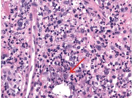
Close examination of the rash revealed bright red plaques studded with sheets of monomorphic pustules. Possible causes other than drug therapy were considered, including folliculitis, acneiform eruption, and pustular psoriasis. Bacterial culture of a pustule was negative, but punch biopsy of a pustule revealed an abundance of round and oval budding yeast forms consistent with Malassezia (formerly called Pityrosporum) “stuffing” the hair follicles and forming cleared spaces in the dermis surrounded by an exuberant lymphohistiocytic infiltrate (Figures 2 and 3). Special stains were negative for bacteria and mycelial forms. The clinicopathologic findings were diagnostic of Malassezia folliculitis with formation of dermal abscesses.

FIGURE 2
  • Download figure
  • Open in new tab
  • Download powerpoint
FIGURE 2

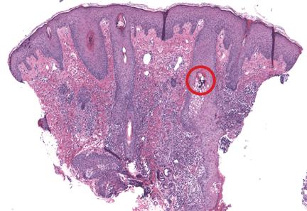
Histologic sections reveal Malassezia folliculitis and abscess formation within the dermis. The red circle highlights yeast forms within the follicular epithelium (hematoxylin and eosin, × 4).

FIGURE 3
  • Download figure
  • Open in new tab
  • Download powerpoint
FIGURE 3

Histologic sections of a perifollicular dermal abscess show mixed inflammatory infiltrate including neutrophils and histiocytes centered on cleared-out spaces with small, grey-blue yeast forms (red arrow) (hematoxylin and eosin, × 100).

The patient was treated with intravenous itraconazole 200 mg daily for 7 days and topical benzoyl peroxide 5% gel, with ketoconazole 2% cream for maintenance therapy. The treatment brought improvement of the rash.

THE DIFFERENTIAL DIAGNOSIS

The appearance of sterile pustules after starting a new antibiotic raised suspicion for acute localized exanthematous pustulosis, a variant of acute generalized exanthematous pustulosis. It is a serious but uncommon adverse drug reaction, with a frequency of one to five cases per million per year. The eruption of erythematous plaques studded with sterile pustules classically appears 1 to 5 days after starting a drug.1 Piperacillin-tazobactam has been infrequently reported in association with acute exanthematous pustulosis, but antibiotics in general are among the most commonly reported culprits.2–4

Clues to the correct diagnosis

Although our concern for acute localized exanthematous pustulosis was warranted, the morphology and distribution of this patient’s exanthem also raised suspicion of fungal folliculitis, which is more common.

Malassezia folliculitis appears as a monomorphic papular and pustular eruption on the chest, back, and face,5 as in our patient. Differentiating fungal folliculitis from pustulosis is important, as each condition is treated differently: Malassezia folliculitis is treated with antifungals,5 and acute localized exanthematous pustulosis is managed with cessation of the offending drug, supportive care, and systemic or topical steroids.4

Take-home point

Our experience with this patient was a reminder to consider fungal folliculitis in the differential diagnosis of a pustular eruption, so as to allow appropriate management and to avert discontinuation of potentially lifesaving medications.

  • Copyright © 2016 The Cleveland Clinic Foundation. All Rights Reserved.

REFERENCES

  1. ↵
    1. Fernando SL
    . Acute generalised exanthematous pustulosis. Australas J Dermatol 2012; 53:87–92.
    OpenUrlCrossRefPubMed
  2. ↵
    1. Talati S,
    2. Lala M,
    3. Kapupara H,
    4. Thet Z
    . Acute generalized exanthematous pustulosis: a rare clinical entity with use of piperacillin/tazobactam. Am J Ther 2009; 16:591–592.
    OpenUrlPubMed
    1. Sidoroff A,
    2. Dunant A,
    3. Viboud C,
    4. et al
    . Risk factors for acute generalized exanthematous pustulosis (AGEP)- results of a multinational case-control study (EuroSCAR). Br J Dermatol 2007; 157:989–996.
    OpenUrlCrossRefPubMed
  3. ↵
    1. Huilaja L,
    2. Kallioinen M,
    3. Soronen M,
    4. Riekki R,
    5. Tasanen K
    . Acute localized exanthematous pustulosis on inguinal area secondary to piperacillin/tazobactam. Acta Derm Venereol 2014; 94:106–107.
    OpenUrl
  4. ↵
    1. Rubenstein RM,
    2. Malerich SA
    . Malassezia (pityrosporum) folliculitis. J Clin Aesthet Dermatol 2014; 73:37–41.
    OpenUrl
PreviousNext
Back to top

In this issue

Cleveland Clinic Journal of Medicine: 83 (3)
Cleveland Clinic Journal of Medicine
Vol. 83, Issue 3
1 Mar 2016
  • Table of Contents
  • Table of Contents (PDF)
  • Index by author
Print
Download PDF
Article Alerts
Sign In to Email Alerts with your Email Address
Email Article

Thank you for your interest in spreading the word on Cleveland Clinic Journal of Medicine.

NOTE: We only request your email address so that the person you are recommending the page to knows that you wanted them to see it, and that it is not junk mail. We do not capture any email address.

Enter multiple addresses on separate lines or separate them with commas.
Fungal folliculitis masquerading as acute exanthematous pustulosis
(Your Name) has sent you a message from Cleveland Clinic Journal of Medicine
(Your Name) thought you would like to see the Cleveland Clinic Journal of Medicine web site.
CAPTCHA
This question is for testing whether or not you are a human visitor and to prevent automated spam submissions.
Citation Tools
Fungal folliculitis masquerading as acute exanthematous pustulosis
Timmie Sharma, Alok Vij, Apra Sood
Cleveland Clinic Journal of Medicine Mar 2016, 83 (3) 179-180; DOI: 10.3949/ccjm.83a.15028

Citation Manager Formats

  • BibTeX
  • Bookends
  • EasyBib
  • EndNote (tagged)
  • EndNote 8 (xml)
  • Medlars
  • Mendeley
  • Papers
  • RefWorks Tagged
  • Ref Manager
  • RIS
  • Zotero
Share
Fungal folliculitis masquerading as acute exanthematous pustulosis
Timmie Sharma, Alok Vij, Apra Sood
Cleveland Clinic Journal of Medicine Mar 2016, 83 (3) 179-180; DOI: 10.3949/ccjm.83a.15028
Twitter logo Facebook logo Mendeley logo
  • Tweet Widget
  • Facebook Like
  • Linkedin Share Button

Jump to section

  • Article
    • THE DIFFERENTIAL DIAGNOSIS
    • REFERENCES
  • Figures & Data
  • Info & Metrics
  • PDF

Related Articles

  • No related articles found.
  • PubMed
  • Google Scholar

Cited By...

  • No citing articles found.
  • Google Scholar

More in this TOC Section

  • Tinea incognito
  • Prolonged venous filling time and dependent rubor in a patient with peripheral artery disease
  • Sarcoidosis with diffuse purplish erythematous plaques on the hands
Show more The Clinical Picture

Similar Articles

Subjects

  • Dermatology
  • Drug Therapy
  • Infectious Diseases

Navigate

  • Current Issue
  • Past Issues
  • Supplements
  • Article Type
  • Specialty
  • CME/MOC Articles
  • CME/MOC Calendar
  • Media Kit

Authors & Reviewers

  • Manuscript Submission
  • Authors & Reviewers
  • Subscriptions
  • About CCJM
  • Contact Us
  • Cleveland Clinic Center for Continuing Education
  • Consult QD

Share your suggestions!

Copyright © 2025 The Cleveland Clinic Foundation. All rights reserved. The information provided is for educational purposes only. Use of this website is subject to the website terms of use and privacy policy. 

Powered by HighWire