
Main menu

  • Home
  • Content
    • Current Issue
    • Ahead of Print
    • Past Issues
    • Supplements
    • Article Type
  • Specialty
    • Articles by Specialty
  • CME/MOC
    • Articles
    • Calendar
  • Info For
    • Manuscript Submission
    • Authors & Reviewers
    • Subscriptions
    • About CCJM
    • Contact Us
    • Media Kit
  • Conversations with Leaders
  • Conference Coverage
    • ACC / WCC 2023
    • AAAAI Meeting 2023
    • ACR Convergence 2022
    • Kidney Week 2022
    • AIDS 2022
    • CHEST 2021
    • IDWeek 2021
    • IAS 2021
    • ADA 2021
    • ATS 2021
    • ACC 2021
    • ACP 2021
    • AAN 2021
  • Other Publications
    • www.clevelandclinic.org

User menu

  • Register
  • Log in

Search

  • Advanced search
Cleveland Clinic Journal of Medicine
  • Other Publications
    • www.clevelandclinic.org
  • Register
  • Log in
Cleveland Clinic Journal of Medicine

Advanced Search

  • Home
  • Content
    • Current Issue
    • Ahead of Print
    • Past Issues
    • Supplements
    • Article Type
  • Specialty
    • Articles by Specialty
  • CME/MOC
    • Articles
    • Calendar
  • Info For
    • Manuscript Submission
    • Authors & Reviewers
    • Subscriptions
    • About CCJM
    • Contact Us
    • Media Kit
  • Conversations with Leaders
  • Conference Coverage
    • ACC / WCC 2023
    • AAAAI Meeting 2023
    • ACR Convergence 2022
    • Kidney Week 2022
    • AIDS 2022
    • CHEST 2021
    • IDWeek 2021
    • IAS 2021
    • ADA 2021
    • ATS 2021
    • ACC 2021
    • ACP 2021
    • AAN 2021
The Clinical Picture

Sarcoidosis mimicking lytic osseous metastases

Anahy M. Brandy-García, Ivan Cabezas-Rodriguez, Luis Caminal-Montero, Carlos Suarez-Cuervo and Pilar Redondo-Buil
Cleveland Clinic Journal of Medicine October 2017, 84 (10) 753-754; DOI: https://doi.org/10.3949/ccjm.84a.16108
Anahy M. Brandy-García
Servicio de Reumatología, Hospital Universitario Central de Asturias Oviedo, Spain
  • Find this author on Google Scholar
  • Find this author on PubMed
  • Search for this author on this site
  • For correspondence: anahymbg@gmail.com
Ivan Cabezas-Rodriguez
Servicio de Reumatología, Hospital Universitario Central de Asturias, Oviedo, Spain
  • Find this author on Google Scholar
  • Find this author on PubMed
  • Search for this author on this site
Luis Caminal-Montero
Consulta-Unidad Enfermedades Autoinmunes Sistémicas, Unidad Clínica de Medicina Interna Hospital Universitario Central de Asturias, Oviedo, Spain
  • Find this author on Google Scholar
  • Find this author on PubMed
  • Search for this author on this site
Carlos Suarez-Cuervo
Department of General Medicine, Borders General Hospital, Melrose, Scotland, UK
  • Find this author on Google Scholar
  • Find this author on PubMed
  • Search for this author on this site
Pilar Redondo-Buil
Servicio de Radiología, Hospital Universitario Central de Asturias, Oviedo, Spain
  • Find this author on Google Scholar
  • Find this author on PubMed
  • Search for this author on this site
  • Article
  • Figures & Data
  • Info & Metrics
  • PDF
Loading

A 41-year-old woman presented with coughing, wheezing, and painful subcutaneous nodules on her legs. She had presented 6 years ago with similar nodules and enlarged retroauricular, occipital, and maxillary lymph nodes. At that time, biopsy study of submaxillary lymph nodes and skin showed nonnecrotizing granulomas, with negative microbiology studies. Chest radiography and spirometry were normal. A diagnosis of sarcoidosis was made. Treatment was offered but refused.

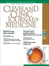
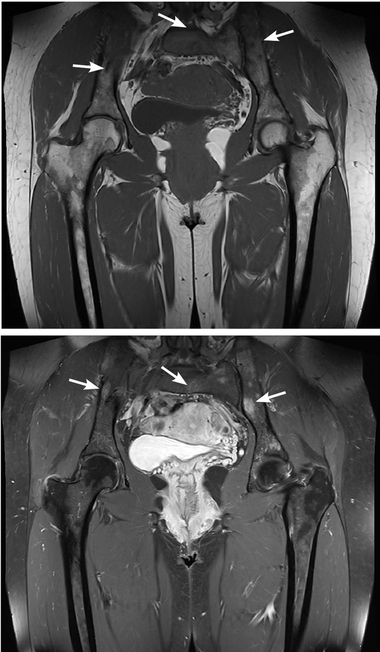
Based on this history, computed tomography of the thorax and abdomen was performed and showed mediastinal and hilar lymphadenopathy, small bilateral lung nodules, and osseous cystic areas in both iliac blades. Magnetic resonance imaging (MRI) showed numerous discrete lesions in both iliac bones (Figure 1). Biopsy study of iliac bone revealed preserved architecture with no evidence of malignancy or granulomas.

FIGURE 1
  • Download figure
  • Open in new tab
  • Download powerpoint
FIGURE 1

Magnetic resonance imaging revealed low-intensity lesions (arrows) on T1-weighted sequences (upper panel) and high-intensity lesions on short tau inversion recovery sequences (lower panel). The lesions have defined borders, no cortical disruption, and no mass effect, features suggesting that the lesions are benign. Lack of signal homogeneity suggests chronic lesions.

Radiography of the hands showed an osseous lytic lesion in the third proximal phalanx of the right hand. No other radiographic abnormalities were noted.

The clinical and radiographic features and the patient’s clinical course were consistent with osseous sarcoidosis. She was started on methotrexate and a low-dose corticosteroid and was symptom-free at 12-month follow-up. Follow-up MRI showed reduction in the lymphadenopathies and stabilization of the bone lesions.

SARCOIDOSIS AND BONE

Sarcoidosis is a systemic granulomatous disease that involves the lung in more than 90% of cases. Skeletal involvement has been reported in 1% to 14% of patients.1,2 Typical osseous involvement is cystic osteitis of the phalangeal bones of the hands and feet, but any part of the skeleton may be involved.3

Bone sarcoidosis is usually asymptomatic and is discovered incidentally. The diagnosis of sarcoidosis has usually been established clinically before bone lesions are detected on MRI. However, sarcoidosis-related bone lesions resembling bone metastases on MRI may be the initial presentation. The presence of intralesional fat has been described as a feature that excludes malignancy.

No treatment has been shown to be of benefit.4 Sarcoidosis is a diagnosis of exclusion and radiographic lytic bone features are not specific, so a neoplastic cause (such as primary osteoblastoma, metastasis, or multiple myeloma) must always be ruled out, as well as other bone conditions such as osteomyelitis or bone cyst.

  • Copyright © 2017 The Cleveland Clinic Foundation. All Rights Reserved.

REFERENCES

  1. ↵
    1. Valeyre D,
    2. Prasse A,
    3. Nunes H,
    4. Uzunhan Y,
    5. Brillet PY,
    6. Müller-Quernheim J
    . Sarcoidosis. Lancet 2014; 383:1155–1167.
    OpenUrlCrossRefPubMed
  2. ↵
    1. James DG,
    2. Neville E,
    3. Carstairs LS
    . Bone and joint sarcoidosis. Semin Arthritis Rheum 1976; 6:53–81.
    OpenUrlCrossRefPubMed
  3. ↵
    1. Moore SL,
    2. Kransdorf MJ,
    3. Schweitzer ME,
    4. Murphey MD,
    5. Babb JS
    . Can sarcoidosis and metastatic bone lesions be reliably differentiated on routine MRI? AJR Am J Roentgenol 2012; 198:1387–1393.
    OpenUrlCrossRefPubMed
  4. ↵
    1. Hamoud S,
    2. Srour S,
    3. Fruchter O,
    4. Vlodavsky E,
    5. Hayek T
    . Lytic bone lesion: presenting finding of sarcoidosis. Isr Med Assoc J 2010; 12:59–60.
    OpenUrlPubMed
PreviousNext
Back to top

In this issue

Cleveland Clinic Journal of Medicine: 84 (10)
Cleveland Clinic Journal of Medicine
Vol. 84, Issue 10
1 Oct 2017
  • Table of Contents
  • Table of Contents (PDF)
  • Index by author
Print
Download PDF
Article Alerts
Sign In to Email Alerts with your Email Address
Email Article

Thank you for your interest in spreading the word on Cleveland Clinic Journal of Medicine.

NOTE: We only request your email address so that the person you are recommending the page to knows that you wanted them to see it, and that it is not junk mail. We do not capture any email address.

Enter multiple addresses on separate lines or separate them with commas.
Sarcoidosis mimicking lytic osseous metastases
(Your Name) has sent you a message from Cleveland Clinic Journal of Medicine
(Your Name) thought you would like to see the Cleveland Clinic Journal of Medicine web site.
CAPTCHA
This question is for testing whether or not you are a human visitor and to prevent automated spam submissions.
Citation Tools
Sarcoidosis mimicking lytic osseous metastases
Anahy M. Brandy-García, Ivan Cabezas-Rodriguez, Luis Caminal-Montero, Carlos Suarez-Cuervo, Pilar Redondo-Buil
Cleveland Clinic Journal of Medicine Oct 2017, 84 (10) 753-754; DOI: 10.3949/ccjm.84a.16108

Citation Manager Formats

  • BibTeX
  • Bookends
  • EasyBib
  • EndNote (tagged)
  • EndNote 8 (xml)
  • Medlars
  • Mendeley
  • Papers
  • RefWorks Tagged
  • Ref Manager
  • RIS
  • Zotero
Share
Sarcoidosis mimicking lytic osseous metastases
Anahy M. Brandy-García, Ivan Cabezas-Rodriguez, Luis Caminal-Montero, Carlos Suarez-Cuervo, Pilar Redondo-Buil
Cleveland Clinic Journal of Medicine Oct 2017, 84 (10) 753-754; DOI: 10.3949/ccjm.84a.16108
Reddit logo Twitter logo Facebook logo Mendeley logo
  • Tweet Widget
  • Facebook Like
  • Linkedin Share Button

Jump to section

  • Article
    • SARCOIDOSIS AND BONE
    • REFERENCES
  • Figures & Data
  • Info & Metrics
  • PDF

Related Articles

  • No related articles found.
  • PubMed
  • Google Scholar

Cited By...

  • No citing articles found.
  • Google Scholar

More in this TOC Section

  • Mass under the lip
  • Oral leukoplakia and oral cancer
  • Median rhomboid glossitis caused by tongue-brushing
Show more The Clinical Picture

Similar Articles

Subjects

  • Rheumatology
  • Pulmonology
  • Imaging

Navigate

  • Current Issue
  • Past Issues
  • Supplements
  • Article Type
  • Specialty
  • CME/MOC Articles
  • CME/MOC Calendar
  • Media Kit

Authors & Reviewers

  • Manuscript Submission
  • Authors & Reviewers
  • Subscriptions
  • About CCJM
  • Contact Us
  • Cleveland Clinic Center for Continuing Education
  • Consult QD

Share your suggestions!

Copyright © 2023 The Cleveland Clinic Foundation. All rights reserved. The information provided is for educational purposes only. Use of this website is subject to the website terms of use and privacy policy. 

Powered by HighWire