
Main menu

  • Home
  • Content
    • Current Issue
    • Ahead of Print
    • Past Issues
    • Supplements
    • Article Type
  • Specialty
    • Articles by Specialty
  • CME/MOC
    • Articles
    • Calendar
  • Info For
    • Manuscript Submission
    • Authors & Reviewers
    • Subscriptions
    • About CCJM
    • Contact Us
    • Media Kit
  • Conversations with Leaders
  • Conference Coverage
    • Kidney Week 2024
    • CHEST 2024
    • ACR Convergence 2023
    • Kidney Week 2023
    • ObesityWeek 2023
    • IDWeek 2023
    • CHEST 2023
    • MDS 2023
    • IAS 2023
    • ACP 2023
    • AAN 2023
    • ACC / WCC 2023
    • AAAAI Meeting 2023
    • ACR Convergence 2022
    • Kidney Week 2022
    • AIDS 2022
  • Other Publications
    • www.clevelandclinic.org

User menu

  • Register
  • Log in

Search

  • Advanced search
Cleveland Clinic Journal of Medicine
  • Other Publications
    • www.clevelandclinic.org
  • Register
  • Log in
Cleveland Clinic Journal of Medicine

Advanced Search

  • Home
  • Content
    • Current Issue
    • Ahead of Print
    • Past Issues
    • Supplements
    • Article Type
  • Specialty
    • Articles by Specialty
  • CME/MOC
    • Articles
    • Calendar
  • Info For
    • Manuscript Submission
    • Authors & Reviewers
    • Subscriptions
    • About CCJM
    • Contact Us
    • Media Kit
  • Conversations with Leaders
  • Conference Coverage
    • Kidney Week 2024
    • CHEST 2024
    • ACR Convergence 2023
    • Kidney Week 2023
    • ObesityWeek 2023
    • IDWeek 2023
    • CHEST 2023
    • MDS 2023
    • IAS 2023
    • ACP 2023
    • AAN 2023
    • ACC / WCC 2023
    • AAAAI Meeting 2023
    • ACR Convergence 2022
    • Kidney Week 2022
    • AIDS 2022
The Clinical Picture

Iliopsoas abscess

Yu Li, MD, Hiraku Funakoshi, MD, MPH, PhD, Takashi Shiga, MD, MPH and Shigeki Fujitani, MD, PhD
Cleveland Clinic Journal of Medicine November 2017, 84 (11) 833-834; DOI: https://doi.org/10.3949/ccjm.84a.17002
Yu Li
Department of Emergency and Critical Care Medicine, Tokyo Bay Urayasu Ichikawa Medical Center, Chiba, Japan
  • Find this author on Google Scholar
  • Find this author on PubMed
  • Search for this author on this site
  • For correspondence: [email protected]
Hiraku Funakoshi
Department of Emergency and Critical Care Medicine, Tokyo Bay Urayasu Ichikawa Medical Center, Chiba, Japan
  • Find this author on Google Scholar
  • Find this author on PubMed
  • Search for this author on this site
Takashi Shiga
Department of Emergency and Critical Care Medicine, Tokyo Bay Urayasu Ichikawa Medical Center, Chiba, Japan
  • Find this author on Google Scholar
  • Find this author on PubMed
  • Search for this author on this site
Shigeki Fujitani
Department of Emergency and Critical Care Medicine, Tokyo Bay Urayasu Ichikawa Medical Center, Chiba, Japan
  • Find this author on Google Scholar
  • Find this author on PubMed
  • Search for this author on this site
  • Article
  • Figures & Data
  • Info & Metrics
  • PDF
Loading

A 52-year-old woman with diabetes mellitus presented with a 1-month history of pain in the right lower abdomen and right back. Although she had a fever when the pain started and her pain was aggravated by walking, her pain and fever had gotten better after taking antibiotics prescribed earlier.

On physical examination, a tender mass with slight warmth was felt in the right lower quadrant. Laboratory testing revealed an active inflammatory reaction: the white blood cell count was 54.8 × 10⁹/L (reference range 4.5–11.0), and the C-reactive protein level was 35.40 mg/dL (reference range < 0.9). Computed tomography showed an abscess in the iliopsoas muscle (Figures 1 and 2), with no evidence of pyogenic spondylitis or other vertebral involvement.

FIGURE 1
  • Download figure
  • Open in new tab
  • Download powerpoint
FIGURE 1

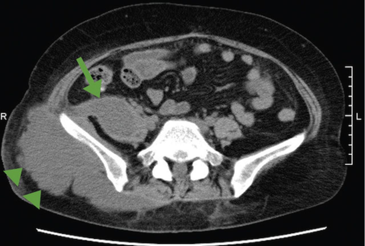
Computed tomography (horizontal plane) showed a low-density area 7 × 4 cm in the right psoas muscle (arrow) and a low-density area 16 × 6 cm in subcutaneous tissue connected to the psoas muscle (arrowheads).

FIGURE 2
  • Download figure
  • Open in new tab
  • Download powerpoint
FIGURE 2

Computed tomography (coronal section) showed a low-density area 16 × 4 cm in the right psoas muscle (arrow), and a low-density area 11 × 7 cm in subcutaneous tissue connected to the psoas muscle (arrowheads).

The patient was admitted to the hospital for percutaneous drainage, which produced 26 mL of pus on the first day and 320 mL on the next day; culture was positive for Escherichia coli. Urine culture was also positive for E coli; blood culture was not. We concluded that these results were secondary to pyelonephritis.

We started intravenous piperacillin-tazobactam 2.25 g every 8 hours for empiric therapy. We changed this to oral ampicillin-cloxacillin 2 g/day after E coli was cultured and pyelonephritis was suspected. The patient was discharged after a 2-week hospital stay, with no significant complications.

ILIOPSOAS ABSCESS: DIAGNOSTIC CLUES

Iliopsoas abscess can occur at any age.1–3 Pain is the most common symptom, occurring in more than 90% of patients.1 Fever with temperatures over 38°C is less common at first, found in less than half of patients.1,2

Only 13% of patients with iliopsoas abscess may have a palpable mass on physical examination.1 The psoas sign—a worsening of lower abdominal pain on the affected side with passive extension of the thigh while supine—has a sensitivity of only 24% for iliopsoas abscess; it can also indicate inflammation to the iliopsoas muscle in other conditions such as retrocecal appendicitis.3

Hip flexion deformity can be a helpful diagnostic feature, as 96% of patients with iliopsoas abscess hold the hip in flexion to relieve pain.4 But pain on hip flexion can also occur in conditions such as septic arthritis.4

Inflammatory markers such as erythrocyte sedimentation rate and C-reactive protein may be elevated in all patients with iliopsoas abscess, so if those markers are not elevated, we may have to consider other conditions such as cancer.1 Computed tomography is nearly 100% sensitive for iliopsoas abscess and is the gold standard for diagnosis.3

TREATMENT

Inadequate treatment of iliopsoas abscess raises the risk of relapse and death.3 Drainage and appropriate antibiotic therapy have been shown to be effective.1,3

Iliopsoas abscess can also be secondary to a number of conditions, eg, Crohn disease, appendicitis, intra-abdominal infection, and cancer,5 and the primary condition needs to be addressed. In addition, culture of a secondary abscess is more likely to grow mixed organisms.5

The average size of the abscess is 6 cm. Percutaneous drainage is required if the mass is larger than 3.5 cm.1

TAKE-HOME MESSAGES

Iliopsoas abscess is difficult to diagnose because patients have few specific complaints. Checking for hip flexion deformity and inflammatory markers may help rule out the disease. When iliopsoas abscess is suspected, computed tomography is necessary to confirm the diagnosis. Drainage and appropriate antibiotics are effective treatment.

  • Copyright © 2017 The Cleveland Clinic Foundation. All Rights Reserved.

REFERENCES

  1. ↵
    1. Tabrizian P,
    2. Nguyen SQ,
    3. Greenstein A,
    4. Rajhbeharrysingh U,
    5. Divino CM
    . Management and treatment of iliopsoas abscess. Arch Surg 2009; 144:946–949.
    OpenUrlCrossRefPubMed
  2. ↵
    1. Shields D,
    2. Robinson P,
    3. Crowley TP
    . Iliopsoas abscess—a review and update on the literature. Int J Surg 2012; 10:466–469.
    OpenUrlCrossRefPubMed
  3. ↵
    1. Huang JJ,
    2. Ruaan MK,
    3. Lan RR,
    4. Wang MC
    . Acute pyogenic iliopsoas abscess in Taiwan: clinical features, diagnosis, treatments and out come. J Infect 2000; 40:248–255.
    OpenUrlCrossRefPubMed
  4. ↵
    1. Stefanich RJ,
    2. Moskowitz A
    . Hip flexion deformity secondary to acute pyogenic psoas abscess. Orthop Rev 1987; 16:67–77.
    OpenUrlPubMed
  5. ↵
    1. Ricci MA,
    2. Rose FB,
    3. Meyer KK
    . Pyogenic psoas abscess: worldwide variations in etiology. World J Surg 1986; 10:834–843.
    OpenUrlCrossRefPubMed
PreviousNext
Back to top

In this issue

Cleveland Clinic Journal of Medicine: 84 (11)
Cleveland Clinic Journal of Medicine
Vol. 84, Issue 11
1 Nov 2017
  • Table of Contents
  • Table of Contents (PDF)
  • Index by author
Print
Download PDF
Article Alerts
Sign In to Email Alerts with your Email Address
Email Article

Thank you for your interest in spreading the word on Cleveland Clinic Journal of Medicine.

NOTE: We only request your email address so that the person you are recommending the page to knows that you wanted them to see it, and that it is not junk mail. We do not capture any email address.

Enter multiple addresses on separate lines or separate them with commas.
Iliopsoas abscess
(Your Name) has sent you a message from Cleveland Clinic Journal of Medicine
(Your Name) thought you would like to see the Cleveland Clinic Journal of Medicine web site.
CAPTCHA
This question is for testing whether or not you are a human visitor and to prevent automated spam submissions.
Citation Tools
Iliopsoas abscess
Yu Li, Hiraku Funakoshi, Takashi Shiga, Shigeki Fujitani
Cleveland Clinic Journal of Medicine Nov 2017, 84 (11) 833-834; DOI: 10.3949/ccjm.84a.17002

Citation Manager Formats

  • BibTeX
  • Bookends
  • EasyBib
  • EndNote (tagged)
  • EndNote 8 (xml)
  • Medlars
  • Mendeley
  • Papers
  • RefWorks Tagged
  • Ref Manager
  • RIS
  • Zotero
Share
Iliopsoas abscess
Yu Li, Hiraku Funakoshi, Takashi Shiga, Shigeki Fujitani
Cleveland Clinic Journal of Medicine Nov 2017, 84 (11) 833-834; DOI: 10.3949/ccjm.84a.17002
Twitter logo Facebook logo Mendeley logo
  • Tweet Widget
  • Facebook Like
  • Linkedin Share Button

Jump to section

  • Article
    • ILIOPSOAS ABSCESS: DIAGNOSTIC CLUES
    • TREATMENT
    • TAKE-HOME MESSAGES
    • REFERENCES
  • Figures & Data
  • Info & Metrics
  • PDF

Related Articles

  • No related articles found.
  • PubMed
  • Google Scholar

Cited By...

  • No citing articles found.
  • Google Scholar

More in this TOC Section

  • Tinea incognito
  • Prolonged venous filling time and dependent rubor in a patient with peripheral artery disease
  • Sarcoidosis with diffuse purplish erythematous plaques on the hands
Show more The Clinical Picture

Similar Articles

Subjects

  • Diabetes
  • Imaging
  • Infectious Diseases

Navigate

  • Current Issue
  • Past Issues
  • Supplements
  • Article Type
  • Specialty
  • CME/MOC Articles
  • CME/MOC Calendar
  • Media Kit

Authors & Reviewers

  • Manuscript Submission
  • Authors & Reviewers
  • Subscriptions
  • About CCJM
  • Contact Us
  • Cleveland Clinic Center for Continuing Education
  • Consult QD

Share your suggestions!

Copyright © 2025 The Cleveland Clinic Foundation. All rights reserved. The information provided is for educational purposes only. Use of this website is subject to the website terms of use and privacy policy. 

Powered by HighWire