
Main menu

  • Home
  • Content
    • Current Issue
    • Ahead of Print
    • Past Issues
    • Supplements
    • Article Type
  • Specialty
    • Articles by Specialty
  • CME/MOC
    • Articles
    • Calendar
  • Info For
    • Manuscript Submission
    • Authors & Reviewers
    • Subscriptions
    • About CCJM
    • Contact Us
    • Media Kit
  • Conversations with Leaders
  • Conference Coverage
    • Kidney Week 2024
    • CHEST 2024
    • ACR Convergence 2023
    • Kidney Week 2023
    • ObesityWeek 2023
    • IDWeek 2023
    • CHEST 2023
    • MDS 2023
    • IAS 2023
    • ACP 2023
    • AAN 2023
    • ACC / WCC 2023
    • AAAAI Meeting 2023
    • ACR Convergence 2022
    • Kidney Week 2022
    • AIDS 2022
  • Other Publications
    • www.clevelandclinic.org

User menu

  • Register
  • Log in

Search

  • Advanced search
Cleveland Clinic Journal of Medicine
  • Other Publications
    • www.clevelandclinic.org
  • Register
  • Log in
Cleveland Clinic Journal of Medicine

Advanced Search

  • Home
  • Content
    • Current Issue
    • Ahead of Print
    • Past Issues
    • Supplements
    • Article Type
  • Specialty
    • Articles by Specialty
  • CME/MOC
    • Articles
    • Calendar
  • Info For
    • Manuscript Submission
    • Authors & Reviewers
    • Subscriptions
    • About CCJM
    • Contact Us
    • Media Kit
  • Conversations with Leaders
  • Conference Coverage
    • Kidney Week 2024
    • CHEST 2024
    • ACR Convergence 2023
    • Kidney Week 2023
    • ObesityWeek 2023
    • IDWeek 2023
    • CHEST 2023
    • MDS 2023
    • IAS 2023
    • ACP 2023
    • AAN 2023
    • ACC / WCC 2023
    • AAAAI Meeting 2023
    • ACR Convergence 2022
    • Kidney Week 2022
    • AIDS 2022
The Clinical Picture

Scapular rash and endocrine neoplasia

Muhammed Razmi T, MD, DNB, Debajyoti Chatterjee, MD and Davinder Parsad, MD
Cleveland Clinic Journal of Medicine November 2017, 84 (11) 831-832; DOI: https://doi.org/10.3949/ccjm.84a.16121
Muhammed Razmi T
Senior Resident, Department of Dermatology, Venereology, and Leprology, Postgraduate Institute of Medical Education and Research, Chandigarh, India
  • Find this author on Google Scholar
  • Find this author on PubMed
  • Search for this author on this site
Debajyoti Chatterjee
Senior Resident, Department of Histopathology, Postgraduate Institute of Medical Education and Research, Chandigarh, India
  • Find this author on Google Scholar
  • Find this author on PubMed
  • Search for this author on this site
Davinder Parsad
Professor, Department of Dermatology Venereology, and Leprology, Postgraduate Institute of Medical Education and Research, Chandigarh, India
  • Find this author on Google Scholar
  • Find this author on PubMed
  • Search for this author on this site
  • For correspondence: [email protected]
  • Article
  • Figures & Data
  • Info & Metrics
  • PDF
Loading

A woman in her 30s presented with an itchy skin-colored rash over her left scapular region that had first appeared 8 years earlier. It had started as itchy skin-colored papules that coalesced to a patch and later became hyperpigmented because of repeated scratching.

She had undergone total thyroidectomy for medullary thyroid carcinoma 1 year ago, and the rash had been diagnosed at that time as lichen planus. She was referred to us by her physician for histopathologic confirmation of the lesions. She denied any history of episodic headache or palpitation.

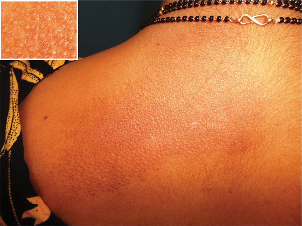
Her blood pressure was 134/86 mm Hg. On physical examination, groups of small hyperpigmented papules were noted over the left scapula (Figure 1).

FIGURE 1
  • Download figure
  • Open in new tab
  • Download powerpoint
FIGURE 1

As seen in the inset, the skin-colored to hyperpigmented plane-topped papules coalesced to form a plaque over the left scapular area.

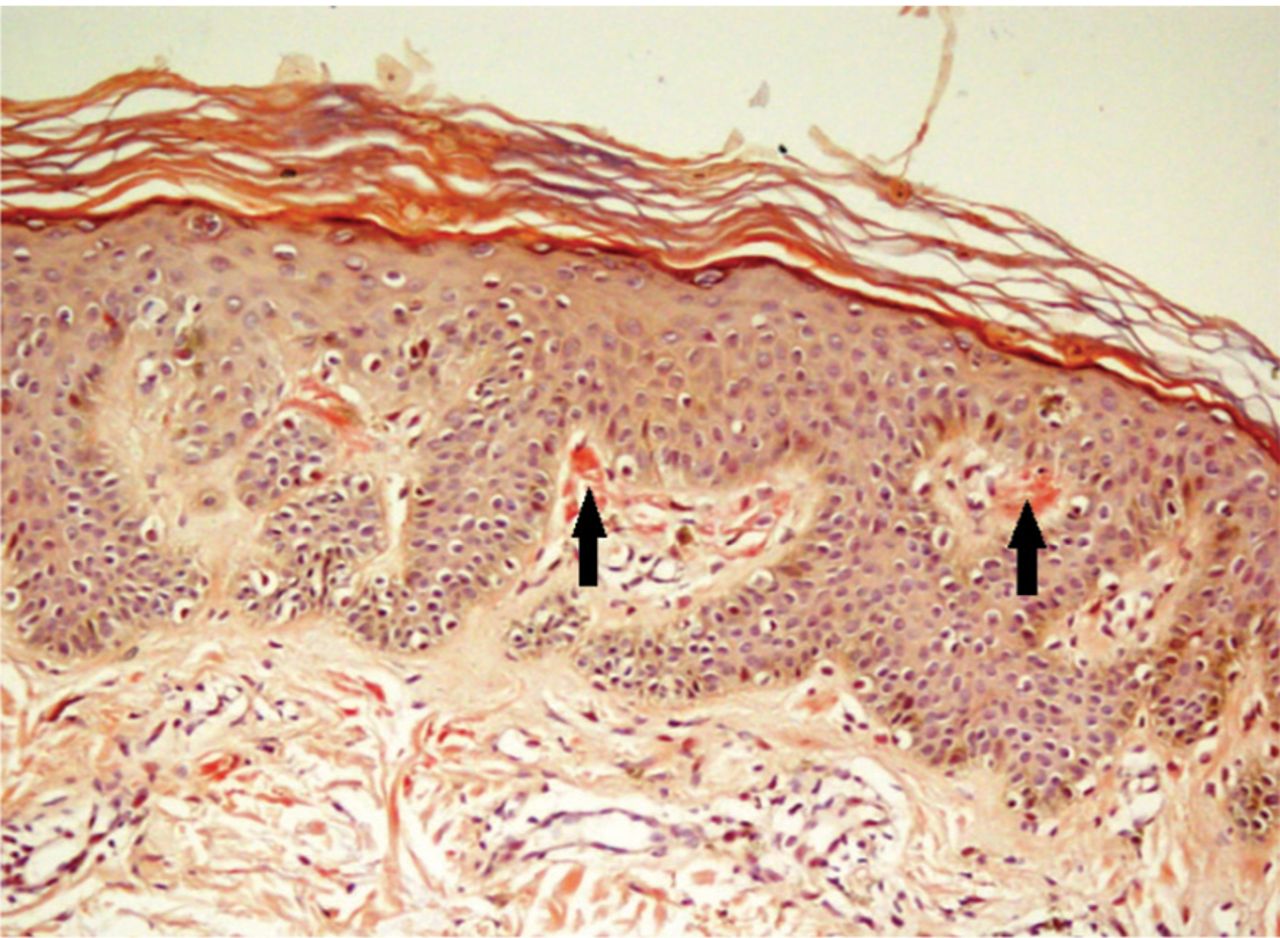
Histopathologic study of a biopsy sample revealed focal degeneration of the basal cell layer with pigment incontinence and deposition of eosinophilic hyaline material along the tip of the papillary dermis, which was confirmed to be amyloid (Figure 2).

FIGURE 2
  • Download figure
  • Open in new tab
  • Download powerpoint
FIGURE 2

Biopsy study of the rash showed congophilic hyaline material along the tip of the papillary dermis (arrows). The apple-green birefringence on polarization confirmed the material to be amyloid (Congo red, × 200).

Her urine normetanephrine excretion was elevated at 1,425 μg/day (reference range 148–560), and her metanephrine excretion was also high at 2,024 μg/day (reference range 44–261).

Contrast-enhanced computed tomography of the abdomen revealed a right adrenal mass (Figure 3). Biopsy study of the mass confirmed pheochromocytoma, a manifestation of multiple endocrine neoplasia (MEN) type 2A.

FIGURE 3
  • Download figure
  • Open in new tab
  • Download powerpoint
FIGURE 3

Computed tomography of the abdomen with contrast showed a heterogeneously enhancing lesion (arrows) in the right suprarenal area that measured 6.5 × 5.5 × 3.5 cm and displaced the inferior vena cava anteriorly.

At a 3-month follow-up visit, the woman’s skin lesions had improved with twice-a-day application of mometasone 0.1% cream; she was lost to follow-up after that visit.

MULTIPLE ENDOCRINE NEOPLASIA

Our patient’s scapular lesions and first-degree family history of MEN type 2A confirmed the diagnosis of the newly recognized variant, MEN type 2A-related cutaneous lichen amyloidosis, in which the characteristic pigmented scapular rash typically predates the first diagnosis of neoplasia.1 The dermal amyloidosis is caused by deposition of keratinlike peptides rather than calcitoninlike peptides.2

A recent systematic review on MEN type 2A with cutaneous lichen amyloidosis showed a female preponderance and a high penetrance of cutaneous lichen amyloidosis, which was the second most frequent manifestation of the syndrome, preceded only by medullary thyroid carcinoma.1

As in our patient’s case, scapular rash and a history of medullary thyroid carcinoma should prompt an investigation for MEN type 2A. These patients should be closely followed for underlying MEN type 2A-related neoplasms.

The mucosal neuromas and skin lipomas seen in MEN type 1 and MEN type 2B are absent in MEN type 2A.3 Cutaneous lichen amyloidosis is the only dermatologic marker for MEN type 2A. Owing to a similar genetic background, cutaneous lichen amyloidosis is also associated with familial medullary thyroid carcinoma, another rare variant of MEN type 2A.4

DIFFERENTIAL DIAGNOSIS

Notalgia paresthetica is a unilateral chronic neuropathic pruritus on the back, mostly located between the shoulders and corresponding to the second and the sixth thoracic nerves. It is mostly attributed to compression of spinal nerves by an abnormality of the thoracic spine.5 In our patient, this was ruled out by the radiologic evaluation.

Before MEN type 2A with cutaneous lichen amyloidosis was recognized as a variant of MEN type 2A, lesions suggestive of notalgia paresthetica were reported with MEN type 2A.3 The classic infrascapular location, history of painful neck muscle spasms, touch hyperesthesia of the lesions, and absence of amyloid deposits on histopathologic study help to differentiate notalgia paresthetica from cutaneous lichen amyloidosis. However, later phases of notalgia paresthetica may show amyloid deposits on histopathologic study, while detection of a scant amount of amyloid is difficult in the early stages of cutaneous lichen amyloidosis.

TAKE-HOME POINT

Cutaneous lichen amyloidosis is usually seen on the extensor surfaces of the extremities. It is considered benign, caused by filamentous degeneration of keratinocytes from repeated scratching. But cutaneous lichen amyloidosis at an early age in the scapular area of women warrants a detailed family history for endocrine neoplasia, blood pressure monitoring, thyroid palpation, and blood testing for serum calcium, calcitonin, and parathyroid hormone.

  • Copyright © 2017 The Cleveland Clinic Foundation. All Rights Reserved.

REFERENCES

  1. ↵
    1. Scapineli JO,
    2. Ceolin L,
    3. Puñales MK,
    4. Dora JM,
    5. Maia AL
    . MEN 2A-related cutaneous lichen amyloidosis: report of three kindred and systematic literature review of clinical, biochemical and molecular characteristics. Fam Cancer 2016; 15:625–633.
    OpenUrlPubMed
  2. ↵
    1. Donovan DT,
    2. Levy ML,
    3. Furst EJ,
    4. et al
    . Familial cutaneous lichen amyloidosis in association with multiple endocrine neoplasia type 2A: a new variant. Henry Ford Hosp Med J 1989; 37:147–150.
    OpenUrlPubMed
  3. ↵
    1. Burns T,
    2. Breathnach S,
    3. Cox N,
    4. Griffiths C
    1. Cox NH,
    2. Coulson IH
    . Systemic disease and the skin. In: Burns T, , Breathnach S, , Cox N, , Griffiths C, eds. Rook’s Textbook of Dermatology. 8th ed. Chichester, UK: John Wiley and Sons Ltd; 2010:62.24.
  4. ↵
    1. Moline J,
    2. Eng C
    . Multiple endocrine neoplasia type 2: an overview. Genet Med 2011; 13:755–764.
    OpenUrlCrossRefPubMed
  5. ↵
    1. Savk O,
    2. Savk E
    . Investigation of spinal pathology in notalgia paresthetica. J Am Acad Dermatol 2005; 52:1085–1087.
    OpenUrlCrossRefPubMed
PreviousNext
Back to top

In this issue

Cleveland Clinic Journal of Medicine: 84 (11)
Cleveland Clinic Journal of Medicine
Vol. 84, Issue 11
1 Nov 2017
  • Table of Contents
  • Table of Contents (PDF)
  • Index by author
Print
Download PDF
Article Alerts
Sign In to Email Alerts with your Email Address
Email Article

Thank you for your interest in spreading the word on Cleveland Clinic Journal of Medicine.

NOTE: We only request your email address so that the person you are recommending the page to knows that you wanted them to see it, and that it is not junk mail. We do not capture any email address.

Enter multiple addresses on separate lines or separate them with commas.
Scapular rash and endocrine neoplasia
(Your Name) has sent you a message from Cleveland Clinic Journal of Medicine
(Your Name) thought you would like to see the Cleveland Clinic Journal of Medicine web site.
CAPTCHA
This question is for testing whether or not you are a human visitor and to prevent automated spam submissions.
Citation Tools
Scapular rash and endocrine neoplasia
Muhammed Razmi T, Debajyoti Chatterjee, Davinder Parsad
Cleveland Clinic Journal of Medicine Nov 2017, 84 (11) 831-832; DOI: 10.3949/ccjm.84a.16121

Citation Manager Formats

  • BibTeX
  • Bookends
  • EasyBib
  • EndNote (tagged)
  • EndNote 8 (xml)
  • Medlars
  • Mendeley
  • Papers
  • RefWorks Tagged
  • Ref Manager
  • RIS
  • Zotero
Share
Scapular rash and endocrine neoplasia
Muhammed Razmi T, Debajyoti Chatterjee, Davinder Parsad
Cleveland Clinic Journal of Medicine Nov 2017, 84 (11) 831-832; DOI: 10.3949/ccjm.84a.16121
Twitter logo Facebook logo Mendeley logo
  • Tweet Widget
  • Facebook Like
  • Linkedin Share Button

Jump to section

  • Article
    • MULTIPLE ENDOCRINE NEOPLASIA
    • DIFFERENTIAL DIAGNOSIS
    • TAKE-HOME POINT
    • REFERENCES
  • Figures & Data
  • Info & Metrics
  • PDF

Related Articles

  • No related articles found.
  • PubMed
  • Google Scholar

Cited By...

  • No citing articles found.
  • Google Scholar

More in this TOC Section

  • Tinea incognito
  • Prolonged venous filling time and dependent rubor in a patient with peripheral artery disease
  • Sarcoidosis with diffuse purplish erythematous plaques on the hands
Show more The Clinical Picture

Similar Articles

Subjects

  • Dermatology
  • Endocrinology
  • Imaging
  • Oncology
  • Women's Health

Navigate

  • Current Issue
  • Past Issues
  • Supplements
  • Article Type
  • Specialty
  • CME/MOC Articles
  • CME/MOC Calendar
  • Media Kit

Authors & Reviewers

  • Manuscript Submission
  • Authors & Reviewers
  • Subscriptions
  • About CCJM
  • Contact Us
  • Cleveland Clinic Center for Continuing Education
  • Consult QD

Share your suggestions!

Copyright © 2025 The Cleveland Clinic Foundation. All rights reserved. The information provided is for educational purposes only. Use of this website is subject to the website terms of use and privacy policy. 

Powered by HighWire