
Main menu

  • Home
  • Content
    • Current Issue
    • Ahead of Print
    • Past Issues
    • Supplements
    • Article Type
  • Specialty
    • Articles by Specialty
  • CME/MOC
    • Articles
    • Calendar
  • Info For
    • Manuscript Submission
    • Authors & Reviewers
    • Subscriptions
    • About CCJM
    • Contact Us
    • Media Kit
  • Conversations with Leaders
  • Conference Coverage
    • Kidney Week 2024
    • CHEST 2024
    • ACR Convergence 2023
    • Kidney Week 2023
    • ObesityWeek 2023
    • IDWeek 2023
    • CHEST 2023
    • MDS 2023
    • IAS 2023
    • ACP 2023
    • AAN 2023
    • ACC / WCC 2023
    • AAAAI Meeting 2023
    • ACR Convergence 2022
    • Kidney Week 2022
    • AIDS 2022
  • Other Publications
    • www.clevelandclinic.org

User menu

  • Register
  • Log in

Search

  • Advanced search
Cleveland Clinic Journal of Medicine
  • Other Publications
    • www.clevelandclinic.org
  • Register
  • Log in
Cleveland Clinic Journal of Medicine

Advanced Search

  • Home
  • Content
    • Current Issue
    • Ahead of Print
    • Past Issues
    • Supplements
    • Article Type
  • Specialty
    • Articles by Specialty
  • CME/MOC
    • Articles
    • Calendar
  • Info For
    • Manuscript Submission
    • Authors & Reviewers
    • Subscriptions
    • About CCJM
    • Contact Us
    • Media Kit
  • Conversations with Leaders
  • Conference Coverage
    • Kidney Week 2024
    • CHEST 2024
    • ACR Convergence 2023
    • Kidney Week 2023
    • ObesityWeek 2023
    • IDWeek 2023
    • CHEST 2023
    • MDS 2023
    • IAS 2023
    • ACP 2023
    • AAN 2023
    • ACC / WCC 2023
    • AAAAI Meeting 2023
    • ACR Convergence 2022
    • Kidney Week 2022
    • AIDS 2022
Review

Cardiopulmonary exercise testing: A contemporary and versatile clinical tool

Kenneth Leclerc, MD, FACC, FACSM
Cleveland Clinic Journal of Medicine February 2017, 84 (2) 161-168; DOI: https://doi.org/10.3949/ccjm.84a.15013
Kenneth Leclerc
Department of Cardiology, Legacy Medical Group, Meridian Park, Tualatin, OR
  • Find this author on Google Scholar
  • Find this author on PubMed
  • Search for this author on this site
  • For correspondence: [email protected]
  • Article
  • Figures & Data
  • Info & Metrics
  • PDF
Loading

ABSTRACT

Cardiopulmonary exercise testing (CPET) helps in detecting disorders of the cardiovascular, pulmonary, and skeletal muscle systems. It has a class I (indicated) recommendation from the American College of Cardiology and American Heart Association for evaluating exertional dyspnea of uncertain cause and for evaluating cardiac patients being considered for heart transplant. Advances in hardware and software and ease of use have brought its application into the clinical arena to the point that providers should become familiar with it and consider it earlier in the evaluation of their patients.

KEY POINTS
  • Technological advances and ease of use have brought CPET out of specialized centers and into the realm of daily clinical practice.

  • CPET is a versatile test that has unique ability to assess cardiopulmonary and metabolic responses to exercise that can reflect underlying pathology.

  • CPET has established value in assessing patients with exertional dyspnea and can guide clinical decision-making and help streamline patient management by focusing on the cause or excluding pathology.

  • CPET has useful prognostic capabilities in patients with heart failure to guide medical treatment or referral for advanced therapies.

Cardiopulmonary exercise testing (CPET) is a versatile tool that can be useful in patient management and clinical decision-making. Many physicians are unfamiliar with it, in part because historically it was cumbersome, done mostly in research or exercise physiology centers, and used mostly in assessing athletic fitness rather than pathologic conditions. In addition, medical schools provide little instruction about it, and hands-on use has typically been relegated to pulmonologists.

Improvements in hardware and software and ease of use have brought this test into the clinical arena to the point that clinicians should consider it earlier in the evaluation of appropriate patients. It now has a class I recommendation (ie, the test is indicated) from the American College of Cardiology and American Heart Association for evaluating exertional dyspnea of uncertain cause and for evaluating cardiac patients being considered for transplant.1 It also is a powerful prognosticator of outcomes in heart failure patients.

CARDIOPULMONARY EXERCISE TESTING MADE SIMPLE

CPET is the analysis of gas exchange during exercise. Modern systems measure, breath-by-breath, the volume of oxygen taken up (Vo2), and the volumes of carbon dioxide (Vco2) and air expired (VE).

Testing can be done with nearly any kind of exercise (treadmill, cycle, arm ergometry), thus accommodating patient or provider preference. Most exercise protocols involve a gradual increase in work rather than increasing stages of work for smooth data collection, and graphical display for optimal test interpretation.

After undergoing baseline screening spirometry, the patient rides a stationary bicycle or walks on a treadmill while breathing through a nonrebreathing mask and wearing electrocardiographic leads, a blood pressure cuff, and a pulse oximeter. The test starts out easy and gets progressively harder until the patient fatigues, reaches his or her predicted peak Vo2, or, as in any stress test, experiences any other clinical indication for stopping, such as arrhythmias, hypotension, or symptoms (rare). We advise patients to wear comfortable workout clothes, and we ask them to try as hard as they can. The test takes about 10 to 15 minutes. Patients are instructed to take all of their usual medications, including beta-blockers, unless advised otherwise at the discretion of the supervising physician.

What the numbers mean

Table 1 lists common CPET variables; Table 2 lists common patterns of results and what they suggest. Other reviews further discuss disease-specific CPET patterns.2–5

View this table:
  • View inline
  • View popup
TABLE 1

Selected cardiopulmonary exercise testing variables

View this table:
  • View inline
  • View popup
TABLE 2

What cardiopulmonary exercise test patterns suggest

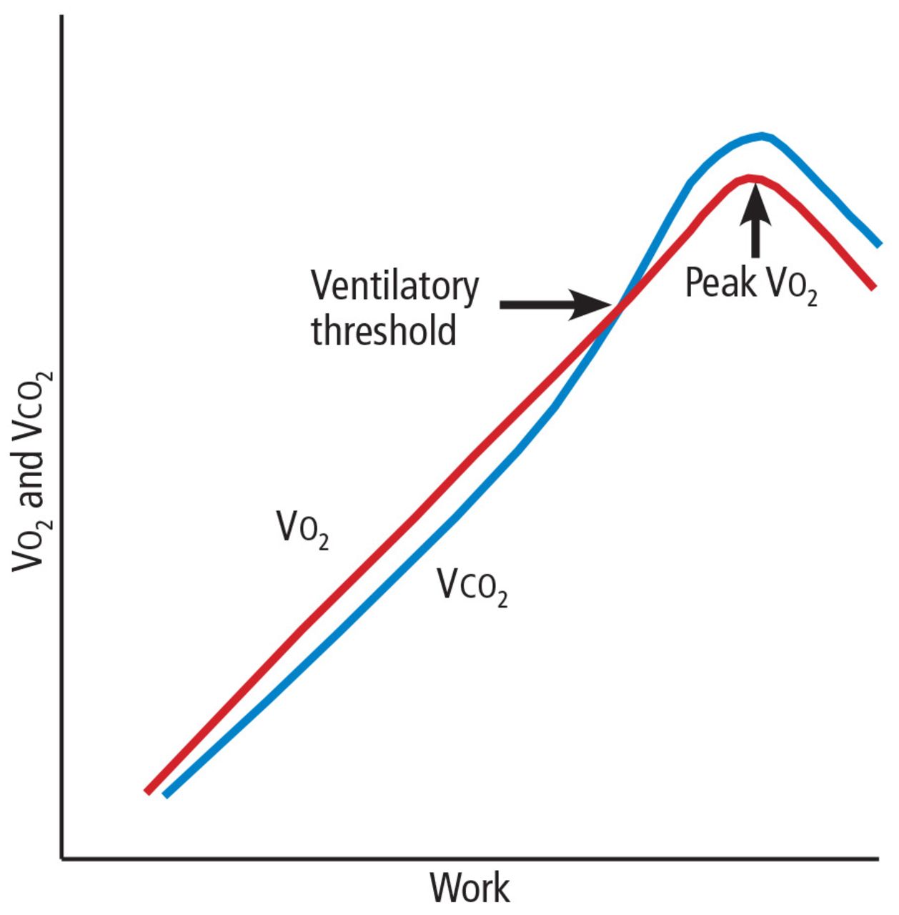
Peak Vo2. As the level of work increases, the body needs more oxygen, and oxygen consumption (Vo2) increases in a linear fashion up to a peak value (Figure 1). Peak Vo2 is the central variable in CPET. Whereas elite athletes have high peak Vo2 values, patients with exercise impairment from any cause have lower values, and average adults typically have results in the middle. Peak Vo2 can be expressed in absolute terms as liters of oxygen per minute, in indexed terms as milliliters of oxygen per kilogram of body weight per minute, and as a percentage of the predicted value.

FIGURE 1
  • Download figure
  • Open in new tab
  • Download powerpoint
FIGURE 1

Diagram of response to work. Impairment from any cause will lower the peak Vo2 and ventilatory threshold.

Ventilatory threshold. Before people reach their peak Vo2, they reach a point where the work demand on the muscles exceeds the oxygen that is being delivered to them, and their metabolism becomes more anaerobic. This point is called the anaerobic threshold, or more precisely the ventilatory threshold. In states of deconditioning or disease, this threshold is often lower than predicted. It can be detected either directly by measuring blood lactate levels or, more often, indirectly from the Vo2, Vco2, and Ve data (Figure 2).

FIGURE 2
  • Download figure
  • Open in new tab
  • Download powerpoint
FIGURE 2

One method of determining the ventilatory threshold is to determine the intersection of the Ve/Vo2 and Vco2 curves.

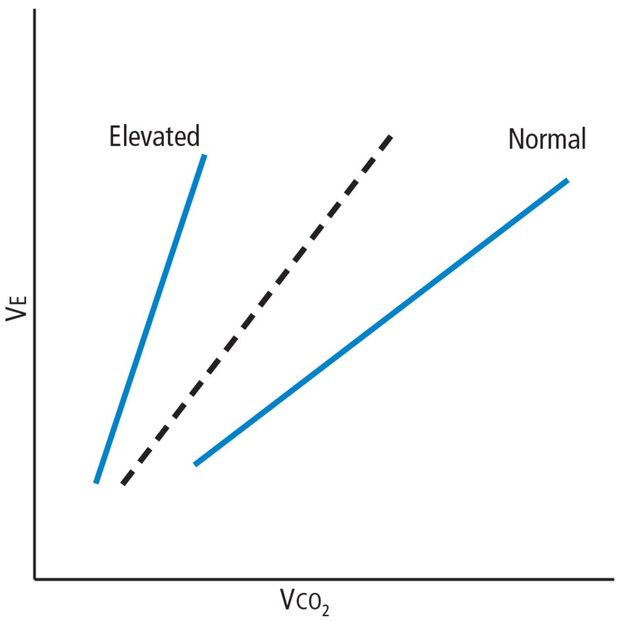
Ve/Vco2 slope. As exercise impairment advances, ventilatory efficiency worsens. Put simply, the demands of exercise result in greater ventilatory effort at any given level of work. This is a consequence of ventilation-perfusion mismatching from a milieu of metabolic, ventilatory, and cardiac dysregulation that accompanies advanced cardiopulmonary or metabolic disease.6,7 The most validated CPET variable reflecting this is the minute ventilation-carbon dioxide relationship (Ve/ Vco2 slope) (Figure 3).

FIGURE 3
  • Download figure
  • Open in new tab
  • Download powerpoint
FIGURE 3

The Ve/Vco2 slope is elevated in advanced heart failure and other hemodynamically significant cardiopulmonary conditions.

Coupled with other common CPET variables and measures such as screening spirometry, electrocardiography, heart and respiratory rate responses, pulse oximetry, and blood pressure, the Ve/Vco2 allows for a detailed and integrated assessment of exercise performance.

USING CPET TO EVALUATE EXERTIONAL DYSPNEA

Shortness of breath, particularly with exertion, is a common reason patients are referred to internists, pulmonologists, and cardiologists. It is a nonspecific symptom for which a precise cause can be elusive. Possible causes range from physical deconditioning due to obesity to new or progressive cardiopulmonary or muscular disease.

If conventional initial studies such as standard exercise testing, echocardiography, or spirometry do not definitively identify the problem, CPET can help guide additional investigation or management. Any abnormal patterns seen, together with the patient’s clinical context and other test results, can give direction to additional evaluation.

Table 2 outlines various CPET patterns that can suggest clinically significant cardiac, pulmonary, or muscle disorders.8–13 Alternatively, normal responses reassure the patient and clinician, since they suggest the patient does not have clinically significant disease.

Case 1: Obesity and dyspnea

You evaluate a 53-year-old mildly obese man for dyspnea. Cardiology evaluation 1 year earlier included normal transthoracic and stress echocardiograms. He is referred for CPET.

His peak Vo2 is low in indexed terms (22.3 mL/kg/min; 74% of predicted) but 90% of predicted in absolute terms (2.8 L/min), reflecting the contribution of his obesity. His ventilatory threshold is near the lower end of normal (50% of peak Vo2), and all other findings are normal. You conclude his dyspnea is due to deconditioning and obesity.

Case 2: Diastolic dysfunction

You follow a normal-weight 65-year-old woman who has long-standing exertional dyspnea. Evaluation 1 year ago included an echocardiogram showing a normal left ventricular ejection fraction and grade II (moderate) diastolic dysfunction, a normal exercise stress test (details were not provided), normal pulmonary function testing, and high-resolution computed tomography of the chest. She too is referred for CPET.

The findings include mild sinus tachycardia at rest and low peak Vo2 (23.7 mL/kg/ min; 69% of predicted). The Ve/Vco2 slope is substantially elevated at 43. Other measures of cardiopulmonary impairment and ventilatory inefficiency such as the end-tidal Pco2 response, oxygen uptake efficiency slope, and oxygen-pulse relationship (O2-pulse, a surrogate for stroke volume) are also abnormal. In clinical context this suggests diastolic dysfunction or unappreciated pulmonary hypertension. You refer her for right heart catheterization, which confirms findings consistent with diastolic dysfunction.

Case 3: Systemic sclerosis

A 64-year-old woman with systemic sclerosis, hypertension, diabetes, and sleep apnea is referred for CPET evaluation of dyspnea. Echocardiography 6 months ago showed a normal left ventricular ejection fraction and moderate diastolic dysfunction.

She undergoes screening spirometry. Results are abnormal and suggest restrictive disease, borderline-low breathing reserve, and low peak Vo2 (20 mL/kg/min; 71% of predicted). She also has chronotropic incompetence (peak heart rate 105 beats per minute; 67% of predicted). These findings are thought to be manifestations of her systemic sclerosis. You refer her for both pulmonary and electrophysiology consultation.

Case 4: Mitral valve prolapse

A generally healthy 73-year-old woman undergoes echocardiography because of a murmur. Findings reveal mitral valve prolapse and mitral regurgitation, which is difficult to quantify. She is referred for CPET as a noninvasive means of assessing the hemodynamic significance of her mitral regurgitation.

Her overall peak Vo2 is low (15 mL/kg/ min). The Ve/Vco2 slope is elevated at 32 (normal < 30), and end-tidal Pco2 response is also abnormal. The recovery heart rate is also abnormally elevated. Collectively, these findings indicate that her mitral valve regurgitation is hemodynamically significant, and you refer her for mitral valve surgery.

CPET’S ROLE IN HEART FAILURE

Over 2 decades ago, the direct measure of peak Vo2 during exercise was found to be an important prognosticator for patients with advanced heart failure and thus became a conventional measure for stratifying patients most in need of a heart transplant.14 To this day, a peak Vo2 of 14 mL/kg/min remains a prognostic threshold—values this low or less carry a poor prognosis.

Additional CPET variables are prognostically useful, both independently and with each other. Many of them reflect the ventilatory and metabolic inefficiencies that result from the extensive central and peripheral pathophysiology seen in heart failure.7,15–17

An elevated Ve/Vco2 slope is a strong predictor of adverse outcomes for patients with heart failure with either reduced or preserved ejection fraction.18,19 Other recognized prognostic indicators include20–23:

  • Low end-tidal Pco2

  • Exercise oscillatory breathing

Low oxygen uptake efficiency slope. All of these are readily provided in the reports of modern CPET systems. Explanations are in Table 1.

Collectively, these variables are strong predictors of outcomes in heart failure patients in terms of survival, adverse cardiac events, or progression to advanced therapy such as a left ventricular assist device or transplant. A multi-center consortium analyzed CPET results from more than 2,600 systolic heart failure patients and devised a scoring system for predicting outcomes (Table 3). This scoring system is a recommended component of the standard evaluation in patients with advanced heart failure.24

View this table:
  • View inline
  • View popup
TABLE 3

Cardiopulmonary exercise testing scoring system for patients with heart failure

EXERCISE TEST REPORTING

Currently there is no universal reporting format for CPET. Using a systematic approach such as the one proposed by Guazzi et al5 can help assure that abnormal values and patterns in all areas will be identified and incorporated in test interpretation. Table 4 lists suggested components of a CPET report and representative examples.

View this table:
  • View inline
  • View popup
TABLE 4

Suggested components of a cardiopulmonary exercise testing report

OTHER USES OF EXERCISE TESTING

CPET has also been found useful in several other clinical conditions that are beyond the scope of this review. These include pulmonary hypertension,25 differentiation of pathologic vs physiologic hypertrophy of the left ventricle,26 preclinical diastolic dysfunction,27,28 congenital heart disease in adults,29 prediction of postoperative complications in bariatric surgery,30 preoperative evaluation for lung resection and pectus excavatum,31,32 hemodynamic impact of mitral regurgitation,33 and mitochondrial myopathies.34

COST-EFFECTIVENESS UNKNOWN

The Current Procedural Terminology code for billing for CPET is 94621 (complex pulmonary stress test). The technical fee is $1,605, and the professional fee is $250. The allowable charges vary according to insurer, but under Medicare A and B, the charges are $258.93 and $70.65, respectively, of which patients typically must copay 20%. Total relative value units are 4.60, of which 1.95 are work relative value units.

The cost-effectiveness of CPET has not been studied. As illustrated in the case examples, patients often undergo numerous tests before CPET. While one might infer that CPET could streamline testing and management if done sooner in disease evaluation, this hypothesis has not been adequately studied, and further research is needed to determine if and how doing so will affect overall costs.

IMPLICATIONS FOR PRACTICE

Newer hardware and software have made CPET more available to practicing clinicians.

CPET has proven value in evaluating patients with exertional dyspnea. If first-line evaluation has not revealed an obvious cause of a patient’s dyspnea, CPET should be considered. This may avoid additional testing or streamline subsequent evaluation and management. CPET also has an established role in risk stratification of those with heart failure.

The clinical application of CPET continues to evolve. Future research will continue to refine its diagnostic and prognostic abilities in a variety of diseases. Most major hospitals and medical centers have CPET capabilities, and interested practitioners should seek out those experienced in test interpretation to increase personal familiarity and to foster appropriate patient referrals.

Manuscript submitted while the author was stationed at San Antonio Military Medical Center, San Antonio, TX.

  • Copyright © 2017 The Cleveland Clinic Foundation. All Rights Reserved.

REFERENCES

  1. ↵
    1. Gibbons RJ,
    2. Balady GJ,
    3. Bricker JT,
    4. et al
    5. American College of Cardiology/American Heart Association Task Force on Practice Guidelines (Committee to Update the 1997 Exercise Testing Guidelines)
    . ACC/ AHA 2002 guideline update for exercise testing: summary article: a report of the American College of Cardiology/American Heart Association Task Force on Practice Guidelines (Committee to Update the 1997 Exercise Testing Guidelines). Circulation 2002; 106:1883–1892.
    OpenUrlFREE Full Text
  2. ↵
    1. American Thoracic Society
    2. American College of Chest Physicians
    . ATS/ACCP statement on cardiopulmonary exercise testing. Am J Respir Crit Care Med 2003; 167:211–277.
    OpenUrlCrossRefPubMed
    1. Mezzani A,
    2. Agostoni P,
    3. Cohen-Solal A,
    4. et al
    . Standards for the use of cardiopulmonary exercise testing for the functional evaluation of cardiac patients: a report from the exercise physiology section of the European Association for Cardiovascular Prevention and Rehabilitation. Eur J Cardiovasc Prev Rehabil 2009; 16:249–267.
    OpenUrlCrossRefPubMed
    1. Balady GJ,
    2. Arena R,
    3. Sietsema K,
    4. et al
    5. American Heart Association Exercise, Cardiac Rehabilitation, and Prevention Committee of the Council on Clinical Cardiology
    6. Council on Epidemiology and Prevention
    7. Council on Peripheral Vascular Disease
    8. Interdisciplinary Council on Quality of Care and Outcomes Research
    . Clinician’s guide to cardiopulmonary exercise testing in adults: a scientific statement from the American Heart Association. Circulation 2010; 122:191–225.
    OpenUrlFREE Full Text
  3. ↵
    1. Guazzi M,
    2. Adams V,
    3. Conraads V,
    4. et al
    5. European Association for Cardiovascular Prevention & Rehabilitation
    6. American Heart Association
    . EACPR/AHA Scientific Statement. Clinical recommendations for cardiopulmonary exercise testing data assessment in specific patient populations. Circulation 2012; 126:2261–2274.
    OpenUrlFREE Full Text
  4. ↵
    1. Wasserman K,
    2. Hansen JE,
    3. Sue DY,
    4. Whipp BJ,
    5. Casaburi R
    . Principles of Exercise Testing and Interpretation: Including Pathophysiology and Clinical Applications. 3rd ed. Baltimore, MD: Lippincott Williams and Wilkins; 1999.
  5. ↵
    1. Lewis GD,
    2. Shah RV,
    3. Pappagianopolas PP,
    4. Systrom DM,
    5. Semigran MJ
    . Determinants of ventilatory efficiency in heart failure: the role of right ventricular performance and pulmonary vascular tone. Circ Heart Fail 2008; 1:227–233.
    OpenUrlAbstract/FREE Full Text
  6. ↵
    1. Wasserman K
    . Diagnosing cardiovascular and lung pathophysiology from exercise gas exchange. Chest 1997; 112:1091–1101.
    OpenUrlCrossRefPubMed
    1. Killian KJ,
    2. Leblanc P,
    3. Martin DH,
    4. Summers E,
    5. Jones NL,
    6. Campbell EJ
    . Exercise capacity and ventilatory, circulatory, and symptom limitation in patients with chronic airflow limitation. Am Rev Respir Dis 1992; 146:935–940.
    OpenUrlCrossRefPubMed
    1. Chaudhry S,
    2. Arena R,
    3. Wasserman K,
    4. et al
    . Exercise-induced myocardial ischemia detected by cardiopulmonary exercise testing. Am J Cardiol 2009; 103:615–619.
    OpenUrlCrossRefPubMed
    1. Tarnopolsky MA,
    2. Raha S
    . Mitochondrial myopathies: diagnosis, exercise intolerance, and treatment options. Med Sci Sports Exerc 2005; 37:2086–2093.
    OpenUrlCrossRefPubMed
    1. Siciliano G,
    2. Volpi L,
    3. Piazza S,
    4. Ricci G,
    5. Mancuso M,
    6. Murri L
    . Functional diagnostics in mitochondrial diseases. Biosci Rep 2007; 27:53–67.
    OpenUrlAbstract/FREE Full Text
  7. ↵
    1. Lorenzo S,
    2. Babb TG
    . Quantification of cardiorespiratory fitness in healthy nonobese and obese men and women. Chest 2012; 141:1031–1039.
    OpenUrlCrossRefPubMed
  8. ↵
    1. Mancini DM,
    2. Eisen H,
    3. Kussmaul W,
    4. Mull R,
    5. Edmunds LH Jr.,
    6. Wilson JR
    . Value of peak exercise oxygen consumption for optimal timing of cardiac transplantation in ambulatory patients with heart failure. Circulation 1991; 83:778–786.
    OpenUrlAbstract/FREE Full Text
  9. ↵
    1. Ponikowski P,
    2. Francis DP,
    3. Piepoli MF,
    4. et al
    . Enhanced ventilatory response to exercise in patients with chronic heart failure and preserved exercise tolerance. Marker of abnormal cardiorespiratory reflex control and predictor of poor prognosis. Circulation 2001; 103:967–972.
    OpenUrlAbstract/FREE Full Text
    1. Levy WC,
    2. Maichel BA,
    3. Steele NP,
    4. Leclerc KM,
    5. Stratton JR
    . Biomechanical efficiency is decreased in heart failure during low-level steady state and maximal ramp exercise. Eur J Heart Fail 2004; 6:917–926.
    OpenUrlCrossRefPubMed
  10. ↵
    1. Poole DC,
    2. Hirai DM,
    3. Copp SW,
    4. Musch TI
    . Muscle oxygen transport and utilization in heart failure: implications for exercise (in)tolerance. Am J Physiol Heart Circ Physiol 2012; 302:H1050–H1063.
    OpenUrlCrossRefPubMed
  11. ↵
    1. Robbins M,
    2. Francis G,
    3. Pashkow FJ,
    4. et al
    . Ventilatory and heart rate responses to exercise: better predictors of heart failure mortality than peak oxygen consumption. Circulation 1999; 100:2411–2417.
    OpenUrlAbstract/FREE Full Text
  12. ↵
    1. Guazzi M,
    2. Myers J,
    3. Arena R
    . Cardiopulmonary exercise testing in the clinical and prognostic assessment of diastolic heart failure. J Am Coll Cardiol 2005; 46:1883–1890.
    OpenUrlFREE Full Text
  13. ↵
    1. Arena R,
    2. Guazzi M,
    3. Myers J
    . Prognostic value of end-tidal carbon dioxide during exercise testing in heart failure. Int J Cardiol 2007; 117:103–108.
    OpenUrlCrossRefPubMed
    1. Leite JJ,
    2. Mansur AJ,
    3. de Freitas HF,
    4. et al
    . Periodic breathing during incremental exercise predicts mortality in patients with chronic heart failure evaluated for cardiac transplantation. J Am Coll Cardiol 2003; 41:2175–2181.
    OpenUrlFREE Full Text
    1. Guazzi M,
    2. Arena R,
    3. Ascione A,
    4. Piepoli M,
    5. Guazzi MD,
    6. Gruppo di Studio Fisiologia dell’Esercizio, Cardiologia dello Sport e Riabilitazione Cardiovascolare of the Italian Society of Cardiology
    . Exercise oscillatory breathing and increased ventilation to carbon dioxide production slope in heart failure: an unfavorable combination with high prognostic value. Am Heart J 2007; 153:859–867.
    OpenUrlCrossRefPubMed
  14. ↵
    1. Davies LC,
    2. Wensel R,
    3. Georgiadou P,
    4. et al
    . Enhanced prognostic value from cardiopulmonary exercise testing in chronic heart failure by non-linear analysis: oxygen uptake efficiency slope. Eur Heart J 2006; 27:684–690.
    OpenUrlCrossRefPubMed
  15. ↵
    1. Myers J,
    2. Oliveira R,
    3. Dewey F,
    4. et al
    . Validation of a cardiopulmonary exercise test score in heart failure. Circ Heart Fail 2013; 6:211–218.
    OpenUrlAbstract/FREE Full Text
  16. ↵
    1. Arena R,
    2. Lavie CJ,
    3. Milani RV,
    4. Myers J,
    5. Guazzi M
    . Cardiopulmonary exercise testing in patients with pulmonary arterial hypertension: an evidence-based review. J Heart Lung Transplant 2010; 29:159–173.
    OpenUrlCrossRefPubMed
  17. ↵
    1. Whyte GP,
    2. Sharma S,
    3. George K,
    4. McKenna WJ
    . Exercise gas exchange responses in the differentiation of pathologic and physiologic left ventricular hypertrophy. Med Sci Sports Exerc 1999; 31:1237–1241.
    OpenUrlCrossRefPubMed
  18. ↵
    1. Wan SH,
    2. Vogel MW,
    3. Chen HH
    . Pre-clinical diastolic dysfunction. J Am Coll Cardiol 2014; 63:407–416.
    OpenUrlFREE Full Text
  19. ↵
    1. Ahmadian H,
    2. Sherratt J,
    3. Lochner K,
    4. duBois M,
    5. Leclerc K
    . Cardiopulmonary exercise testing responses and pro-BNP values in adults with mild degrees of diastolic dysfunction. JARCP J Aging Res Clin Practice 2014; 4:1–3.
    OpenUrl
  20. ↵
    1. Inuzuka R,
    2. Diller GP,
    3. Borgia F,
    4. et al
    . Comprehensive use of cardio pulmonary exercise testing identifies adults with congenital heart disease at increased mortality risk in the medium term. Circulation 2012; 125:250–259.
    OpenUrlAbstract/FREE Full Text
  21. ↵
    1. McCullough PA,
    2. Gallagher MJ,
    3. Dejong AT,
    4. et al
    . Cardiorespiratory fitness and short-term complications after bariatric surgery. Chest 2006; 130:517–525.
    OpenUrlCrossRefPubMed
  22. ↵
    1. Kallianos A,
    2. Rapti A,
    3. Tsimpoukis S,
    4. et al
    . Cardiopulmonary exercise testing (CPET) as preoperative test before lung resection. In Vivo 2014; 28:1013–1020.
    OpenUrlAbstract/FREE Full Text
  23. ↵
    1. Cavestri B,
    2. Wurtz A,
    3. Bart F,
    4. Neviere R,
    5. Augilaniu B,
    6. Wallaert B
    . Cardiopulmonary exercise testing in patients with pectus excavatum. Rev Mal Respir 2010; 27:717–723. French.
    OpenUrlPubMed
  24. ↵
    1. Messika-Zeitoun D,
    2. Johnson BD,
    3. Nkomo V,
    4. et al
    . Cardiopulmonary exercise testing determination of functional capacity in mitral regurgitation. J Am Coll Cardiol 2006; 47:2521–2527.
    OpenUrlFREE Full Text
  25. ↵
    1. Testa M,
    2. Navazio FM,
    3. Neugebauer J
    . Recognition, diagnosis, and treatment of mitochondrial myopathies in endurance athletes. Curr Sports Med Rep 2005; 4:282–287.
    OpenUrlPubMed
PreviousNext
Back to top

In this issue

Cleveland Clinic Journal of Medicine: 84 (2)
Cleveland Clinic Journal of Medicine
Vol. 84, Issue 2
1 Feb 2017
  • Table of Contents
  • Table of Contents (PDF)
  • Index by author
Print
Download PDF
Article Alerts
Sign In to Email Alerts with your Email Address
Email Article

Thank you for your interest in spreading the word on Cleveland Clinic Journal of Medicine.

NOTE: We only request your email address so that the person you are recommending the page to knows that you wanted them to see it, and that it is not junk mail. We do not capture any email address.

Enter multiple addresses on separate lines or separate them with commas.
Cardiopulmonary exercise testing: A contemporary and versatile clinical tool
(Your Name) has sent you a message from Cleveland Clinic Journal of Medicine
(Your Name) thought you would like to see the Cleveland Clinic Journal of Medicine web site.
CAPTCHA
This question is for testing whether or not you are a human visitor and to prevent automated spam submissions.
Citation Tools
Cardiopulmonary exercise testing: A contemporary and versatile clinical tool
Kenneth Leclerc
Cleveland Clinic Journal of Medicine Feb 2017, 84 (2) 161-168; DOI: 10.3949/ccjm.84a.15013

Citation Manager Formats

  • BibTeX
  • Bookends
  • EasyBib
  • EndNote (tagged)
  • EndNote 8 (xml)
  • Medlars
  • Mendeley
  • Papers
  • RefWorks Tagged
  • Ref Manager
  • RIS
  • Zotero
Share
Cardiopulmonary exercise testing: A contemporary and versatile clinical tool
Kenneth Leclerc
Cleveland Clinic Journal of Medicine Feb 2017, 84 (2) 161-168; DOI: 10.3949/ccjm.84a.15013
Twitter logo Facebook logo Mendeley logo
  • Tweet Widget
  • Facebook Like
  • Linkedin Share Button

Jump to section

  • Article
    • ABSTRACT
    • CARDIOPULMONARY EXERCISE TESTING MADE SIMPLE
    • USING CPET TO EVALUATE EXERTIONAL DYSPNEA
    • CPET’S ROLE IN HEART FAILURE
    • EXERCISE TEST REPORTING
    • OTHER USES OF EXERCISE TESTING
    • COST-EFFECTIVENESS UNKNOWN
    • IMPLICATIONS FOR PRACTICE
    • REFERENCES
  • Figures & Data
  • Info & Metrics
  • PDF

Related Articles

  • No related articles found.
  • PubMed
  • Google Scholar

Cited By...

  • No citing articles found.
  • Google Scholar

More in this TOC Section

  • Alpha-gal syndrome: Recognizing and managing a tick-bite–related meat allergy
  • Managing right ventricular failure in the setting of pulmonary embolism
  • Psychedelics, spirituality, and existential distress in patients at the end of life
Show more Review

Similar Articles

Subjects

  • Cardiology
  • Obesity
  • Pulmonology

Navigate

  • Current Issue
  • Past Issues
  • Supplements
  • Article Type
  • Specialty
  • CME/MOC Articles
  • CME/MOC Calendar
  • Media Kit

Authors & Reviewers

  • Manuscript Submission
  • Authors & Reviewers
  • Subscriptions
  • About CCJM
  • Contact Us
  • Cleveland Clinic Center for Continuing Education
  • Consult QD

Share your suggestions!

Copyright © 2025 The Cleveland Clinic Foundation. All rights reserved. The information provided is for educational purposes only. Use of this website is subject to the website terms of use and privacy policy. 

Powered by HighWire