
Main menu

  • Home
  • Content
    • Current Issue
    • Ahead of Print
    • Past Issues
    • Supplements
    • Article Type
  • Specialty
    • Articles by Specialty
  • CME/MOC
    • Articles
    • Calendar
  • Info For
    • Manuscript Submission
    • Authors & Reviewers
    • Subscriptions
    • About CCJM
    • Contact Us
    • Media Kit
  • Conversations with Leaders
  • Conference Coverage
    • Kidney Week 2024
    • CHEST 2024
    • ACR Convergence 2023
    • Kidney Week 2023
    • ObesityWeek 2023
    • IDWeek 2023
    • CHEST 2023
    • MDS 2023
    • IAS 2023
    • ACP 2023
    • AAN 2023
    • ACC / WCC 2023
    • AAAAI Meeting 2023
    • ACR Convergence 2022
    • Kidney Week 2022
    • AIDS 2022
  • Other Publications
    • www.clevelandclinic.org

User menu

  • Register
  • Log in

Search

  • Advanced search
Cleveland Clinic Journal of Medicine
  • Other Publications
    • www.clevelandclinic.org
  • Register
  • Log in
Cleveland Clinic Journal of Medicine

Advanced Search

  • Home
  • Content
    • Current Issue
    • Ahead of Print
    • Past Issues
    • Supplements
    • Article Type
  • Specialty
    • Articles by Specialty
  • CME/MOC
    • Articles
    • Calendar
  • Info For
    • Manuscript Submission
    • Authors & Reviewers
    • Subscriptions
    • About CCJM
    • Contact Us
    • Media Kit
  • Conversations with Leaders
  • Conference Coverage
    • Kidney Week 2024
    • CHEST 2024
    • ACR Convergence 2023
    • Kidney Week 2023
    • ObesityWeek 2023
    • IDWeek 2023
    • CHEST 2023
    • MDS 2023
    • IAS 2023
    • ACP 2023
    • AAN 2023
    • ACC / WCC 2023
    • AAAAI Meeting 2023
    • ACR Convergence 2022
    • Kidney Week 2022
    • AIDS 2022
The Clinical Picture

Dysmorphic red blood cell formation

José Lucas Daza, MD, Marcelo De Rosa, MD and Graciela De Rosa, MD
Cleveland Clinic Journal of Medicine January 2018, 85 (1) 12-13; DOI: https://doi.org/10.3949/ccjm.85a.17029
José Lucas Daza
Division of Nephrology, Department of Internal Medicine, Hospital de Clinicas, University of Buenos Aires, Argentina
  • Find this author on Google Scholar
  • Find this author on PubMed
  • Search for this author on this site
Marcelo De Rosa
Division of Nephrology, Department of Internal Medicine, Hospital de Clinicas, University of Buenos Aires, Argentina
  • Find this author on Google Scholar
  • Find this author on PubMed
  • Search for this author on this site
  • For correspondence: [email protected]
Graciela De Rosa
Department of Pathology, Hospital de Clinicas, University of Buenos Aires, Argentina
  • Find this author on Google Scholar
  • Find this author on PubMed
  • Search for this author on this site
  • Article
  • Figures & Data
  • Info & Metrics
  • PDF
Loading

A 23-year-old woman presented with hematuria. Her blood pressure was normal, and she had no rash, joint pain, or other symptoms. Urinalysis was positive for proteinuria and hematuria, and urinary sediment analysis showed dysmorphic red blood cells (RBCs) and red cell casts, leading to a diagnosis of glomerulonephritis. She had proteinuria of 1.2 g/24 hours. Laboratory tests for systemic diseases were negative. Renal biopsy study revealed stage III immunoglobulin A (IgA) nephropathy.

See related editorial, page 22

GLOMERULAR HEMATURIA

Glomerular hematuria may represent an immune-mediated injury to the glomerular capillary wall, but it can also be present in noninflammatory glomerulopathies.1

The type of dysmorphic RBCs (crenated or misshapen cells, acanthocytes) may be of diagnostic importance. In particular, dysmorphic red cells alone may be predictive of only renal bleeding, while acanthocytes (ring-shaped RBCs with vesicle-shaped protrusions best seen on phase-contrast microscopy) appear to be most predictive of glomerular disease.2 For example, in 1 study,3 the presence of acanthocytes comprising at least 5% of excreted RBCs had a sensitivity of 52% for glomerular disease and a specificity of 98%.3

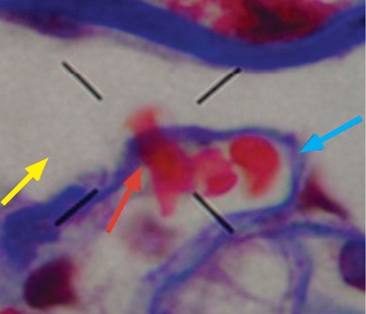
How erythrocytes become dysmorphic is not entirely known.4 In vitro, changes in osmolality or pH do not produce dysmorphism, but it can occur along the different tubular segments.5,6 In addition, RBCs can lose their shape when crossing the glomerular basement membrane (Figure 1) and during passage along the tubular system.4

FIGURE 1
  • Download figure
  • Open in new tab
  • Download powerpoint
FIGURE 1

A red blood cell (red arrow) is seen crossing the basement membrane (blue arrow) and losing its shape, one of the mechanisms proposed for acanthocyte formation; the yellow arrow points to the urinary space (Masson trichrome stain, × 1,000.

Dysmorphic urinary RBCs have been regarded as an indicator of glomerular pathology,7 and dysmorphic RBCs in urinary sediment (Figure 2) are also considered to represent renal pathology. On electron microscopy, the glomerular basement membrane is usually 300 to 400 nm wide, whereas the diameter of the red blood cell is 7 μm. The change in RBC shape is manifested as blebs, budding, and segmental loss of membrane, resulting in marked variability in shape and in a reduction in mean cell size. These changes may be due to mechanical trauma as the cells pass through rents in the glomerular basement membrane and to osmotic trauma as cells flow through the nephron.8

FIGURE 2
  • Download figure
  • Open in new tab
  • Download powerpoint
FIGURE 2

Examination of urinary sediment shows typical dysmorphic hematuria with acanthocytes (× 400).

  • Copyright © 2018 The Cleveland Clinic Foundation. All Rights Reserved.

REFERENCES

  1. ↵
    1. Collar JE,
    2. Ladva S,
    3. Cairns TD,
    4. Cattell V
    . Red cell traverse through thin glomerular basement membranes. Kidney Int 2001; 59:2069–2072.
    OpenUrlCrossRefPubMed
  2. ↵
    1. Fogazzi GB,
    2. Ponticelli C,
    3. Ritz E
    . The Urinary Sediment: An Integrated View. 2nd ed. Oxford: Oxford University Press; 1999:30.
  3. ↵
    1. Köhler H,
    2. Wandel E,
    3. Brunck B
    . Acanthocyturia–a characteristic marker for glomerular bleeding. Kidney Int 1991; 40:115–120.
    OpenUrlCrossRefPubMed
  4. ↵
    1. Fogazzi GB
    . The Urinary Sediment: An Integrated View. 3rd ed. France: Elsevier; 2010.
  5. ↵
    1. Briner VA,
    2. Reinhart WH
    . In vitro production of ‘glomerular red cells’: role of pH and osmolality. Nephron 1990; 56:13–18.
    OpenUrlPubMed
  6. ↵
    1. Schramek P,
    2. Moritsch A,
    3. Haschkowitz H,
    4. Binder BR,
    5. Maier M
    . In vitro generation of dysmorphic erythrocytes. Kidney Int 1989; 36:72–77.
    OpenUrlCrossRefPubMed
  7. ↵
    1. Pollock C,
    2. Liu PL,
    3. Györy AZ, et al
    . Dysmorphism of urinary red blood cells–value in diagnosis. Kidney Int 1989; 36:1045–1049.
    OpenUrlCrossRefPubMed
  8. ↵
    1. Shichiri M,
    2. Hosoda K,
    3. Nishio Y, et al
    . Red-cell-volume distribution curves in diagnosis of glomerular and non-glomerular haematuria. Lancet 1988; 1:908–911.
    OpenUrlPubMed
PreviousNext
Back to top

In this issue

Cleveland Clinic Journal of Medicine: 85 (1)
Cleveland Clinic Journal of Medicine
Vol. 85, Issue 1
1 Jan 2018
  • Table of Contents
  • Table of Contents (PDF)
  • Index by author
Print
Download PDF
Article Alerts
Sign In to Email Alerts with your Email Address
Email Article

Thank you for your interest in spreading the word on Cleveland Clinic Journal of Medicine.

NOTE: We only request your email address so that the person you are recommending the page to knows that you wanted them to see it, and that it is not junk mail. We do not capture any email address.

Enter multiple addresses on separate lines or separate them with commas.
Dysmorphic red blood cell formation
(Your Name) has sent you a message from Cleveland Clinic Journal of Medicine
(Your Name) thought you would like to see the Cleveland Clinic Journal of Medicine web site.
CAPTCHA
This question is for testing whether or not you are a human visitor and to prevent automated spam submissions.
Citation Tools
Dysmorphic red blood cell formation
José Lucas Daza, Marcelo De Rosa, Graciela De Rosa
Cleveland Clinic Journal of Medicine Jan 2018, 85 (1) 12-13; DOI: 10.3949/ccjm.85a.17029

Citation Manager Formats

  • BibTeX
  • Bookends
  • EasyBib
  • EndNote (tagged)
  • EndNote 8 (xml)
  • Medlars
  • Mendeley
  • Papers
  • RefWorks Tagged
  • Ref Manager
  • RIS
  • Zotero
Share
Dysmorphic red blood cell formation
José Lucas Daza, Marcelo De Rosa, Graciela De Rosa
Cleveland Clinic Journal of Medicine Jan 2018, 85 (1) 12-13; DOI: 10.3949/ccjm.85a.17029
Twitter logo Facebook logo Mendeley logo
  • Tweet Widget
  • Facebook Like
  • Linkedin Share Button

Jump to section

  • Article
    • GLOMERULAR HEMATURIA
    • REFERENCES
  • Figures & Data
  • Info & Metrics
  • PDF

Related Articles

  • Quality in urine microscopy: The eyes of the beholder
  • PubMed
  • Google Scholar

Cited By...

  • Quality in urine microscopy: The eyes of the beholder
  • Google Scholar

More in this TOC Section

  • Tinea incognito
  • Prolonged venous filling time and dependent rubor in a patient with peripheral artery disease
  • Sarcoidosis with diffuse purplish erythematous plaques on the hands
Show more The Clinical Picture

Similar Articles

Subjects

  • Allergy/Immunology
  • Nephrology

Navigate

  • Current Issue
  • Past Issues
  • Supplements
  • Article Type
  • Specialty
  • CME/MOC Articles
  • CME/MOC Calendar
  • Media Kit

Authors & Reviewers

  • Manuscript Submission
  • Authors & Reviewers
  • Subscriptions
  • About CCJM
  • Contact Us
  • Cleveland Clinic Center for Continuing Education
  • Consult QD

Share your suggestions!

Copyright © 2025 The Cleveland Clinic Foundation. All rights reserved. The information provided is for educational purposes only. Use of this website is subject to the website terms of use and privacy policy. 

Powered by HighWire