
Main menu

  • Home
  • Content
    • Current Issue
    • Ahead of Print
    • Past Issues
    • Supplements
    • Article Type
  • Specialty
    • Articles by Specialty
  • CME/MOC
    • Articles
    • Calendar
  • Info For
    • Manuscript Submission
    • Authors & Reviewers
    • Subscriptions
    • About CCJM
    • Contact Us
    • Media Kit
  • Conversations with Leaders
  • Conference Coverage
    • ACC / WCC 2023
    • AAAAI Meeting 2023
    • ACR Convergence 2022
    • Kidney Week 2022
    • AIDS 2022
    • CHEST 2021
    • IDWeek 2021
    • IAS 2021
    • ADA 2021
    • ATS 2021
    • ACC 2021
    • ACP 2021
    • AAN 2021
  • Other Publications
    • www.clevelandclinic.org

User menu

  • Register
  • Log in

Search

  • Advanced search
Cleveland Clinic Journal of Medicine
  • Other Publications
    • www.clevelandclinic.org
  • Register
  • Log in
Cleveland Clinic Journal of Medicine

Advanced Search

  • Home
  • Content
    • Current Issue
    • Ahead of Print
    • Past Issues
    • Supplements
    • Article Type
  • Specialty
    • Articles by Specialty
  • CME/MOC
    • Articles
    • Calendar
  • Info For
    • Manuscript Submission
    • Authors & Reviewers
    • Subscriptions
    • About CCJM
    • Contact Us
    • Media Kit
  • Conversations with Leaders
  • Conference Coverage
    • ACC / WCC 2023
    • AAAAI Meeting 2023
    • ACR Convergence 2022
    • Kidney Week 2022
    • AIDS 2022
    • CHEST 2021
    • IDWeek 2021
    • IAS 2021
    • ADA 2021
    • ATS 2021
    • ACC 2021
    • ACP 2021
    • AAN 2021
The Clinical Picture

When stroke runs in the family

Nellowe Candelario, MD and Kevin Bryan Lo, MD
Cleveland Clinic Journal of Medicine October 2018, 85 (10) 743-744; DOI: https://doi.org/10.3949/ccjm.85a.18055
Nellowe Candelario
Department of Internal Medicine, Einstein Medical Center, Philadelphia, PA
  • Find this author on Google Scholar
  • Find this author on PubMed
  • Search for this author on this site
  • For correspondence: candelan@einstein.edu
Kevin Bryan Lo
Department of Internal Medicine, Einstein Medical Center, Philadelphia, PA
  • Find this author on Google Scholar
  • Find this author on PubMed
  • Search for this author on this site
  • Article
  • Figures & Data
  • Info & Metrics
  • PDF
Loading

A 54-year-old man presented to our hospital with acute-onset left-sided weakness and right facial droop. Three days earlier he had also had migraine-like headaches, which he had never experienced before. He also reported a change in behavior during the past week, which his family had described as inappropriate laughter.

He had no history of hypertension, diabetes, or dyslipidemia. He did not smoke or drink alcohol. However, he had an extensive family history of stroke. His mother had a stroke at age 50, his brother a stroke at age 57, and his sister had been admitted for a stroke 1 month earlier at the age of 52.

On examination, he had weakness of the left arm and leg, right facial droop, and hyperactive reflexes on the left side. He had no sensory or cerebellar deficits. He had episodes of laughter during the examination.

Behavioral changes such as inappropriate laughter are nonspecific and can be associated with any subcortical frontoparietal and temporal infarction.

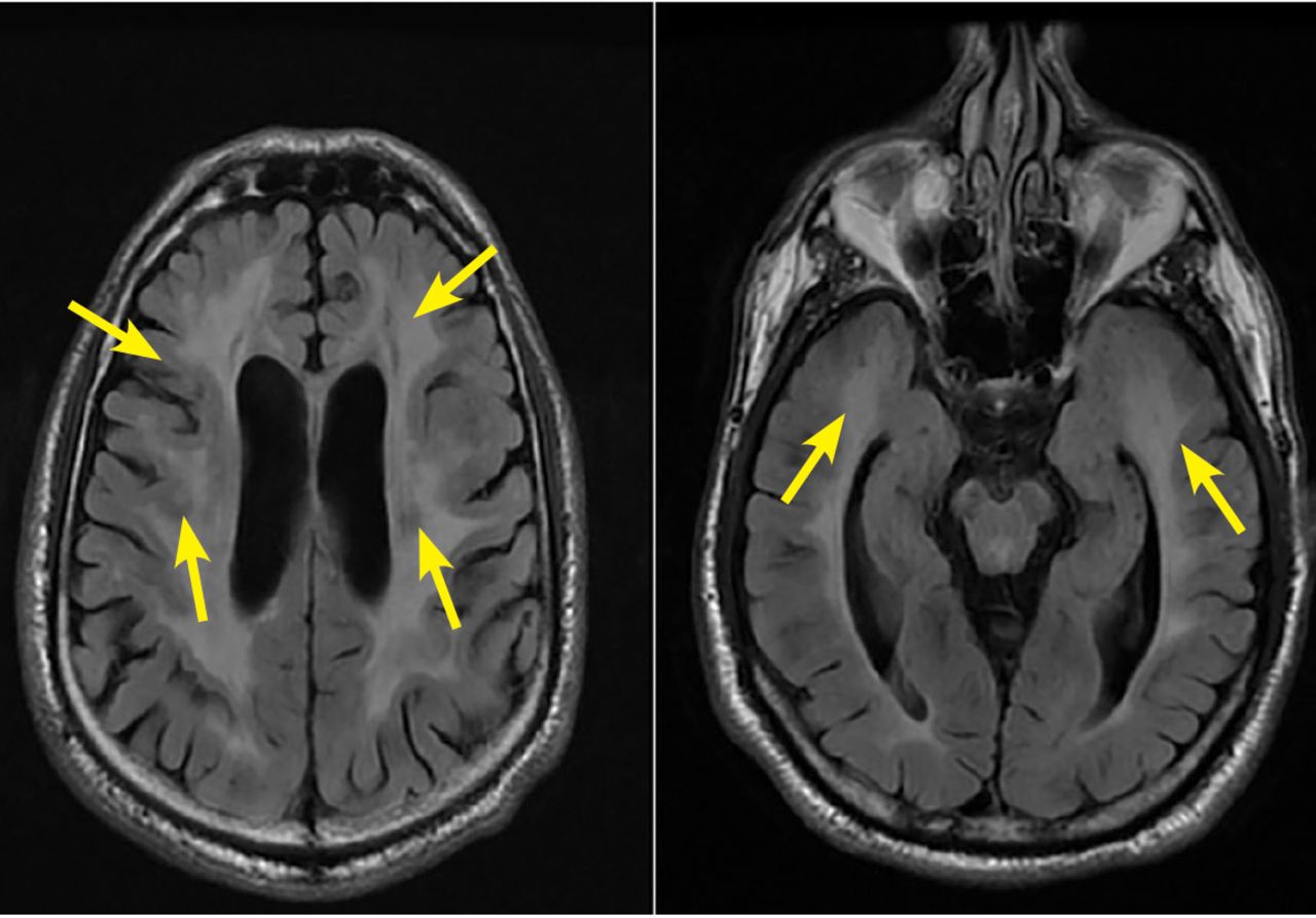
Computed tomography of the brain without contrast (Figure 1) showed patchy confluent areas of low attenuation in both cerebral hemispheres. Magnetic resonance imaging (MRI) showed an 8-mm focus of mild restricted diffusion in the right lateral thalamus, indicating acute infarction, and axial T2 fluid-attenuated inversion recovery images (Figure 2) showed confluent and symmetric white matter hyperintensities in the frontoparietotemporal region. The extent of these white matter changes, especially in the temporal lobe, were not typical of the microvascular disease changes seen in elderly individuals with risk factors for stroke. Microvascular disease may present the same way, but not as extensively and this was not consistent with the patient’s relatively young age. The imaging findings suggested cerebral autosomal dominant arteriopathy with subcortical infarcts and leukoencephalopathy (CADASIL).

Figure 1
  • Download figure
  • Open in new tab
  • Download powerpoint
Figure 1

Computed tomography of the brain without contrast showed patchy confluent areas of low attenuation in both cerebral hemispheres (arrows).

Figure 2
  • Download figure
  • Open in new tab
  • Download powerpoint
Figure 2

Magnetic resonance imaging with axial T2 fluid-attenuated inversion recovery showed confluent and symmetric white matter hyperintensities in the frontoparietotemporal region (arrows). These changes, especially in the temporal lobe, were atypical of microvascular disease given the patient’s age.

We learned that the patient’s sister had undergone a workup showing mutations in the NOTCH3 gene and a skin biopsy study consistent with CADASIL.

Our patient was started on antiplatelet and high-intensity statin therapy. His symptoms improved, and he was discharged to an acute inpatient rehabilitation facility. He was referred to a CADASIL registry.

STROKE AND HEREDITY

CADASIL is a rare hereditary vascular disor der inherited in an autosomal dominant manner. It is the most common inherited form of small-vessel disease and results from a mutation in the NOTCH3 gene that leads to degeneration of smooth muscle in cerebral blood vessels. It can manifest as migraine with aura, vascular dementia, cognitive impairment, or ischemic stroke.

The diagnosis is based on a clinical picture that typically includes stroke at a young age (age 40 to 50) in the absence of stroke risk factors, or frequent lacunar infarction episodes that can manifest as migraine, lacunar infarct, or dementia.1 Some patients, such as ours, may have subtle nonspecific behavioral changes such as inappropriate laughter, which may herald the development of an infarct.

Characteristic findings on MRI are white matter hyperintensities that tend to be bilateral and symmetrical in the periventricular areas. Symmetrical involvement in the temporal lobes has high sensitivity and specificity for CADASIL.2 Biopsy study of the skin, muscle, or sural nerve shows small-vessel changes that include thickening of the media, granular material positive on periodic acid-Schiff staining, and narrowing of the lumen. However, the gold standard for diagnosis is confirmation of the NOTCH3 mutation on chromosome 19.1,2

There is no known treatment for CADASIL.

  • Copyright © 2018 The Cleveland Clinic Foundation. All Rights Reserved.

REFERENCES

  1. ↵
    1. Davous P
    . CADASIL: a review with proposed diagnostic criteria. Eur J Neurol 1998; 5(3):219–233. pmid:10210836
    OpenUrlCrossRefPubMed
  2. ↵
    1. Stojanov D,
    2. Vojinovic S,
    3. Aracki-Trenkic A,
    4. et al
    . Imaging characteristics of cerebral autosomal dominant arteriopathy with subcortical infarcts and leucoencephalopathy (CADASIL). Bosn J Basic Med Sci 2015; 15(1):1–8. doi:10.17305/bjbms.2015.247
    OpenUrlCrossRef
PreviousNext
Back to top

In this issue

Cleveland Clinic Journal of Medicine: 85 (10)
Cleveland Clinic Journal of Medicine
Vol. 85, Issue 10
1 Oct 2018
  • Table of Contents
  • Table of Contents (PDF)
  • Index by author
Print
Download PDF
Article Alerts
Sign In to Email Alerts with your Email Address
Email Article

Thank you for your interest in spreading the word on Cleveland Clinic Journal of Medicine.

NOTE: We only request your email address so that the person you are recommending the page to knows that you wanted them to see it, and that it is not junk mail. We do not capture any email address.

Enter multiple addresses on separate lines or separate them with commas.
When stroke runs in the family
(Your Name) has sent you a message from Cleveland Clinic Journal of Medicine
(Your Name) thought you would like to see the Cleveland Clinic Journal of Medicine web site.
CAPTCHA
This question is for testing whether or not you are a human visitor and to prevent automated spam submissions.
Citation Tools
When stroke runs in the family
Nellowe Candelario, Kevin Bryan Lo
Cleveland Clinic Journal of Medicine Oct 2018, 85 (10) 743-744; DOI: 10.3949/ccjm.85a.18055

Citation Manager Formats

  • BibTeX
  • Bookends
  • EasyBib
  • EndNote (tagged)
  • EndNote 8 (xml)
  • Medlars
  • Mendeley
  • Papers
  • RefWorks Tagged
  • Ref Manager
  • RIS
  • Zotero
Share
When stroke runs in the family
Nellowe Candelario, Kevin Bryan Lo
Cleveland Clinic Journal of Medicine Oct 2018, 85 (10) 743-744; DOI: 10.3949/ccjm.85a.18055
Reddit logo Twitter logo Facebook logo Mendeley logo
  • Tweet Widget
  • Facebook Like
  • Linkedin Share Button

Jump to section

  • Article
    • STROKE AND HEREDITY
    • REFERENCES
  • Figures & Data
  • Info & Metrics
  • PDF

Related Articles

  • No related articles found.
  • PubMed
  • Google Scholar

Cited By...

  • No citing articles found.
  • Google Scholar

More in this TOC Section

  • Mass under the lip
  • Oral leukoplakia and oral cancer
  • Median rhomboid glossitis caused by tongue-brushing
Show more The Clinical Picture

Similar Articles

Subjects

  • Neurology
  • Vascular Medicine
  • Imaging
  • Genetics

Navigate

  • Current Issue
  • Past Issues
  • Supplements
  • Article Type
  • Specialty
  • CME/MOC Articles
  • CME/MOC Calendar
  • Media Kit

Authors & Reviewers

  • Manuscript Submission
  • Authors & Reviewers
  • Subscriptions
  • About CCJM
  • Contact Us
  • Cleveland Clinic Center for Continuing Education
  • Consult QD

Share your suggestions!

Copyright © 2023 The Cleveland Clinic Foundation. All rights reserved. The information provided is for educational purposes only. Use of this website is subject to the website terms of use and privacy policy. 

Powered by HighWire