
Main menu

  • Home
  • Content
    • Current Issue
    • Ahead of Print
    • Past Issues
    • Supplements
    • Article Type
  • Specialty
    • Articles by Specialty
  • CME/MOC
    • Articles
    • Calendar
  • Info For
    • Manuscript Submission
    • Authors & Reviewers
    • Subscriptions
    • About CCJM
    • Contact Us
    • Media Kit
  • Conversations with Leaders
  • Conference Coverage
    • Kidney Week 2024
    • CHEST 2024
    • ACR Convergence 2023
    • Kidney Week 2023
    • ObesityWeek 2023
    • IDWeek 2023
    • CHEST 2023
    • MDS 2023
    • IAS 2023
    • ACP 2023
    • AAN 2023
    • ACC / WCC 2023
    • AAAAI Meeting 2023
    • ACR Convergence 2022
    • Kidney Week 2022
    • AIDS 2022
  • Other Publications
    • www.clevelandclinic.org

User menu

  • Register
  • Log in

Search

  • Advanced search
Cleveland Clinic Journal of Medicine
  • Other Publications
    • www.clevelandclinic.org
  • Register
  • Log in
Cleveland Clinic Journal of Medicine

Advanced Search

  • Home
  • Content
    • Current Issue
    • Ahead of Print
    • Past Issues
    • Supplements
    • Article Type
  • Specialty
    • Articles by Specialty
  • CME/MOC
    • Articles
    • Calendar
  • Info For
    • Manuscript Submission
    • Authors & Reviewers
    • Subscriptions
    • About CCJM
    • Contact Us
    • Media Kit
  • Conversations with Leaders
  • Conference Coverage
    • Kidney Week 2024
    • CHEST 2024
    • ACR Convergence 2023
    • Kidney Week 2023
    • ObesityWeek 2023
    • IDWeek 2023
    • CHEST 2023
    • MDS 2023
    • IAS 2023
    • ACP 2023
    • AAN 2023
    • ACC / WCC 2023
    • AAAAI Meeting 2023
    • ACR Convergence 2022
    • Kidney Week 2022
    • AIDS 2022
Im Board Review

A 75-year-old with abdominal pain, hypoxia, and weak pulses in the left leg

Maya Serhal, MD, Natalie Evans, MD, RPVI and Heather L. Gornik, MD, RVT, RPVI
Cleveland Clinic Journal of Medicine February 2018, 85 (2) 145-154; DOI: https://doi.org/10.3949/ccjm.85a.16069
Gregory W. Rutecki
Vascular Medicine Section, Department of Cardiovascular Medicine, Cleveland Clinic
  • Find this author on Google Scholar
  • Find this author on PubMed
  • Search for this author on this site
Maya Serhal
  • Find this author on Google Scholar
  • Find this author on PubMed
  • Search for this author on this site
  • For correspondence: [email protected]
Natalie Evans
  • Find this author on Google Scholar
  • Find this author on PubMed
  • Search for this author on this site
Heather L. Gornik
  • Find this author on Google Scholar
  • Find this author on PubMed
  • Search for this author on this site
  • Article
  • Figures & Data
  • Info & Metrics
  • PDF
Loading

Article Figures & Data

Figures

  • Tables
  • Figure 1
    • Download figure
    • Open in new tab
    • Download powerpoint
    Figure 1

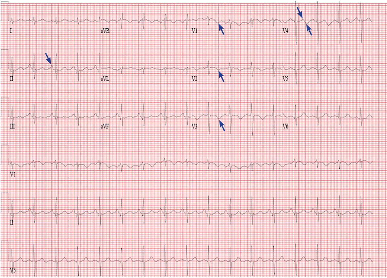
    The patient’s electrocardiogram on presentation. Arrows point to notable features (see text).

  • Figure 2
    • Download figure
    • Open in new tab
    • Download powerpoint
    Figure 2

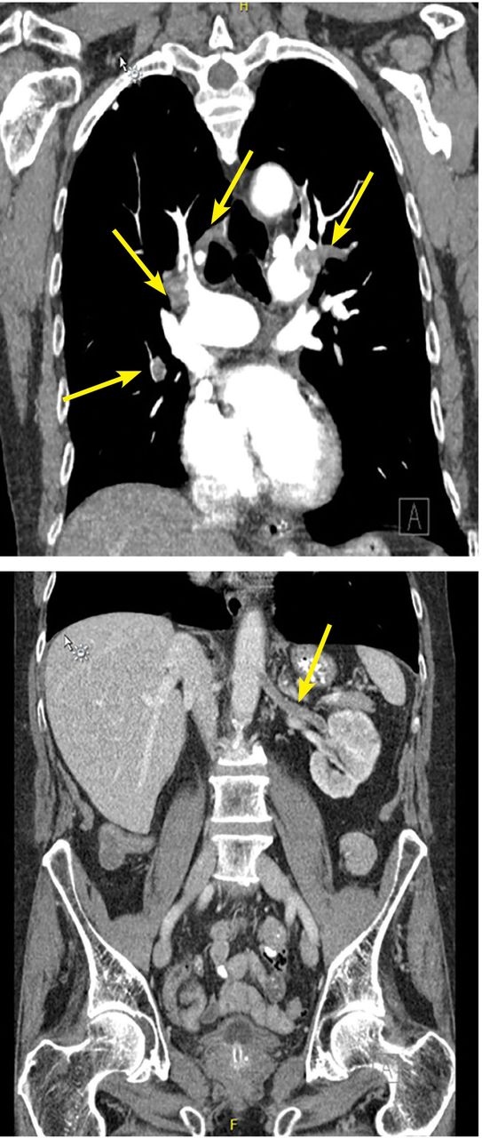
    Computed tomography (CT) with a chest pulmonary embolism protocol (top) showed filling defects (arrows). CT of the abdomen and pelvis showed renal artery thrombosis (arrow).
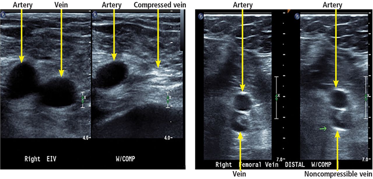
  • Figure 3
    • Download figure
    • Open in new tab
    • Download powerpoint
    Figure 3

    Example of lower-extremity duplex ultrasonography. Arteries and veins are labeled. Veins with-out deep vein thrombosis are compressible, as seen on the left. A vein is shown that is not compressible, suggesting deep vein thrombosis.

  • Figure 4
    • Download figure
    • Open in new tab
    • Download powerpoint
    Figure 4

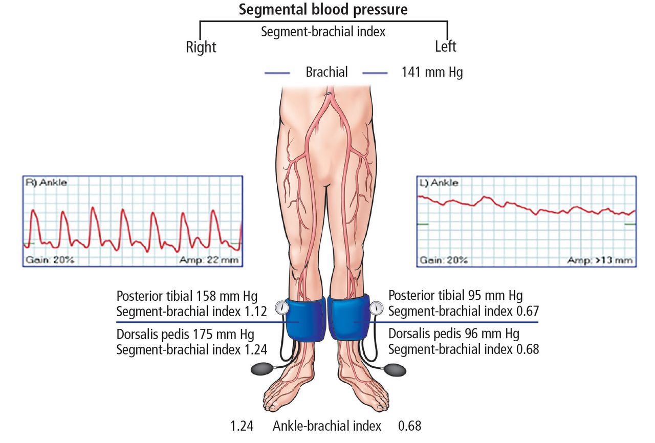
    The patient’s ankle-brachial index and pulse-volume recordings. Right side 1.24, left side 0.68. This suggests moderate disease on the left and normal vessels on the right.
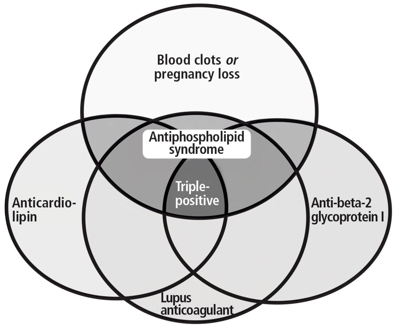
  • Figure 5
    • Download figure
    • Open in new tab
    • Download powerpoint
    Figure 5

    Antiphospholipid antibody tests and terminology.

    From Houghton DE, Moll S. Antiphospholipid antibodies. Vasc Med 2017; 22:545–550.

Tables

  • Figures
    • View popup
    TABLE 1

    The patient’s laboratory data on presentation

    TestValueaReference range
    Complete blood cell count
    White blood cell count15.373.70–11.00 × 10⁹/L
    Red blood cell count4.454.20–6.00 × 10⁹/L
    Hemoglobin13.713.0–17.0 g/dL
    Hematocrit41.239.0%–51.0%
    Mean corpuscular volume92.680.0–100.0 fL
    Mean corpuscular hemoglobin30.826.0–34.0 pg
    Mean corpuscular hemoglobin concentration33.330.5–36.0 g/dL
    Red blood cell distribution width13.811.5%–15.0%
    Platelet count204150–400 × 109/L
    Mean platelet volume11.89.0–12.7 fL
    Complete metabolic panel
    Protein, total6.76.0–8.4 g/dL
    Albumin3.13.5–5.0 g/dL
    Calcium8.48.5–10.5 mg/dL
    Bilirubin, total0.20.0–1.5 mg/dL
    Alkaline phosphatase11840–150 U/L
    Aspartate aminotransferase707–40 U/L
    Glucose11265–100 mg/dL
    Blood urea nitrogen2410–25 mg/dL
    Creatinine1.670.70–1.40 mg/dL
    Sodium140135–146 mmol/L
    Potassium4.93.5–5.0 mmol/L
    Chloride10098–110 mmol/L
    Carbon dioxide2723–32 mmol/L
    Anion gap130–15 mmol/L
    Alanine aminotransferase575–50 U/L
    Estimated glomerular filtration rate40> 90 mL/min
    Cardiac biomarkers
    NT-proBNP11,336< 450 pg/mL
    Troponin T0.0810–0.029 ng/mL
    Creatine kinase9430–220 U/L
    Creatine kinase MB6.40.0–8.8 ng/mL
    Coagulation profile
    Activated partial thromboplastin time27.223.0–32.4 sec
    Prothrombin time10.78.4–13.0 sec
    Prothrombin time INRx1.10.8–1.2
    Urinalysis
    ColorYellowYellow
    ClaritySlightly cloudyClear
    Glucose, urineNegativeNegative
    Bilirubin, urineNegativeNegative
    KetonesNegativeNegative
    Specific gravity< 1.0051.005–1.030
    Hemoglobin/blood2+Negative
    pH5.04.5–8.0
    Protein300 mg/dL
    UribilinogenNormalNormal
    NitritesNegativeNegative
    Leukocyte esteraseNegativeNegative
    White blood cells0–50–5/high-power field
    Red blood cells6–100–3/high-power field
    • ↵a Abnormal values are shown in bold. INR = international normalized ratio; NT-proBNP = N-terminal pro-B-type natriuretic peptide

    • View popup
    TABLE 2

    Classification of pulmonary embolism

    Massive
    Sustained hypotension: systolic blood pressure < 90 mm Hg for at least 15 minutes OR requiring inotropes (cannot be due to another cause)
    Pulseless
    Persistent bradycardia (≤ 40 beats per minute with signs or symptoms of shock)
    Submassive
    Systolic blood pressure ≥ 90 mm Hg
    Right ventricular dysfunction or myocardial necrosis
    Low risk
    Normal blood pressure
    Normal biomarker levels
    No right ventricular dysfunction
    • Adapted from information in reference 3.

PreviousNext
Back to top

In this issue

Cleveland Clinic Journal of Medicine: 85 (2)
Cleveland Clinic Journal of Medicine
Vol. 85, Issue 2
1 Feb 2018
  • Table of Contents
  • Table of Contents (PDF)
  • Index by author
Print
Download PDF
Article Alerts
Sign In to Email Alerts with your Email Address
Email Article

Thank you for your interest in spreading the word on Cleveland Clinic Journal of Medicine.

NOTE: We only request your email address so that the person you are recommending the page to knows that you wanted them to see it, and that it is not junk mail. We do not capture any email address.

Enter multiple addresses on separate lines or separate them with commas.
A 75-year-old with abdominal pain, hypoxia, and weak pulses in the left leg
(Your Name) has sent you a message from Cleveland Clinic Journal of Medicine
(Your Name) thought you would like to see the Cleveland Clinic Journal of Medicine web site.
CAPTCHA
This question is for testing whether or not you are a human visitor and to prevent automated spam submissions.
Citation Tools
A 75-year-old with abdominal pain, hypoxia, and weak pulses in the left leg
Maya Serhal, Natalie Evans, Heather L. Gornik
Cleveland Clinic Journal of Medicine Feb 2018, 85 (2) 145-154; DOI: 10.3949/ccjm.85a.16069

Citation Manager Formats

  • BibTeX
  • Bookends
  • EasyBib
  • EndNote (tagged)
  • EndNote 8 (xml)
  • Medlars
  • Mendeley
  • Papers
  • RefWorks Tagged
  • Ref Manager
  • RIS
  • Zotero
Share
A 75-year-old with abdominal pain, hypoxia, and weak pulses in the left leg
Maya Serhal, Natalie Evans, Heather L. Gornik
Cleveland Clinic Journal of Medicine Feb 2018, 85 (2) 145-154; DOI: 10.3949/ccjm.85a.16069
Twitter logo Facebook logo Mendeley logo
  • Tweet Widget
  • Facebook Like
  • Linkedin Share Button

Jump to section

  • Article
    • PREVIOUS HOSPITALIZATION
    • CURRENT PRESENTATION
    • WHAT DOES HIS ELECTROCARDIOGRAM SHOW?
    • CLASSIFICATION OF ACUTE PULMONARY EMBOLISM
    • ULTRASONOGRAPHY FOR DIAGNOSIS OF DEEP VEIN THROMBOSIS
    • RISK STRATIFICATION IN ACUTE PULMONARY EMBOLISM
    • ASSESSING PERIPHERAL ARTERY DISEASE
    • WHAT CAN CAUSE BOTH ARTERIAL AND VENOUS THROMBOSIS?
    • ANTIPHOSPHOLIPID ANTIBODY SYNDROME
    • ANTIPHOSPHOLIPID ANTIBODIES ARE NOT ALL THE SAME
    • PARADOXICAL EMBOLISM
    • SUMMARY OF CASE
    • Footnotes
    • REFERENCES
  • Figures & Data
  • Info & Metrics
  • PDF

Related Articles

  • No related articles found.
  • PubMed
  • Google Scholar

Cited By...

  • 'Non-criteria antiphospholipid antibodies and thrombosis (February 2018)
  • Google Scholar

More in this TOC Section

  • A 67-year-old woman with bilateral hand numbness
  • A 50-year-old woman with new-onset seizure
Show more Im Board Review

Similar Articles

Subjects

  • Emergency Medicine
  • Hematology
  • Imaging
  • Nephrology
  • Vascular Medicine

Navigate

  • Current Issue
  • Past Issues
  • Supplements
  • Article Type
  • Specialty
  • CME/MOC Articles
  • CME/MOC Calendar
  • Media Kit

Authors & Reviewers

  • Manuscript Submission
  • Authors & Reviewers
  • Subscriptions
  • About CCJM
  • Contact Us
  • Cleveland Clinic Center for Continuing Education
  • Consult QD

Share your suggestions!

Copyright © 2025 The Cleveland Clinic Foundation. All rights reserved. The information provided is for educational purposes only. Use of this website is subject to the website terms of use and privacy policy. 

Powered by HighWire