
Main menu

  • Home
  • Content
    • Current Issue
    • Ahead of Print
    • Past Issues
    • Supplements
    • Article Type
  • Specialty
    • Articles by Specialty
  • CME/MOC
    • Articles
    • Calendar
  • Info For
    • Manuscript Submission
    • Authors & Reviewers
    • Subscriptions
    • About CCJM
    • Contact Us
    • Media Kit
  • Conversations with Leaders
  • Conference Coverage
    • Kidney Week 2024
    • CHEST 2024
    • ACR Convergence 2023
    • Kidney Week 2023
    • ObesityWeek 2023
    • IDWeek 2023
    • CHEST 2023
    • MDS 2023
    • IAS 2023
    • ACP 2023
    • AAN 2023
    • ACC / WCC 2023
    • AAAAI Meeting 2023
    • ACR Convergence 2022
    • Kidney Week 2022
    • AIDS 2022
  • Other Publications
    • www.clevelandclinic.org

User menu

  • Register
  • Log in

Search

  • Advanced search
Cleveland Clinic Journal of Medicine
  • Other Publications
    • www.clevelandclinic.org
  • Register
  • Log in
Cleveland Clinic Journal of Medicine

Advanced Search

  • Home
  • Content
    • Current Issue
    • Ahead of Print
    • Past Issues
    • Supplements
    • Article Type
  • Specialty
    • Articles by Specialty
  • CME/MOC
    • Articles
    • Calendar
  • Info For
    • Manuscript Submission
    • Authors & Reviewers
    • Subscriptions
    • About CCJM
    • Contact Us
    • Media Kit
  • Conversations with Leaders
  • Conference Coverage
    • Kidney Week 2024
    • CHEST 2024
    • ACR Convergence 2023
    • Kidney Week 2023
    • ObesityWeek 2023
    • IDWeek 2023
    • CHEST 2023
    • MDS 2023
    • IAS 2023
    • ACP 2023
    • AAN 2023
    • ACC / WCC 2023
    • AAAAI Meeting 2023
    • ACR Convergence 2022
    • Kidney Week 2022
    • AIDS 2022
The Clinical Picture

Gas under the right diaphragm

Hiroki Matsuura, MD and Hidenori Hata, MD, PhD
Cleveland Clinic Journal of Medicine February 2018, 85 (2) 98-100; DOI: https://doi.org/10.3949/ccjm.85a.16139
Hiroki Matsuura
Department of General Internal Medicine, Mitoyo General Hospital, Kagawa, Japan
  • Find this author on Google Scholar
  • Find this author on PubMed
  • Search for this author on this site
  • For correspondence: [email protected]
Hidenori Hata
Department of Gastroenterology, Mitoyo General Hospital, Kagawa, Japan
  • Find this author on Google Scholar
  • Find this author on PubMed
  • Search for this author on this site
  • Article
  • Figures & Data
  • Info & Metrics
  • PDF
Loading

This article has a correction. Please see:

  • Gas under the right diagphragm (February 2018) - April 01, 2018

A 66-year-old man presented to the hospital with 3 days of nausea, vomiting, and abdominal pain. He had come to the emergency department several times during this period, but the cause of his symptoms had not been determined.

He had no significant medical history and no previous hospital admissions. He had never undergone colonoscopy and had never taken anticoagulant agents, steroids, laxatives, or nonsteroidal anti-inflammatory drugs.

His abdomen was mildly tender without guarding or rigidity. The standing chest radiograph showed gas under the right diaphragm (Figure 1), and computed tomography (CT) revealed the Chilaiditi sign, with volvulus of the cecum between the diaphragm and liver and a closed-loop obstruction (Figure 2).

Figure 1
  • Download figure
  • Open in new tab
  • Download powerpoint
Figure 1

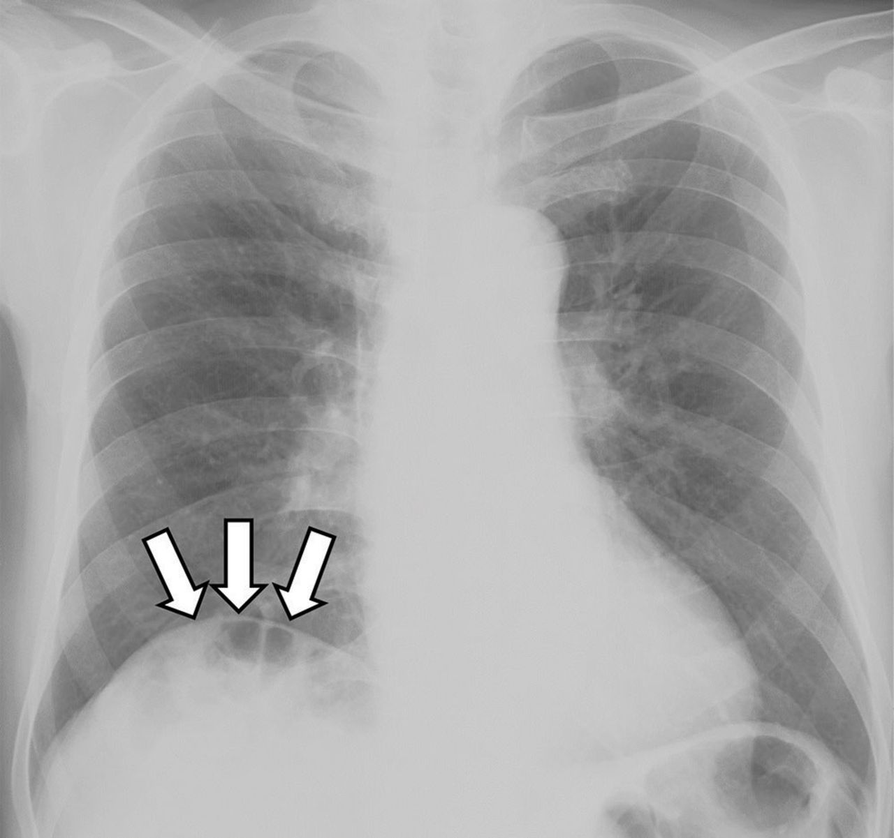
Radiography of the chest with the patient standing showed gas under the right diaphragm (arrows).

Figure 2
  • Download figure
  • Open in new tab
  • Download powerpoint
Figure 2

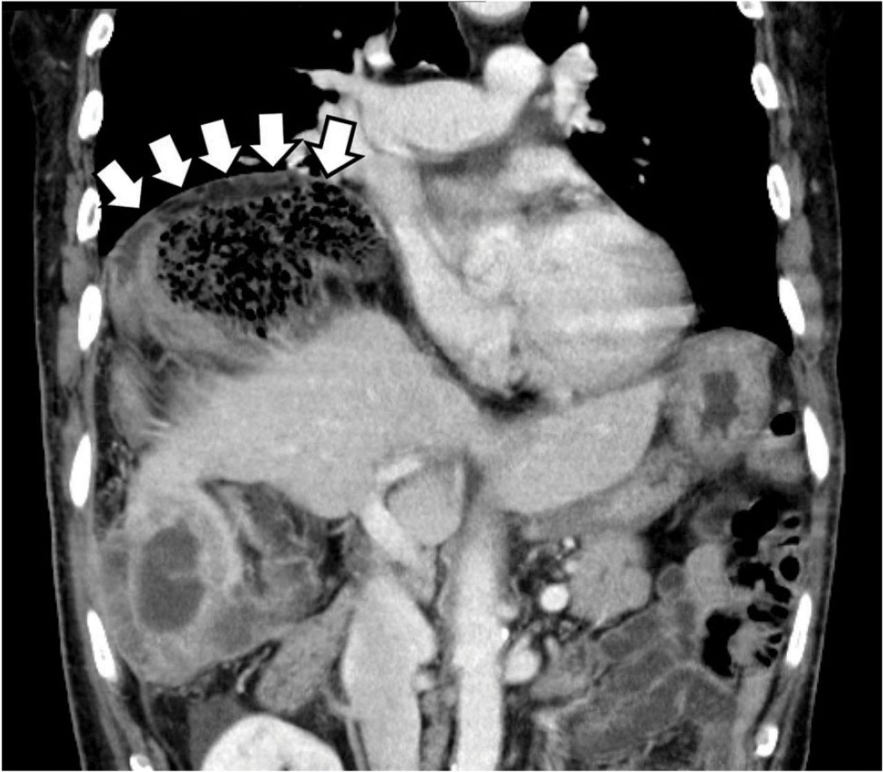
The Chilaiditi sign on computed tomography, with volvulus of the cecum between the diaphragm and liver and a closed-loop obstruction (arrows).

The patient was successfully treated with urgent right hemicolectomy.

THE CHILAIDITI SIGN AND SYNDROME

The Chilaiditi sign is an infrequent anomaly found incidentally on chest or abdominal radiography as a colonic interposition between the liver and right hemidiaphragm.1 It is often asymptomatic but is sometimes accompanied by nausea, vomiting, abdominal pain, and constipation, ie, Chilaiditi syndrome.

Generally, after conservative treatment with fasting and pain control, symptoms may subside and follow-up should be sufficient. However, nasogastric decompression and laxatives are occasionally needed and are often effective in patients with Chilaiditi syndrome. Urgent abdominal surgery is indicated for patients with symptoms of volvulus of the colon, stomach, or small intestine.2

DISTINGUISHING CHARACTERISTICS

The Chilaiditi sign is often confused with pneumoperitoneum, which usually requires urgent abdominal surgery. But the presence of haustration or valvulae conniventes (folds in the small bowel mucosa) in the hepatodiaphragmatic space helps distinguish between intraluminal gas and free air. If the patient presents with abdominal pain without signs of peritonitis, and if imaging indicates the Chilaiditi sign, then supplementary imaging (eg, decubitus radiography, chest CT, abdominal CT) is recommended to make the definitive diagnosis and to avoid unnecessary surgery.

Gas under the diaphragm on standing chest radiography without signs of peritonitis may also be seen after laparotomy and after scuba diving as well as in cases of biliary enteric fistula, incompetent sphincter of Oddi, gallstone ileus, and pneumatosis cystoides intestinalis. The incidence rate of the Chilaiditi sign detected by radiography is between 0.025% and 0.28%.3

PREDISPOSING FACTORS

The cause of the Chilaiditi sign remains unknown. Predisposing factors can be categorized as diaphragmatic (diaphragmatic thinning, phrenic nerve injury, expanded thoracic cavity), intestinal (megacolon, increased intra-abdominal pressure), and hepatic (hepatic atrophy, cirrhosis, ascites).

In healthy people, Chilaiditi syndrome is usually attributed to a congenital abnormal lengthening of the colon or to undue looseness of ligaments of the colon and liver.

Recognizing the Chilaiditi sign is particularly important in patients scheduled to undergo a percutaneous transhepatic procedure or colonoscopic examination, as these procedures increase the risk of perforation.

  • Copyright © 2018 The Cleveland Clinic Foundation. All Rights Reserved.

REFERENCES

  1. ↵
    1. Chilaiditi D
    . Zur Frage der Hepatoptose und Ptose im allegemeinen im Anschluss an drei Fälle von temporärer, partieller Leberverlagerung. Fortschritte auf dem Gebiete der Röntgenstrahlen 1910; 16:173–208.
    OpenUrl
  2. ↵
    1. Williams A,
    2. Cox R,
    3. Palaniappan B,
    4. Woodward A
    . Chilaiditi’s syndrome associated with colonic volvulus and intestinal malrotation—a rare case. Int J Surg Case Rep 2014; 5:335–338.
    OpenUrl
  3. ↵
    1. Orangio GR,
    2. Fazio VW,
    3. Winkelman E,
    4. McGonagle BA
    . The Chilaiditi syndrome and associated volvulus of the transverse colon. An indication for surgical therapy. Dis Colon Rectum 1986; 29:653–656.
    OpenUrlCrossRefPubMed
PreviousNext
Back to top

In this issue

Cleveland Clinic Journal of Medicine: 85 (2)
Cleveland Clinic Journal of Medicine
Vol. 85, Issue 2
1 Feb 2018
  • Table of Contents
  • Table of Contents (PDF)
  • Index by author
Print
Download PDF
Article Alerts
Sign In to Email Alerts with your Email Address
Email Article

Thank you for your interest in spreading the word on Cleveland Clinic Journal of Medicine.

NOTE: We only request your email address so that the person you are recommending the page to knows that you wanted them to see it, and that it is not junk mail. We do not capture any email address.

Enter multiple addresses on separate lines or separate them with commas.
Gas under the right diaphragm
(Your Name) has sent you a message from Cleveland Clinic Journal of Medicine
(Your Name) thought you would like to see the Cleveland Clinic Journal of Medicine web site.
CAPTCHA
This question is for testing whether or not you are a human visitor and to prevent automated spam submissions.
Citation Tools
Gas under the right diaphragm
Hiroki Matsuura, Hidenori Hata
Cleveland Clinic Journal of Medicine Feb 2018, 85 (2) 98-100; DOI: 10.3949/ccjm.85a.16139

Citation Manager Formats

  • BibTeX
  • Bookends
  • EasyBib
  • EndNote (tagged)
  • EndNote 8 (xml)
  • Medlars
  • Mendeley
  • Papers
  • RefWorks Tagged
  • Ref Manager
  • RIS
  • Zotero
Share
Gas under the right diaphragm
Hiroki Matsuura, Hidenori Hata
Cleveland Clinic Journal of Medicine Feb 2018, 85 (2) 98-100; DOI: 10.3949/ccjm.85a.16139
Twitter logo Facebook logo Mendeley logo
  • Tweet Widget
  • Facebook Like
  • Linkedin Share Button

Jump to section

  • Article
    • THE CHILAIDITI SIGN AND SYNDROME
    • DISTINGUISHING CHARACTERISTICS
    • PREDISPOSING FACTORS
    • REFERENCES
  • Figures & Data
  • Info & Metrics
  • PDF

Related Articles

  • Gas under the right diagphragm (February 2018)
  • PubMed
  • Google Scholar

Cited By...

  • No citing articles found.
  • Google Scholar

More in this TOC Section

  • Tinea incognito
  • Prolonged venous filling time and dependent rubor in a patient with peripheral artery disease
  • Sarcoidosis with diffuse purplish erythematous plaques on the hands
Show more The Clinical Picture

Similar Articles

Subjects

  • Emergency Medicine
  • Gastroenterology
  • Imaging

Navigate

  • Current Issue
  • Past Issues
  • Supplements
  • Article Type
  • Specialty
  • CME/MOC Articles
  • CME/MOC Calendar
  • Media Kit

Authors & Reviewers

  • Manuscript Submission
  • Authors & Reviewers
  • Subscriptions
  • About CCJM
  • Contact Us
  • Cleveland Clinic Center for Continuing Education
  • Consult QD

Share your suggestions!

Copyright © 2025 The Cleveland Clinic Foundation. All rights reserved. The information provided is for educational purposes only. Use of this website is subject to the website terms of use and privacy policy. 

Powered by HighWire