
Main menu

  • Home
  • Content
    • Current Issue
    • Ahead of Print
    • Past Issues
    • Supplements
    • Article Type
  • Specialty
    • Articles by Specialty
  • CME/MOC
    • Articles
    • Calendar
  • Info For
    • Manuscript Submission
    • Authors & Reviewers
    • Subscriptions
    • About CCJM
    • Contact Us
    • Media Kit
  • Conversations with Leaders
  • Conference Coverage
    • Kidney Week 2024
    • CHEST 2024
    • ACR Convergence 2023
    • Kidney Week 2023
    • ObesityWeek 2023
    • IDWeek 2023
    • CHEST 2023
    • MDS 2023
    • IAS 2023
    • ACP 2023
    • AAN 2023
    • ACC / WCC 2023
    • AAAAI Meeting 2023
    • ACR Convergence 2022
    • Kidney Week 2022
    • AIDS 2022
  • Other Publications
    • www.clevelandclinic.org

User menu

  • Register
  • Log in

Search

  • Advanced search
Cleveland Clinic Journal of Medicine
  • Other Publications
    • www.clevelandclinic.org
  • Register
  • Log in
Cleveland Clinic Journal of Medicine

Advanced Search

  • Home
  • Content
    • Current Issue
    • Ahead of Print
    • Past Issues
    • Supplements
    • Article Type
  • Specialty
    • Articles by Specialty
  • CME/MOC
    • Articles
    • Calendar
  • Info For
    • Manuscript Submission
    • Authors & Reviewers
    • Subscriptions
    • About CCJM
    • Contact Us
    • Media Kit
  • Conversations with Leaders
  • Conference Coverage
    • Kidney Week 2024
    • CHEST 2024
    • ACR Convergence 2023
    • Kidney Week 2023
    • ObesityWeek 2023
    • IDWeek 2023
    • CHEST 2023
    • MDS 2023
    • IAS 2023
    • ACP 2023
    • AAN 2023
    • ACC / WCC 2023
    • AAAAI Meeting 2023
    • ACR Convergence 2022
    • Kidney Week 2022
    • AIDS 2022
The Clinical Picture

Paraneoplastic acral vascular syndrome

Satish Maharaj, MBBS, Simone Chang, MBBS, Karan Seegobin, MBBS and Carmen Isache, MD
Cleveland Clinic Journal of Medicine February 2018, 85 (2) 101-102; DOI: https://doi.org/10.3949/ccjm.85a.17007
Satish Maharaj
Department of Internal Medicine, University of Florida College of Medicine, Jacksonville, FL
  • Find this author on Google Scholar
  • Find this author on PubMed
  • Search for this author on this site
  • For correspondence: [email protected]
Simone Chang
Jackson Memorial Hospital, Miami, FL
  • Find this author on Google Scholar
  • Find this author on PubMed
  • Search for this author on this site
Karan Seegobin
Department of Internal Medicine, University of Florida College of Medicine, Jacksonville, FL
  • Find this author on Google Scholar
  • Find this author on PubMed
  • Search for this author on this site
Carmen Isache
Department of Internal Medicine, University of Florida College of Medicine, Jacksonville, FL
  • Find this author on Google Scholar
  • Find this author on PubMed
  • Search for this author on this site
  • Article
  • Figures & Data
  • Info & Metrics
  • PDF
Loading

A 66-year-old woman presented to the emergency room with pain and bluish discoloration of her left great toe for the past 2 days. She had no history of trauma to the toe or of peripheral vascular disease. She also complained of intermittent abdominal pain for the last 5 months and unintentional weight loss of 8 pounds.

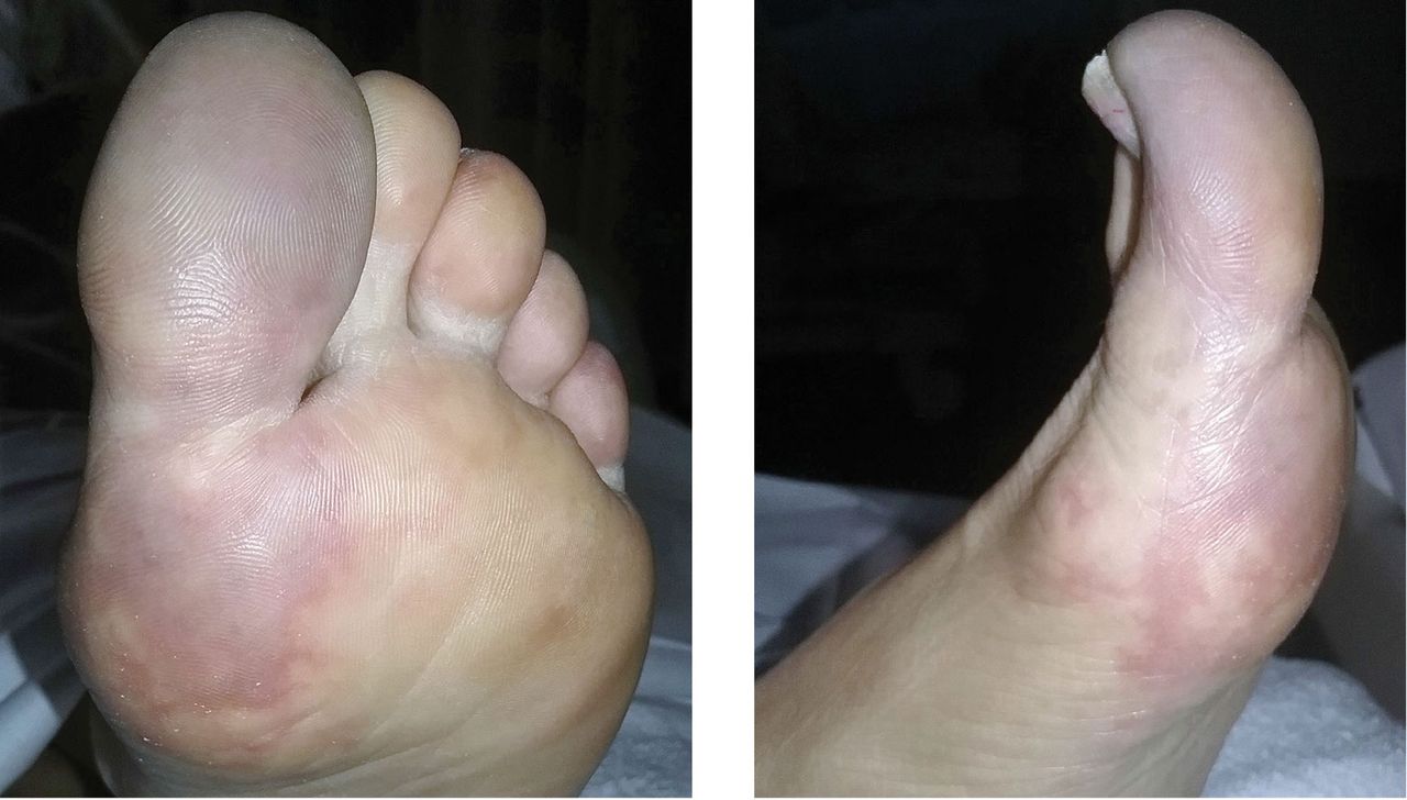
On examination, the left hallux was bluish-purple (Figure 1) with an acral distribution (ie, more affected distally). The other extremities were not affected. There was no difference in temperature between her feet, and distal sensation and peripheral pulses were intact.

Figure 1
  • Download figure
  • Open in new tab
  • Download powerpoint
Figure 1

Acrocyanosis of the left hallux with an acral distribution.

Vascular disease was suspected, and the patient was started on systemic anticoagulation. However, chest computed tomography (CT) and conventional angiography showed no aortic disease; vessels were of normal caliber, and no distal filling defects were noted.

A routine evaluation with complete blood cell count, peripheral smear, renal function testing, and urinalysis with eosinophil smear was also unrevealing. An extensive investigation followed, including serum complement testing, assays for antinuclear, antineutrophil cytoplasmic, and anti-Sjögren syndrome A and B antibodies, cryoglobulin testing, and hepatitis B and C virus serology, as well as screening for syphilis and lupus anticoagulant, anticardiolipin, and beta-2 glycoprotein antibodies. The results for all these tests were also unremarkable.

Results of coagulation testing and venous duplex ultrasonography of the legs were normal, and electrocardiography and echocardiography showed no signs of valvular vegetation, myxoma, or patent foramen ovale.

Given our patient’s age, abdominal pain, and weight loss but negative vascular evaluation, we considered a diagnosis of paraneo-plastic acral vascular syndrome. Abdominal CT revealed a tumor of the pancreatic head with multiple liver lesions, and cytologic study confirmed pancreatic adenocarcinoma.

DISTANT MARKERS OF MALIGNANCY

Causes of blue toe syndrome to consider in the differential diagnosis include arterial thromboembolism, vasoconstrictive drug use or disorders, vasculitis, and venous thrombosis.1 These are common and deserve prompt investigation. However, if they are ruled out, a peripheral acral vascular syndrome should be considered. These syndromes present as Raynaud phenomenon, gangrene, or acrocyanosis of the fingers or toes with underlying neoplasia. Unusual features such as sudden onset in a patient over age 50, an acral distribution, and associated symptoms such as unrelated pain and weight loss should spark concern for underlying malignancy.

Paraneoplastic syndromes are defined as signs and symptoms that present distant from the site of malignancy. Dermatoses as markers of internal malignancy are well-established but perplexing clinical entities whose exact causes remain unknown.2

Paraneoplastic acral vascular syndrome is associated with certain cancers, predominantly adenocarcinoma and metastatic disease.3 As in our patient, it usually precedes or is present at the time of diagnosis of internal malignancy. Paraneoplastic dermatosis as the presenting sign of pancreatic cancer is uncommon, and a search of the literature found 1 case report of acral vascular syndrome.4 Paraneoplastic dermatoses also include necrolytic migratory erythema, Leser-Trélat syndrome, and acrokeratosis paraneoplastica (Table 1).

View this table:
  • View inline
  • View popup
TABLE 1

Paraneoplastic dermatoses associated with internal malignancy

Paraneoplastic dermatoses are well recognized as harbingers of metastatic disease.5,6 Our patient’s story demonstrates need for a thorough diagnostic investigation.

  • Copyright © 2018 The Cleveland Clinic Foundation. All Rights Reserved.

REFERENCES

  1. ↵
    1. Hirschmann JV,
    2. Raugi GJ
    . Blue (or purple) toe syndrome. J Am Acad Dermatol 2009; 60:1–20.
    OpenUrlCrossRefPubMed
  2. ↵
    1. Naschitz JE,
    2. Rosner I,
    3. Rozenbaum M,
    4. Zuckerman E,
    5. Yeshurun D
    . Rheumatic syndromes: clues to occult neoplasia. Semin Arthritis Rheum 1999; 29:43–55.
    OpenUrlCrossRefPubMed
  3. ↵
    1. Poszepczynska-Guigné E,
    2. Viguier M,
    3. Chosidow O,
    4. Orcel B,
    5. Emmerich J,
    6. Dubertret L
    . Paraneoplastic acral vascular syndrome: epidemiologic features, clinical manifestations, and disease sequelae. J Am Acad Dermatol 2002; 47:47–52.
    OpenUrlCrossRefPubMed
  4. ↵
    1. DeCross AJ,
    2. Sahasrabudhe DM
    . Paraneoplastic Raynaud’s phenomenon. Am J Med 1992; 92:571–572.
    OpenUrlCrossRefPubMed
  5. ↵
    1. Ramos-E-Silva M,
    2. Carvalho JC,
    3. Carneiro SC
    . Cutaneous paraneoplasia. Clin Dermatol 2011; 29:541–547.
    OpenUrlPubMed
  6. ↵
    1. Chung VQ,
    2. Moschella SL,
    3. Zembowicz A,
    4. Liu V
    . Clinical and pathologic findings of paraneoplastic dermatoses. J Am Acad Dermatol 2006; 54:745–762.
    OpenUrlCrossRefPubMed
PreviousNext
Back to top

In this issue

Cleveland Clinic Journal of Medicine: 85 (2)
Cleveland Clinic Journal of Medicine
Vol. 85, Issue 2
1 Feb 2018
  • Table of Contents
  • Table of Contents (PDF)
  • Index by author
Print
Download PDF
Article Alerts
Sign In to Email Alerts with your Email Address
Email Article

Thank you for your interest in spreading the word on Cleveland Clinic Journal of Medicine.

NOTE: We only request your email address so that the person you are recommending the page to knows that you wanted them to see it, and that it is not junk mail. We do not capture any email address.

Enter multiple addresses on separate lines or separate them with commas.
Paraneoplastic acral vascular syndrome
(Your Name) has sent you a message from Cleveland Clinic Journal of Medicine
(Your Name) thought you would like to see the Cleveland Clinic Journal of Medicine web site.
CAPTCHA
This question is for testing whether or not you are a human visitor and to prevent automated spam submissions.
Citation Tools
Paraneoplastic acral vascular syndrome
Satish Maharaj, Simone Chang, Karan Seegobin, Carmen Isache
Cleveland Clinic Journal of Medicine Feb 2018, 85 (2) 101-102; DOI: 10.3949/ccjm.85a.17007

Citation Manager Formats

  • BibTeX
  • Bookends
  • EasyBib
  • EndNote (tagged)
  • EndNote 8 (xml)
  • Medlars
  • Mendeley
  • Papers
  • RefWorks Tagged
  • Ref Manager
  • RIS
  • Zotero
Share
Paraneoplastic acral vascular syndrome
Satish Maharaj, Simone Chang, Karan Seegobin, Carmen Isache
Cleveland Clinic Journal of Medicine Feb 2018, 85 (2) 101-102; DOI: 10.3949/ccjm.85a.17007
Twitter logo Facebook logo Mendeley logo
  • Tweet Widget
  • Facebook Like
  • Linkedin Share Button

Jump to section

  • Article
    • DISTANT MARKERS OF MALIGNANCY
    • REFERENCES
  • Figures & Data
  • Info & Metrics
  • PDF

Related Articles

  • No related articles found.
  • PubMed
  • Google Scholar

Cited By...

  • Paraneoplastic acral vascular syndrome: case presentation and literature review
  • Google Scholar

More in this TOC Section

  • Tinea incognito
  • Prolonged venous filling time and dependent rubor in a patient with peripheral artery disease
  • Sarcoidosis with diffuse purplish erythematous plaques on the hands
Show more The Clinical Picture

Similar Articles

Subjects

  • Dermatology
  • Oncology

Navigate

  • Current Issue
  • Past Issues
  • Supplements
  • Article Type
  • Specialty
  • CME/MOC Articles
  • CME/MOC Calendar
  • Media Kit

Authors & Reviewers

  • Manuscript Submission
  • Authors & Reviewers
  • Subscriptions
  • About CCJM
  • Contact Us
  • Cleveland Clinic Center for Continuing Education
  • Consult QD

Share your suggestions!

Copyright © 2025 The Cleveland Clinic Foundation. All rights reserved. The information provided is for educational purposes only. Use of this website is subject to the website terms of use and privacy policy. 

Powered by HighWire