
Main menu

  • Home
  • Content
    • Current Issue
    • Ahead of Print
    • Past Issues
    • Supplements
    • Article Type
  • Specialty
    • Articles by Specialty
  • CME/MOC
    • Articles
    • Calendar
  • Info For
    • Manuscript Submission
    • Authors & Reviewers
    • Subscriptions
    • About CCJM
    • Contact Us
    • Media Kit
  • Conversations with Leaders
  • Conference Coverage
    • Kidney Week 2024
    • CHEST 2024
    • ACR Convergence 2023
    • Kidney Week 2023
    • ObesityWeek 2023
    • IDWeek 2023
    • CHEST 2023
    • MDS 2023
    • IAS 2023
    • ACP 2023
    • AAN 2023
    • ACC / WCC 2023
    • AAAAI Meeting 2023
    • ACR Convergence 2022
    • Kidney Week 2022
    • AIDS 2022
  • Other Publications
    • www.clevelandclinic.org

User menu

  • Register
  • Log in

Search

  • Advanced search
Cleveland Clinic Journal of Medicine
  • Other Publications
    • www.clevelandclinic.org
  • Register
  • Log in
Cleveland Clinic Journal of Medicine

Advanced Search

  • Home
  • Content
    • Current Issue
    • Ahead of Print
    • Past Issues
    • Supplements
    • Article Type
  • Specialty
    • Articles by Specialty
  • CME/MOC
    • Articles
    • Calendar
  • Info For
    • Manuscript Submission
    • Authors & Reviewers
    • Subscriptions
    • About CCJM
    • Contact Us
    • Media Kit
  • Conversations with Leaders
  • Conference Coverage
    • Kidney Week 2024
    • CHEST 2024
    • ACR Convergence 2023
    • Kidney Week 2023
    • ObesityWeek 2023
    • IDWeek 2023
    • CHEST 2023
    • MDS 2023
    • IAS 2023
    • ACP 2023
    • AAN 2023
    • ACC / WCC 2023
    • AAAAI Meeting 2023
    • ACR Convergence 2022
    • Kidney Week 2022
    • AIDS 2022
The Clinical Picture

Hidden lesion easily missed on chest radiography

Kamolyut Lapumnuaypol, MD and Jessica M. Stempel, MD
Cleveland Clinic Journal of Medicine April 2018, 85 (4) 270-273; DOI: https://doi.org/10.3949/ccjm.85a.17067
Kamolyut Lapumnuaypol
Department of Internal Medicine, Albert Einstein Medical Center, Philadelphia, PA
  • Find this author on Google Scholar
  • Find this author on PubMed
  • Search for this author on this site
  • For correspondence: [email protected]
Jessica M. Stempel
Department of Internal Medicine, Albert Einstein Medical Center, Philadelphia, PA
  • Find this author on Google Scholar
  • Find this author on PubMed
  • Search for this author on this site
  • Article
  • Figures & Data
  • Info & Metrics
  • PDF
Loading

A 46-year-old man with poorly controlled hypertension presented with the sudden onset of chest pain and shortness of breath. His blood pressure was 158/96 mm Hg and his left ventricular ejection fraction was less than 20%. He was admitted to the hospital for newly diagnosed heart failure.

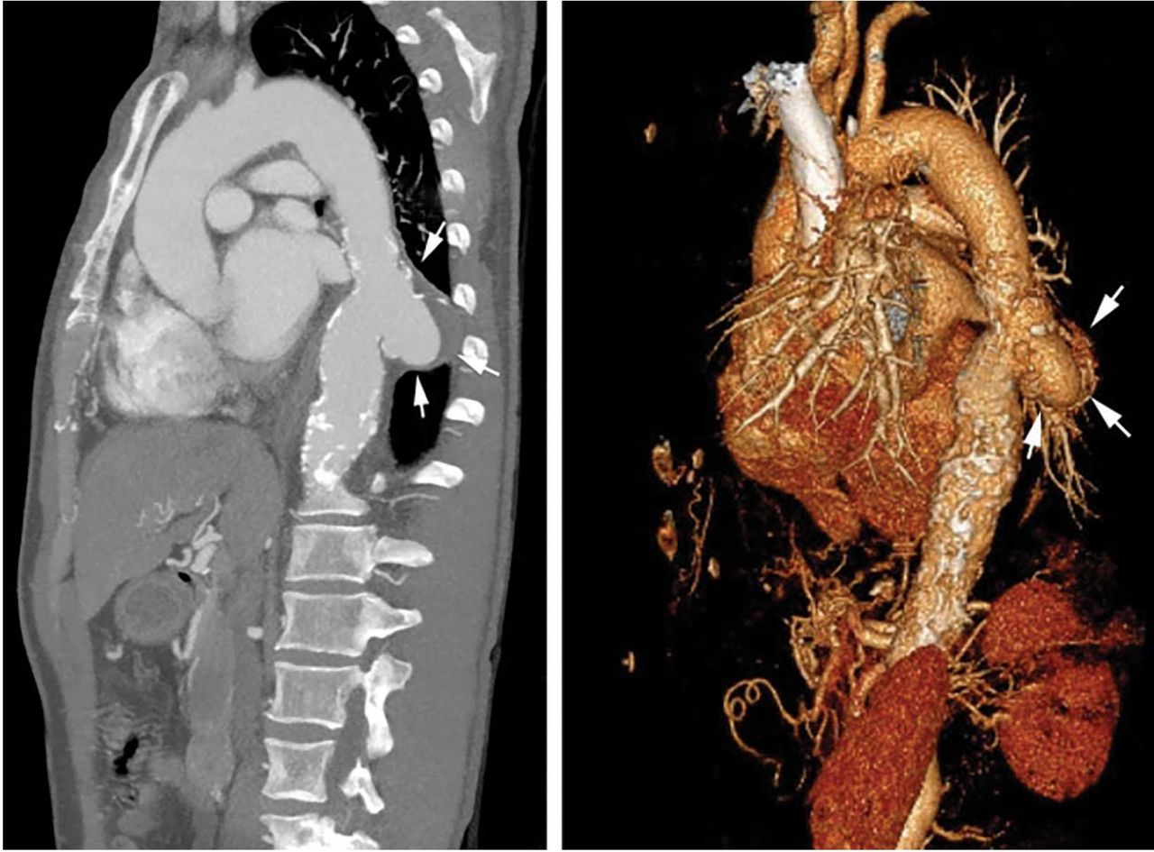
Chest radiography showed cardiomegaly, bilateral pleural effusions, and a retrocardiac masslike opacity (Figure 1). Computed tomographic angiography revealed the opacity to be a saccular aneurysm of the descending thoracic aorta measuring 6.3 × 3.7 cm, with intimal calcifications and chronic intramural hemorrhage (Figure 2). The descending thoracic aorta was heavily calcified.

Figure 1
  • Download figure
  • Open in new tab
  • Download powerpoint
Figure 1

Radiography showed cardiomegaly, bilateral pleural effusions, and a retrocardiac masslike opacity (arrows).

Figure 2
  • Download figure
  • Open in new tab
  • Download powerpoint
Figure 2

Computed tomographic angiography (sagittal view) and a 3-dimensional rendering show an aorta with intimal calcifications and chronic intramural hemorrhage (arrows).

The patient then underwent cardiac catheterization, which revealed triple-vessel coronary artery disease. A cardiothoracic surgeon recommended coronary artery bypass grafting with aneurysm repair. However, the patient declined surgery and instead chose medical therapy. His blood pressure was under control at the time of discharge.

SACCULAR AORTIC ANEURYSMS

This case shows the value of carefully examining the chest radiograph, especially behind the heart.

Saccular aneurysms of the descending thoracic aorta are rare and can be easily missed if asymptomatic. They are less common than fusiform aneurysms and may be more prone to rupture. Shang et al1 identified atherosclerosis as the most frequent cause of saccular aneurysms of the thoracic aorta. However, they can be caused by other inflammatory conditions and infections.1

Without surgical repair, thoracic aortic aneurysms larger than 6 cm have higher rates of expansion and rupture (a 20% 6-year cumulative risk) than smaller ones.2 Mid-descending aortic aneurysms expand faster than those of the ascending aorta.3

Indications for surgery include rupture, severe chest pain, compressive symptoms, large size (eg, ≥ 5.5 cm for asymptomatic descending thoracic aneurysms), and rapid growth rate (≥ 10 mm per year), all of which are associated with a higher mortality rate.4

Endovascular grafting should be strongly considered in patients who have significant comorbidities, but this approach may have poorer long-term outcomes compared with open surgery. Blood pressure should be lowered as far as the patient can tolerate without adverse effects, usually with a beta-blocker along with either an angiotensin-converting enzyme inhibitor or an angiotensin receptor blocker.4

Statin therapy to lower the low-density lipoprotein cholesterol level to less than 70 mg/dL is also needed to reduce the risk of complications and cardiovascular disease.

All patients with saccular aortic aneurysm should be followed closely and be evaluated for surgery.

  • Copyright © 2018 The Cleveland Clinic Foundation. All Rights Reserved.

REFERENCES

  1. ↵
    1. Shang EK,
    2. Nathan DP,
    3. Boonn WW,
    4. et al
    . A modern experience with saccular aortic aneurysms. J Vasc Surg 2013; 57(1):84–88. doi:10.1016/j.jvs.2012.07.002.
    OpenUrlCrossRefPubMed
  2. ↵
    1. Dapunt OE,
    2. Galla JD,
    3. Sadeghi AM,
    4. et al
    . The natural history of thoracic aortic aneurysms. J Thorac Cardiovasc Surg 1994; 107(5):1323–1333.
    OpenUrlPubMed
  3. ↵
    1. Bonser RS,
    2. Pagano D,
    3. Lewis ME,
    4. et al
    . Clinical and pathoanatomical factors affecting expansion of thoracic aortic aneurysms. Heart 2000; 84(3):277–283.
    OpenUrlAbstract/FREE Full Text
  4. ↵
    1. Hiratzka LF,
    2. Bakris GL,
    3. Beckman JA,
    4. et al
    . 2010 ACCF/ AHA/AATS/ACR/ASA/SCA/SCAI/SIR/STS/SVM guidelines for the diagnosis and management of patients with thoracic aortic disease. Circulation 2010; 121(13):e266–e369. doi:10.1161/CIR.0b013e3181d4739e.
    OpenUrlFREE Full Text
PreviousNext
Back to top

In this issue

Cleveland Clinic Journal of Medicine: 85 (4)
Cleveland Clinic Journal of Medicine
Vol. 85, Issue 4
1 Apr 2018
  • Table of Contents
  • Table of Contents (PDF)
  • Index by author
Print
Download PDF
Article Alerts
Sign In to Email Alerts with your Email Address
Email Article

Thank you for your interest in spreading the word on Cleveland Clinic Journal of Medicine.

NOTE: We only request your email address so that the person you are recommending the page to knows that you wanted them to see it, and that it is not junk mail. We do not capture any email address.

Enter multiple addresses on separate lines or separate them with commas.
Hidden lesion easily missed on chest radiography
(Your Name) has sent you a message from Cleveland Clinic Journal of Medicine
(Your Name) thought you would like to see the Cleveland Clinic Journal of Medicine web site.
CAPTCHA
This question is for testing whether or not you are a human visitor and to prevent automated spam submissions.
Citation Tools
Hidden lesion easily missed on chest radiography
Kamolyut Lapumnuaypol, Jessica M. Stempel
Cleveland Clinic Journal of Medicine Apr 2018, 85 (4) 270-273; DOI: 10.3949/ccjm.85a.17067

Citation Manager Formats

  • BibTeX
  • Bookends
  • EasyBib
  • EndNote (tagged)
  • EndNote 8 (xml)
  • Medlars
  • Mendeley
  • Papers
  • RefWorks Tagged
  • Ref Manager
  • RIS
  • Zotero
Share
Hidden lesion easily missed on chest radiography
Kamolyut Lapumnuaypol, Jessica M. Stempel
Cleveland Clinic Journal of Medicine Apr 2018, 85 (4) 270-273; DOI: 10.3949/ccjm.85a.17067
Twitter logo Facebook logo Mendeley logo
  • Tweet Widget
  • Facebook Like
  • Linkedin Share Button

Jump to section

  • Article
    • SACCULAR AORTIC ANEURYSMS
    • REFERENCES
  • Figures & Data
  • Info & Metrics
  • PDF

Related Articles

  • No related articles found.
  • PubMed
  • Google Scholar

Cited By...

  • No citing articles found.
  • Google Scholar

More in this TOC Section

  • Tinea incognito
  • Prolonged venous filling time and dependent rubor in a patient with peripheral artery disease
  • Sarcoidosis with diffuse purplish erythematous plaques on the hands
Show more The Clinical Picture

Similar Articles

Subjects

  • Cardiology
  • Imaging
  • Preventive Care
  • Vascular Medicine

Navigate

  • Current Issue
  • Past Issues
  • Supplements
  • Article Type
  • Specialty
  • CME/MOC Articles
  • CME/MOC Calendar
  • Media Kit

Authors & Reviewers

  • Manuscript Submission
  • Authors & Reviewers
  • Subscriptions
  • About CCJM
  • Contact Us
  • Cleveland Clinic Center for Continuing Education
  • Consult QD

Share your suggestions!

Copyright © 2025 The Cleveland Clinic Foundation. All rights reserved. The information provided is for educational purposes only. Use of this website is subject to the website terms of use and privacy policy. 

Powered by HighWire