
Main menu

  • Home
  • Content
    • Current Issue
    • Ahead of Print
    • Past Issues
    • Supplements
    • Article Type
  • Specialty
    • Articles by Specialty
  • CME/MOC
    • Articles
    • Calendar
  • Info For
    • Manuscript Submission
    • Authors & Reviewers
    • Subscriptions
    • About CCJM
    • Contact Us
    • Media Kit
  • Conversations with Leaders
  • Conference Coverage
    • Kidney Week 2024
    • CHEST 2024
    • ACR Convergence 2023
    • Kidney Week 2023
    • ObesityWeek 2023
    • IDWeek 2023
    • CHEST 2023
    • MDS 2023
    • IAS 2023
    • ACP 2023
    • AAN 2023
    • ACC / WCC 2023
    • AAAAI Meeting 2023
    • ACR Convergence 2022
    • Kidney Week 2022
    • AIDS 2022
  • Other Publications
    • www.clevelandclinic.org

User menu

  • Register
  • Log in

Search

  • Advanced search
Cleveland Clinic Journal of Medicine
  • Other Publications
    • www.clevelandclinic.org
  • Register
  • Log in
Cleveland Clinic Journal of Medicine

Advanced Search

  • Home
  • Content
    • Current Issue
    • Ahead of Print
    • Past Issues
    • Supplements
    • Article Type
  • Specialty
    • Articles by Specialty
  • CME/MOC
    • Articles
    • Calendar
  • Info For
    • Manuscript Submission
    • Authors & Reviewers
    • Subscriptions
    • About CCJM
    • Contact Us
    • Media Kit
  • Conversations with Leaders
  • Conference Coverage
    • Kidney Week 2024
    • CHEST 2024
    • ACR Convergence 2023
    • Kidney Week 2023
    • ObesityWeek 2023
    • IDWeek 2023
    • CHEST 2023
    • MDS 2023
    • IAS 2023
    • ACP 2023
    • AAN 2023
    • ACC / WCC 2023
    • AAAAI Meeting 2023
    • ACR Convergence 2022
    • Kidney Week 2022
    • AIDS 2022
The Clinical Picture

Eyes of the mimicker

Tamara A. Sussman, MD, Nabin K. Shrestha, MD and Maria Miklowski, MD
Cleveland Clinic Journal of Medicine May 2018, 85 (5) 356-357; DOI: https://doi.org/10.3949/ccjm.85a.17074
Tamara A. Sussman
Department of Internal Medicine, Cleveland Clinic
  • Find this author on Google Scholar
  • Find this author on PubMed
  • Search for this author on this site
  • For correspondence: [email protected]
Nabin K. Shrestha
  • Find this author on Google Scholar
  • Find this author on PubMed
  • Search for this author on this site
Maria Miklowski
  • Find this author on Google Scholar
  • Find this author on PubMed
  • Search for this author on this site
  • Article
  • Figures & Data
  • Info & Metrics
  • PDF
Loading

A 42-year-old man presented with intermittent fevers, rigors, headache, and myalgia for 6 days, followed by blurred vision in his right eye with central vision loss for the past several hours. He reported no associated pain, flashes of light, or floaters. He was sexually active with 1 male partner for 23 years. He had no history of human immunodeficiency virus (HIV), sexually transmitted infections, or intravenous drug use.

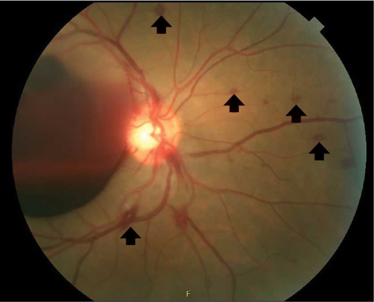
Dilated funduscopy showed a large subhyaloid hemorrhage and retinal hemorrhages with white centers (Roth spots) in the right eye, and disc edema in the left eye (Figures 1–3).

Figure 1
  • Download figure
  • Open in new tab
  • Download powerpoint
Figure 1

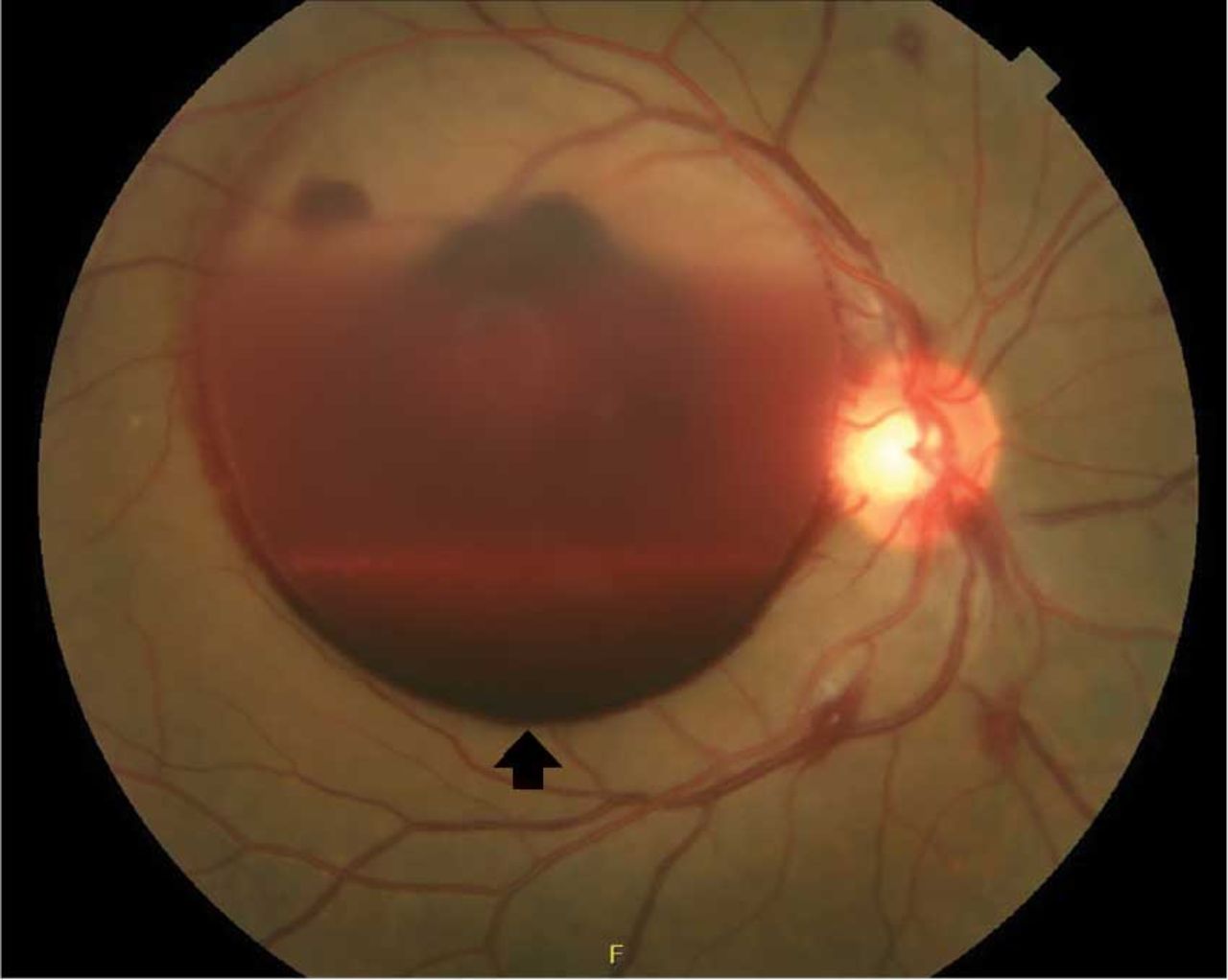
A dilated fundus photograph of the patient’s right eye shows large subhyaloid hemorrhage (arrow).

Figure 2
  • Download figure
  • Open in new tab
  • Download powerpoint
Figure 2

Multiple white-centered intra-retinal hemorrhages, or Roth spots (arrows) were noted in the right eye.

Figure 3
  • Download figure
  • Open in new tab
  • Download powerpoint
Figure 3

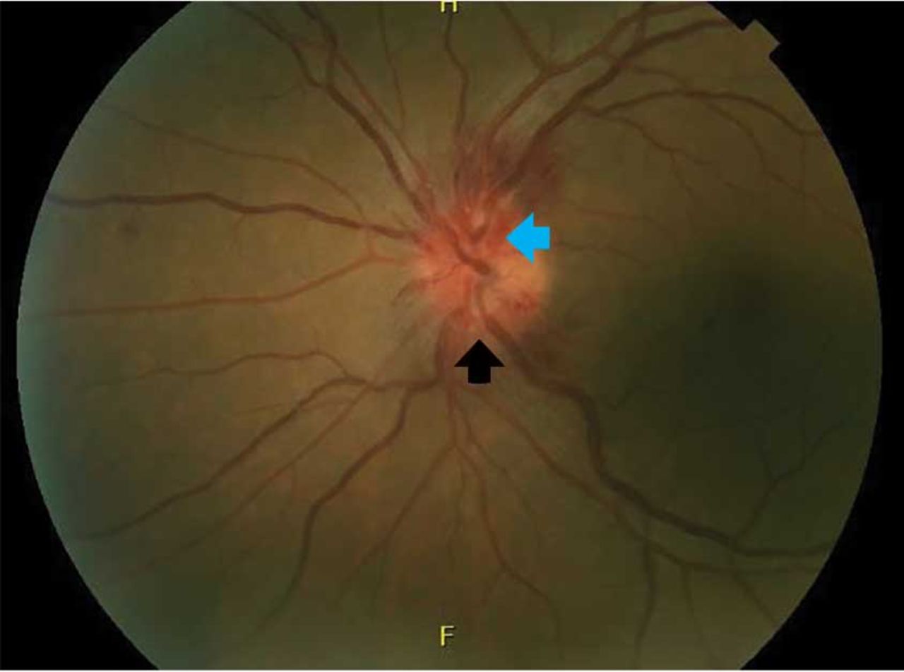
A dilated fundus photograph of the left eye shows optic disc edema (black arrow) with associated optic disc hemorrhages (blue arrow).

On laboratory testing, serum rapid plasma reagin was reactive, Treponema pallidum immunoglobulin G was reactive (antibody index > 8.0), and HIV testing was negative.

Lumbar puncture study revealed 34 nucleated cells/μL (94% lymphocytes), protein 58 mg/dL, and glucose 62 mg/dL. Cerebrospinal fluid Venereal Disease Research Laboratory and fluorescent treponemal antibody absorption tests were reactive, confirming a diagnosis of ocular syphilis.

The patient was admitted to the hospital for treatment with intravenous penicillin G. After 5 days, he was discharged with instructions to complete a 10-day course of intravenous ceftriaxone (chosen for its ease of administration), for a total of 14 days of antibiotic therapy. His vision improved with treatment.

He continued to follow up with ophthalmology and infectious disease. Subsequent dilated fundus examinations showed resolution of pathology in the left eye, resolution of Roth spots in the right eye, and resolution of the subhyaloid hemorrhage. Repeat cerebrospinal fluid study examination was planned if the serum rapid plasma reagin had not become nonreactive 24 months after treatment.

RECOGNIZING AND MANAGING OCULAR SYPHILIS AND NEUROSYPHILIS

In addition to ocular syphilis and neurosyphilis, the differential diagnosis for Roth spots and disc edema on dilated funduscopy includes endocarditis, viral retinitis, and autoimmune or inflammatory conditions such as sarcoidosis and vasculitis.

In our patient, infectious endocarditis was considered, given his history of intermittent fevers and rigors, but it was ultimately ruled out by negative blood cultures and the absence of valvular vegetations on echocardiography.

The large subhyaloid hemorrhage raised suspicion of leukemia, but this was ruled out by the normal total white blood cell count and differential. HIV, herpetic retinitis, and toxoplasmosis were also considered, but laboratory tests for these infections were negative.

Typically, retinal precipitates are more characteristic of syphilitic retinitis and distinguish it from other infectious causes such as herpetic retinitis and toxoplasmosis.1 Additionally, ocular syphilis more commonly manifests as uveitis or panuveitis.1,2 Our patient’s ocular syphilis presented with white-centered retinal hemorrhages, subhyaloid hemorrhage, and optic disc edema.

Who is at highest risk?

About 90% of syphilis cases occur in men, and 81% occur in men who have sex with men. The US Centers for Disease Control and Prevention (CDC) thus recommends annual syphilis testing for men who have sex with men.3

Classically, syphilis was called “the great imitator” because it mimicked manifestations of other diseases. Patients with ocular manifestations of syphilis may not have other neurologic symptoms.4,5 Nevertheless, cerebrospinal fluid examination should be done in all instances of ocular syphilis, as many patients with ocular syphilis have evidence of neurosyphilis on testing.2 The CDC also recommends follow-up cerebrospinal fluid analysis to assess treatment response.2 This was planned in our patient.

  • Copyright © 2018 The Cleveland Clinic Foundation. All Rights Reserved.

REFERENCES

  1. ↵
    1. Fu EX,
    2. Geraets RL,
    3. Dodds EM,
    4. et al
    . Superficial retinal precipitates in patients with syphilitic retinitis. Retina 2010; 30(7):1135–1143. doi:10.1097/IAE.0b013e3181cdf3ae
    OpenUrlCrossRefPubMed
  2. ↵
    1. US Centers for Disease Control and Prevention
    . Sexually Transmitted Diseases. Clinical Advisory: Ocular Syphilis in the United States, March 24, 2016. www.cdc.gov/std/syphilis/clinicaladvisoryos2015.htm.Accessed March 28, 2018.
  3. ↵
    1. US Centers for Disease Control and Prevention
    . Sexually Transmitted Disease Surveillance, 2015. www.cdc.gov/std/stats15/std-surveillance-2015-print.pdf. Accessed March 28, 2018.
  4. ↵
    1. Rishi E,
    2. Govindarajan MV,
    3. Biswas J,
    4. Agarwal M,
    5. Sudharshan S,
    6. Rishi P
    . Syphilitic uveitis as the presenting feature of HIV. Indian J Ophthalmol 2016; 64(2):149–150. doi:10.4103/0301-4738.179714
    OpenUrlCrossRef
  5. ↵
    1. Zhang R,
    2. Qian J,
    3. Guo J,
    4. et al
    . Clinical manifestations and treatment outcomes of syphilitic uveitis in a Chinese population. J Ophthalmol 2016; 2016:2797028. doi:10.1155/2016/2797028
    OpenUrlCrossRef
PreviousNext
Back to top

In this issue

Cleveland Clinic Journal of Medicine: 85 (5)
Cleveland Clinic Journal of Medicine
Vol. 85, Issue 5
1 May 2018
  • Table of Contents
  • Table of Contents (PDF)
  • Index by author
Print
Download PDF
Article Alerts
Sign In to Email Alerts with your Email Address
Email Article

Thank you for your interest in spreading the word on Cleveland Clinic Journal of Medicine.

NOTE: We only request your email address so that the person you are recommending the page to knows that you wanted them to see it, and that it is not junk mail. We do not capture any email address.

Enter multiple addresses on separate lines or separate them with commas.
Eyes of the mimicker
(Your Name) has sent you a message from Cleveland Clinic Journal of Medicine
(Your Name) thought you would like to see the Cleveland Clinic Journal of Medicine web site.
CAPTCHA
This question is for testing whether or not you are a human visitor and to prevent automated spam submissions.
Citation Tools
Eyes of the mimicker
Tamara A. Sussman, Nabin K. Shrestha, Maria Miklowski
Cleveland Clinic Journal of Medicine May 2018, 85 (5) 356-357; DOI: 10.3949/ccjm.85a.17074

Citation Manager Formats

  • BibTeX
  • Bookends
  • EasyBib
  • EndNote (tagged)
  • EndNote 8 (xml)
  • Medlars
  • Mendeley
  • Papers
  • RefWorks Tagged
  • Ref Manager
  • RIS
  • Zotero
Share
Eyes of the mimicker
Tamara A. Sussman, Nabin K. Shrestha, Maria Miklowski
Cleveland Clinic Journal of Medicine May 2018, 85 (5) 356-357; DOI: 10.3949/ccjm.85a.17074
Twitter logo Facebook logo Mendeley logo
  • Tweet Widget
  • Facebook Like
  • Linkedin Share Button

Jump to section

  • Article
    • RECOGNIZING AND MANAGING OCULAR SYPHILIS AND NEUROSYPHILIS
    • REFERENCES
  • Figures & Data
  • Info & Metrics
  • PDF

Related Articles

  • The algorithm less traveled
  • PubMed
  • Google Scholar

Cited By...

  • No citing articles found.
  • Google Scholar

More in this TOC Section

  • Tinea incognito
  • Prolonged venous filling time and dependent rubor in a patient with peripheral artery disease
  • Sarcoidosis with diffuse purplish erythematous plaques on the hands
Show more The Clinical Picture

Similar Articles

Subjects

  • Emergency Medicine
  • Infectious Diseases
  • Neurology

Navigate

  • Current Issue
  • Past Issues
  • Supplements
  • Article Type
  • Specialty
  • CME/MOC Articles
  • CME/MOC Calendar
  • Media Kit

Authors & Reviewers

  • Manuscript Submission
  • Authors & Reviewers
  • Subscriptions
  • About CCJM
  • Contact Us
  • Cleveland Clinic Center for Continuing Education
  • Consult QD

Share your suggestions!

Copyright © 2025 The Cleveland Clinic Foundation. All rights reserved. The information provided is for educational purposes only. Use of this website is subject to the website terms of use and privacy policy. 

Powered by HighWire