
Main menu

  • Home
  • Content
    • Current Issue
    • Ahead of Print
    • Past Issues
    • Supplements
    • Article Type
  • Specialty
    • Articles by Specialty
  • CME/MOC
    • Articles
    • Calendar
  • Info For
    • Manuscript Submission
    • Authors & Reviewers
    • Subscriptions
    • About CCJM
    • Contact Us
    • Media Kit
  • Conversations with Leaders
  • Conference Coverage
    • Kidney Week 2024
    • CHEST 2024
    • ACR Convergence 2023
    • Kidney Week 2023
    • ObesityWeek 2023
    • IDWeek 2023
    • CHEST 2023
    • MDS 2023
    • IAS 2023
    • ACP 2023
    • AAN 2023
    • ACC / WCC 2023
    • AAAAI Meeting 2023
    • ACR Convergence 2022
    • Kidney Week 2022
    • AIDS 2022
  • Other Publications
    • www.clevelandclinic.org

User menu

  • Register
  • Log in

Search

  • Advanced search
Cleveland Clinic Journal of Medicine
  • Other Publications
    • www.clevelandclinic.org
  • Register
  • Log in
Cleveland Clinic Journal of Medicine

Advanced Search

  • Home
  • Content
    • Current Issue
    • Ahead of Print
    • Past Issues
    • Supplements
    • Article Type
  • Specialty
    • Articles by Specialty
  • CME/MOC
    • Articles
    • Calendar
  • Info For
    • Manuscript Submission
    • Authors & Reviewers
    • Subscriptions
    • About CCJM
    • Contact Us
    • Media Kit
  • Conversations with Leaders
  • Conference Coverage
    • Kidney Week 2024
    • CHEST 2024
    • ACR Convergence 2023
    • Kidney Week 2023
    • ObesityWeek 2023
    • IDWeek 2023
    • CHEST 2023
    • MDS 2023
    • IAS 2023
    • ACP 2023
    • AAN 2023
    • ACC / WCC 2023
    • AAAAI Meeting 2023
    • ACR Convergence 2022
    • Kidney Week 2022
    • AIDS 2022
The Clinical Picture

An unusual complication of peritoneal dialysis

Mohamad Hanouneh, MD, Pranav Damera, Mehrdad Fetrat, MBBS, MD and Duvuru Geetha, MD
Cleveland Clinic Journal of Medicine May 2018, 85 (5) 352-354; DOI: https://doi.org/10.3949/ccjm.85a.17090
Mohamad Hanouneh
Nephrology Fellow, Department of Medicine, Division of Nephrology, Johns Hopkins University, Baltimore, MD
  • Find this author on Google Scholar
  • Find this author on PubMed
  • Search for this author on this site
  • For correspondence: [email protected]
Pranav Damera
University of Maryland-College Park, College Park, MD
  • Find this author on Google Scholar
  • Find this author on PubMed
  • Search for this author on this site
Mehrdad Fetrat
Instructor of Radiology and Radiological Science, Department of Radiology, Johns Hopkins University, Baltimore, MD
  • Find this author on Google Scholar
  • Find this author on PubMed
  • Search for this author on this site
Duvuru Geetha
Associate Professor of Medicine, Department of Medicine, Division of Nephrology, Johns Hopkins University, Baltimore, MD
  • Find this author on Google Scholar
  • Find this author on PubMed
  • Search for this author on this site
  • Article
  • Figures & Data
  • Info & Metrics
  • PDF
Loading

A 45-year-old man with end-stage renal disease secondary to hypertension presented with abdominal pain, nausea, vomiting, and fever. He had been on peritoneal dialysis for 15 years.

Results of initial laboratory testing were as follows:

  • Sodium 137 mmol/L (reference range 136– 144)

  • Potassium 3.7 mmol/L (3.5–5.0)

  • Bicarbonate 31 mmol/L (22–30)

  • Creatinine 17.5 mg/dL (0.58–0.96)

  • Blood urea nitrogen 57 mg/dL (7–21)

  • Lactic acid 1.7 mmol/L (0.5–2.2)

  • White blood cell count 14.34 × 10⁹/L (3.70–11.0).

Blood cultures were negative. Peritoneal fluid analysis showed a white blood cell count of 1.2 × 10⁹/L (reference range < 0.5 × 10⁹/L) with 89% neutrophils, and an amylase level less than 3 U/L (reference range < 100). Fluid cultures were positive for coagulase-negative staphylococci and Staphylococcus epidermidis.

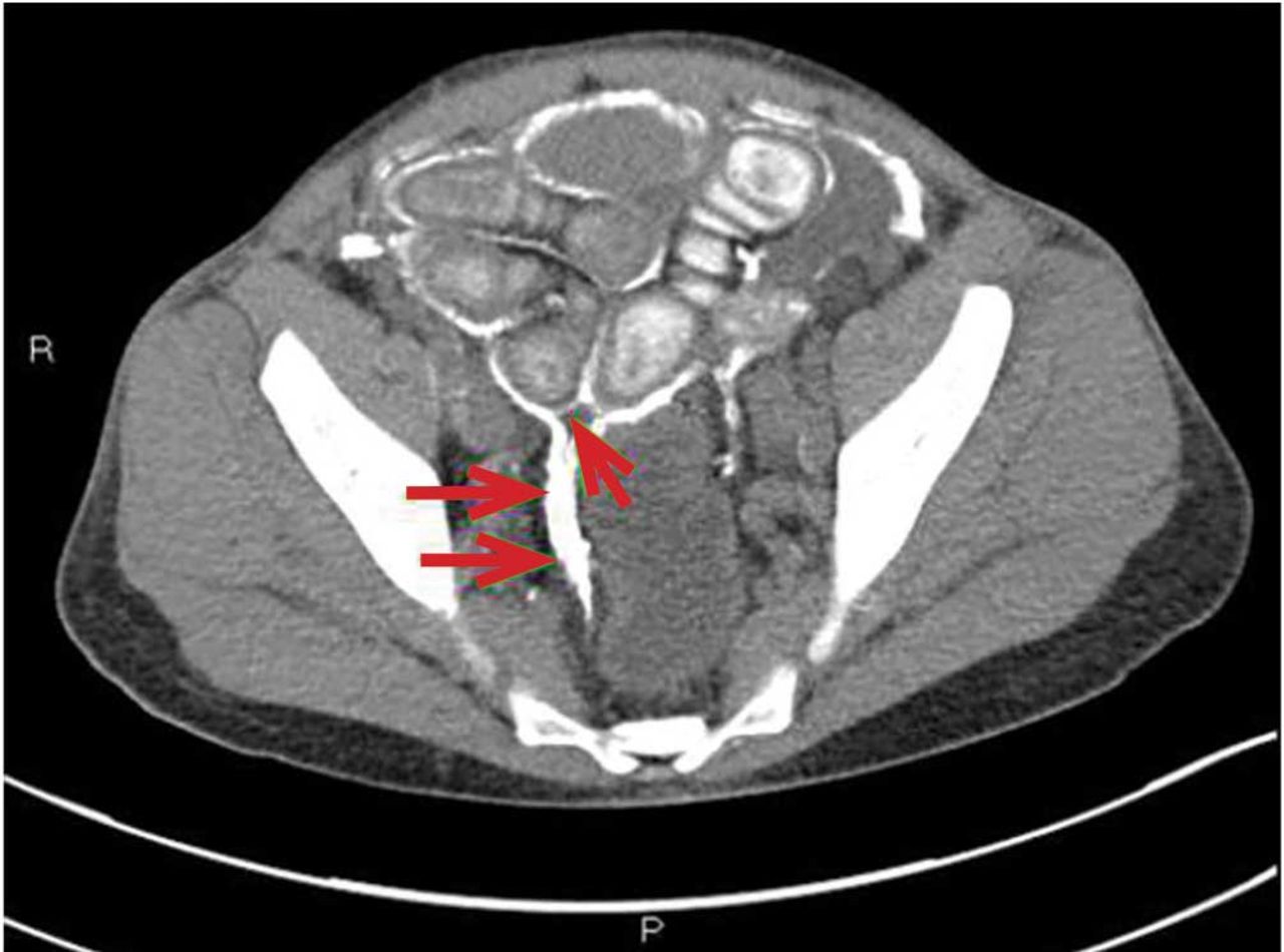
Computed tomography (CT) showed diffuse parietal and visceral peritoneal calcifications encasing the small-bowel loops, consistent with encapsulating peritoneal sclerosis (Figure 1). His peritoneal dialysis catheter was removed, and he was transitioned to hemodialysis. He was discharged on a course of antibiotics and was started on prednisone and tamoxifen.

Figure 1
  • Download figure
  • Open in new tab
  • Download powerpoint
Figure 1

Computed tomography showed diffuse parietal and visceral peritoneal calcifications encasing the small-bowel loops (arrows), consistent with encapsulating peritoneal sclerosis.

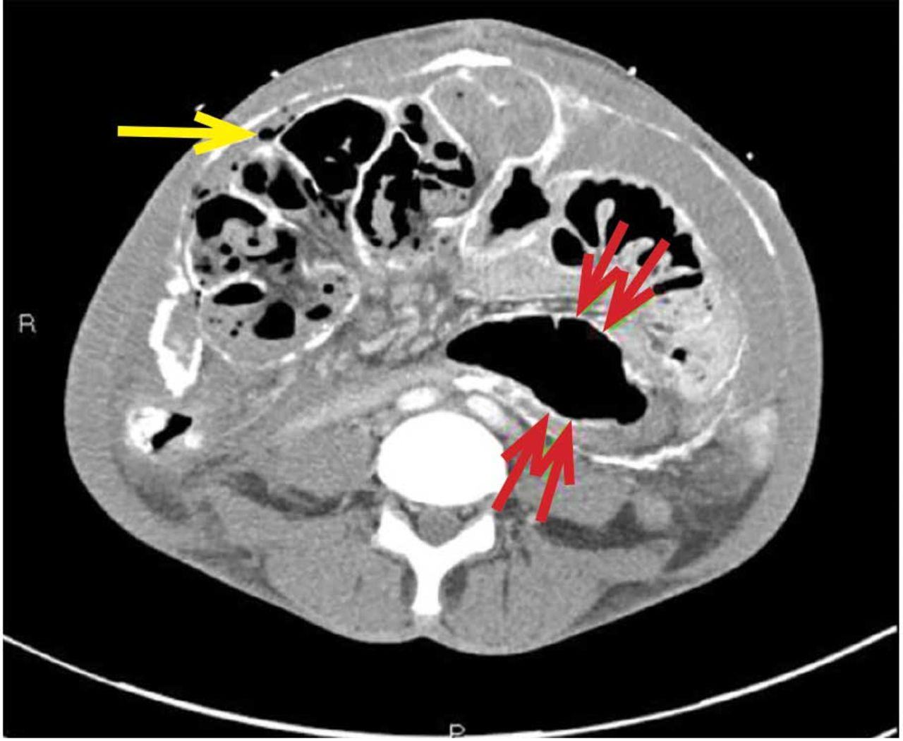
He presented again a few months later with abdominal pain. At that time, CT (Figure 2) demonstrated dilated small-bowel loops indicative of small-bowel obstruction and pneumoperitoneum consistent with bowel perforation. He declined surgical treatment and was referred for hospice care.

Figure 2
  • Download figure
  • Open in new tab
  • Download powerpoint
Figure 2

Several months later, computed tomography showed dilated small-bowel loops indicative of small-bowel obstruction (red arrows) and pneumo-peritoneum (yellow arrow).

CAUSES AND CLINICAL FEATURES

Encapsulating peritoneal sclerosis is a devastating complication of peritoneal dialysis, occurring in 3% of patients on peritoneal dialysis. The mortality rate is above 40%.1,2 It is characterized by an initial inflammatory phase followed by extensive intraperitoneal fibrosis and encasement of bowel. Causes include prolonged exposure to peritoneal dialysis or glucose degradation products, a history of severe peritonitis, use of acetate as a dialysate buffer, and reaction to medications such as beta-blockers.3

Clinical features result from inflammation, ileus, and peritoneal adhesions and include abdominal pain, nausea, and vomiting. A high peritoneal transport rate, which often heralds development of encapsulating peritoneal sclerosis, leads to failure of ultrafiltration and to fluid retention.

CT is recommended for diagnosis and demonstrates peritoneal calcification with bowel thickening and dilation.

TREATMENT

Treatment entails stopping peritoneal dialysis, changing to hemodialysis, bowel rest, and corticosteroids. Successful treatment has been reported with a combination of corticosteroids and azathioprine.4,5 A retrospective study showed that adding the antifibrotic agent tamoxifen was associated with a decrease in the mortality rate.6 Bowel obstruction is a common complication, and surgery may be indicated. Enterolysis is a new surgical technique that has shown improved outcomes.7

Footnotes

  • Dr. Geetha has disclosed consulting for ChemoCentryx and Genentech.

  • Copyright © 2018 The Cleveland Clinic Foundation. All Rights Reserved.

REFERENCES

  1. ↵
    1. Kawaguchi Y,
    2. Saito A,
    3. Kawanishi H,
    4. et al
    . Recommendations on the management of encapsulating peritoneal sclerosis in Japan, 2005: diagnosis, predictive markers, treatment, and preventive measures. Perit Dial Int 2005; 25(suppl 4):S83–S95. pmid:16300277
    OpenUrlAbstract/FREE Full Text
  2. ↵
    1. Lee HY,
    2. Kim BS,
    3. Choi HY,
    4. et al
    . Sclerosing encapsulating peritonitis as a complication of long-term continuous ambulatory peritoneal dialysis in Korea. Nephrology (Carlton) 2003; 8(suppl 2):S33–S39. doi:10.1046/J.1440-1797.8.S.11.X
    OpenUrlCrossRefPubMed
  3. ↵
    1. Kawaguchi Y,
    2. Tranaeus A
    . A historical review of encapsulating peritoneal sclerosis. Perit Dial Int 2005; 25(suppl 4):S7–S13. pmid:16300267
    OpenUrlAbstract/FREE Full Text
  4. ↵
    1. Martins LS,
    2. Rodrigues AS,
    3. Cabrita AN,
    4. Guimaraes S
    . Sclerosing encapsulating peritonitis: a case successfully treated with immunosuppression. Perit Dial Int 1999; 19(5):478–481. pmid:11379862
    OpenUrlFREE Full Text
  5. ↵
    1. Wong CF,
    2. Beshir S,
    3. Khalil A,
    4. Pai P,
    5. Ahmad R
    . Successful treatment of encapsulating peritoneal sclerosis with azathioprine and prednisolone. Perit Dial Int 2005; 25(3):285–287. pmid:15981777
    OpenUrlFREE Full Text
  6. ↵
    1. Korte MR,
    2. Fieren MW,
    3. Sampimon DE,
    4. Lingsma HF,
    5. Weimar W,
    6. Betjes MG,
    7. investigators of the Dutch Multicentre EPS Study
    . Tamoxifen is associated with lower mortality of encapsulating peritoneal sclerosis: results of the Dutch Multicentre EPS Study. Nephrol Dial Transplant 2011; 26(2):691–697. doi:10.1093/ndt/gfq362
    OpenUrlCrossRefPubMed
  7. ↵
    1. Kawanishi H,
    2. Watanabe H,
    3. Moriishi M,
    4. Tsuchiya S
    . Successful surgical management of encapsulating peritoneal sclerosis. Perit Dial Int 2005; 25(suppl 4):S39–S47. pmid:16300271
    OpenUrlAbstract/FREE Full Text
PreviousNext
Back to top

In this issue

Cleveland Clinic Journal of Medicine: 85 (5)
Cleveland Clinic Journal of Medicine
Vol. 85, Issue 5
1 May 2018
  • Table of Contents
  • Table of Contents (PDF)
  • Index by author
Print
Download PDF
Article Alerts
Sign In to Email Alerts with your Email Address
Email Article

Thank you for your interest in spreading the word on Cleveland Clinic Journal of Medicine.

NOTE: We only request your email address so that the person you are recommending the page to knows that you wanted them to see it, and that it is not junk mail. We do not capture any email address.

Enter multiple addresses on separate lines or separate them with commas.
An unusual complication of peritoneal dialysis
(Your Name) has sent you a message from Cleveland Clinic Journal of Medicine
(Your Name) thought you would like to see the Cleveland Clinic Journal of Medicine web site.
CAPTCHA
This question is for testing whether or not you are a human visitor and to prevent automated spam submissions.
Citation Tools
An unusual complication of peritoneal dialysis
Mohamad Hanouneh, Pranav Damera, Mehrdad Fetrat, Duvuru Geetha
Cleveland Clinic Journal of Medicine May 2018, 85 (5) 352-354; DOI: 10.3949/ccjm.85a.17090

Citation Manager Formats

  • BibTeX
  • Bookends
  • EasyBib
  • EndNote (tagged)
  • EndNote 8 (xml)
  • Medlars
  • Mendeley
  • Papers
  • RefWorks Tagged
  • Ref Manager
  • RIS
  • Zotero
Share
An unusual complication of peritoneal dialysis
Mohamad Hanouneh, Pranav Damera, Mehrdad Fetrat, Duvuru Geetha
Cleveland Clinic Journal of Medicine May 2018, 85 (5) 352-354; DOI: 10.3949/ccjm.85a.17090
Twitter logo Facebook logo Mendeley logo
  • Tweet Widget
  • Facebook Like
  • Linkedin Share Button

Jump to section

  • Article
    • CAUSES AND CLINICAL FEATURES
    • TREATMENT
    • Footnotes
    • REFERENCES
  • Figures & Data
  • Info & Metrics
  • PDF

Related Articles

  • No related articles found.
  • PubMed
  • Google Scholar

Cited By...

  • No citing articles found.
  • Google Scholar

More in this TOC Section

  • Tinea incognito
  • Prolonged venous filling time and dependent rubor in a patient with peripheral artery disease
  • Sarcoidosis with diffuse purplish erythematous plaques on the hands
Show more The Clinical Picture

Similar Articles

Subjects

  • Gastroenterology
  • Imaging
  • Nephrology

Navigate

  • Current Issue
  • Past Issues
  • Supplements
  • Article Type
  • Specialty
  • CME/MOC Articles
  • CME/MOC Calendar
  • Media Kit

Authors & Reviewers

  • Manuscript Submission
  • Authors & Reviewers
  • Subscriptions
  • About CCJM
  • Contact Us
  • Cleveland Clinic Center for Continuing Education
  • Consult QD

Share your suggestions!

Copyright © 2025 The Cleveland Clinic Foundation. All rights reserved. The information provided is for educational purposes only. Use of this website is subject to the website terms of use and privacy policy. 

Powered by HighWire